Introduction
Adenosine triphosphate (ATP) is universally seen as an energy exchange factor that connects anabolism and catabolism, while also fueling processes such as motile contraction, phosphorylation, and active transport (Bonora et al., Reference Bonora, Patergnani, Rimessi, de Marchi, Suski, Bononi, Giorgi, Marchi, Missiroli, Poletti, Wieckowski and Pinton2012). In addition, ATP is actively released in the extracellular environment in response to tissue damage and cellular stress (Ribeiro, Reference Ribeiro1978; St John, Reference St John2002; Holzer & Granstein, Reference Holzer and Granstein2004; Brevini et al., Reference Brevini, Cillo, Antonini and Gandolfi2007; Stagg & Smyth, Reference Stagg and Smyth2010).
Once released, ATP is free to bind to receptors on the surface of the plasma membrane on the same cell and on neighboring cells (Dubyak & El-Moatassim, Reference Dubyak and El-Moatassim1993; Schwiebert & Zsembery, Reference Schwiebert and Zsembery2003; Lohman et al., Reference Lohman, Billaud and Isakson2012). As extracellular ATP is degraded rapidly, it is thought of as a local mediator that acts in an autocrine or paracrine manner within tissues and tissue microenvironments to stimulate its receptors before it is chemically modified (Schwiebert & Zsembery, Reference Schwiebert and Zsembery2003). Extracellular ATP binding to the receptors influences many cell events such as secretion, chemoattraction, cell proliferation, differentiation and death during development and so on (Burnstock, Reference Burnstock2009; Burnstock & Ulrich, Reference Burnstock and Ulrich2011).
For gametes, extracellular ATP is required for fertilization in pig sperm (Yi et al., Reference Yi, Park, Kim, Song, Jeong and Sutovsky2009), and enhanced sperm fertilizability with dibutyryl cyclic adenosine monophosphate in the rat (Yamashiro et al., Reference Yamashiro, Toyomizu, Toyama, Aono, Sakurai, Hiradate, Yokoo, Moisyadi and Sato2010). Extracellular ATP induced a depolarizing response in cumulus-cell-enclosed oocytes (Arellano et al., Reference Arellano, Martínez-Torres and Garay2002) and elicited Ca2+ release in cumulus cells that resulted in Ca2+ increase in mouse oocytes (Webb et al., Reference Webb, Bains, Cruttwell and Carroll2002). Ca2+ homeostatic regulation is important for oocyte maturation (Wakai & Fissore, Reference Wakai and Fissore2013), therefore it is suggested that extracellular ATP may influence oocyte maturational events.
The receptors for extracellular ATP can be divided into two groups: firstly, P2X receptors, which are ligand-gated ion channel receptors, and secondly P2Y3 L1 receptors, which are G protein-coupled receptors (Erb et al., Reference Erb, Liao, Seye and Weisman2006; Burnstock, Reference Burnstock2007). P2X receptors are extracellular ATP-gated calcium-permeable non-selective cation channels that are modulated by extracellular Ca2+, Na+, Mg2+, H+ and metal ions such Zn2+ and/or Cu2+. Both receptors are antagonized by pyridoxalphosphate-6-azophenyl-2′,4′-disulfonic acid (PPADS) and reactive blue-2 (RB2), respectively (Ohnishi et al., Reference Ohnishi, Matsumura and Ito2009).
The developmental competence of the oocytes varies according to the follicular size in human and pig (Bagg et al., Reference Bagg, Nottle, Armstrong and Grupen2007; Ito et al., Reference Ito, Iwata, Kitagawa, Kon, Kuwayama and Monji2008; Antosik et al., Reference Antosik, Kempisty, Bukowska, Jackowska, Włdarczyk, Budna, Brūssow, Lianeri, Jagodriński and Jaśkowski2009; Bertoldo et al., Reference Bertoldo, Nadal-Desbarats, Gérard, Dubois, Holyoake and Grupen2013; Mehri et al., Reference Mehri, Setti, Greco, Sakkas, Martinez and Patrizio2014). One of the causes for this variation may be proteins and/or metabolites in the follicular fluid (Ito et al., Reference Ito, Iwata, Kitagawa, Kon, Kuwayama and Monji2008; Bertoldo et al., Reference Bertoldo, Nadal-Desbarats, Gérard, Dubois, Holyoake and Grupen2013). With these, it lets us infer that there may be different ATP concentrations in follicular fluids from various sizes of follicles.
Based on these findings, we undertook measurement of ATP concentrations in the follicular fluids in three sizes of follicle. Then, we researched the relationship in pig between extracellular ATP, the same concentration in porcine follicles, oocyte maturation and P2 receptors during in vitro maturation using two antagonists (PPADS and RB2).
Materials and methods
Chemicals
All chemicals were purchased from Sigma-Aldrich (St Louis, MO, USA) unless otherwise stated.
Oocyte collection
Porcine ovaries, immersed in physiological saline (27–30°C) supplemented with 100 U/ml penicillin G potassium (No. 26239–42, Nacalai Tesque, Kyoto, Japan) and 50 μg/ml streptomycin sulphate (No. 32237–72, Nacalai Tesque), were collected from a local slaughterhouse, and brought to our laboratory within 2 h. The cumulus-cell-enclosed oocytes (COs) were aspirated from superficial follicles (3–6 mm in diameter) using a 18G needle (Terumo Co., Tokyo, Japan) attached to a 5 ml syringe (Terumo Co.), and washed with 2 ml PXM-HEPES buffer (Yoshioka et al., Reference Yoshioka, Suzuki and Onishi2008) on a warm plate (39°C) four times.
Nuclear maturation assay
The COs were washed with porcine oocyte medium (POM) containing 0.3% (w/v) polyvinyl alcohol (Yoshioka et al., Reference Yoshioka, Suzuki and Onishi2008) three times, and incubated (38.5°C, 5% CO2, 95% air, 100% humidity) in POM droplets supplemented with 100 μM PPADS or RB-2 (No. 04352, Wako Pure Chemical Industries, Ltd, Osaka, Japan) (Ohnishi et al., Reference Ohnishi, Matsumura and Ito2009) for 30 min (medium volume 5 μl/CO).
After treatment, the COs were loaded into POM droplets (medium volume 5 μl/CO) with or without 20 nM ATP, and matured for 44 ± 1 h. From onset (0 h) to 21 ± 1 h during maturation, 1 mM dibutyryl adenosine, 10 iu/ml PMSG (ASKA Pharmaceutical. Co., Ltd, Tokyo) and 10 iu/ml hCG (Mochida Pharmaceutical Co., Ltd., Tokyo) were added to the POM. Then, the oocytes were left to mature without these reagents.
After maturation, the COs in each group were denuded of their cumulus cells with 0.1% hyaluronidase and pipetting, and fixed with acetic alcohol for at least 48 h. Then, the oocytes were stained with 1% orcein (No. 7100, Merck, Darmstädt, Germany). To clarify the nucleus stage of the oocytes before maturation, they were also fixed and stained.
To investigate the effect of RB-2 on the cumulus cells and oocytes, the cumulus cells were denuded (DOs) before maturation, and matured in the same medium with or without the 20 nM ATP used for the DOs. Then, the oocytes were fixed, stained and their nuclear stage was observed by the same method.
Measurement of ATP concentration in follicular fluid (pFF)
The pFF were collected from superficial small (less than 3 mm in diameter), medium (3 to 6 mm in diameter) and large follicles (larger than 6 mm in diameter) using an 18G needle attached to a 5 ml syringe. After collection, pFF were stored at –20°C until ATP assay. Thawed pFF were centrifuged for 15 min (10,000 g) at room temperature (RT), and their ATP contents were measured by the method mentioned below.
The 0.5 μM ATP stock solution was thawed and diluted with 10 mM PBS (Conn et al, Reference Conn, Charache and Chappelle1975; Uehara et al., Reference Uehara, Yauchi and Kumasaka2004) to a concentration of 2.5–10 nM. To measure the ATP contents of pFF, a 200 μl sample and the ATP stock solution were introduced into a lumicounter (No. A-237, Advantec Co., Tokyo, Japan), and then pure luciferase (No. 60311, Kikkoman, Noda, Chiba, Japan) was added at room temperature (29–30°C).
Survivability assay for the oocytes
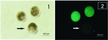
The matured oocytes, treated with RB-2, were stained for 3 min at 38.5°C with 2.5 μg/ml fluorescein diacetate (FDA) dissolved in D-PBS supplemented with 5 mg/ml bovine serum albumin (BSA) (No. 01860–07, Nacalai Tesque). After staining, the oocytes were washed twice with D-PBS BSA, and mounted on a glass slide. In addition, the oocytes before maturation were also stained. The stained oocytes were observed with a fluorescence microscope (E-600, Nikon, Japan) using an ultraviolet (UV) light filter (excitation wavelength: 365 nm, absorption wavelength: 400 nm). The oocytes that glowed green were judged as live, and those that did not glow green were judged as dead (Fig. 1).
FDA-stained immature oocytes with light microscopy (photograph 1) or fluorescence microscopy (photograph 2). Note that the oocytes seen as green were judged as live, and those not seen as green were judged to be dead (arrows).

Parthenogenetic activation of the oocytes
The oocytes were activated by the method of Che et al. (Reference Che, Lalonde and Bordignon2007) with some modifications. In brief, the matured COs treated with or without RB-2 were denuded of their cumulus cells, and immersed into a 15 μM ionophore in TCM-199 for 5 min on a warm plate (39°C). Then, the oocytes were transferred to modified porcine zygote medium-5 (mPZM-5; Yamanaka et al., Reference Yamanaka, Sugimura, Wakai, Kawahara and Sato2009) supplemented with 7.5 μg/ml cytochalasin B and 10 μg/ml cycloheximide and incubated for 6 h. Thereafter, the oocytes were transferred to mPZM-5 without both reagents, and incubated for 7 days.
To observe the pronuclear formation rates in each group, the oocytes were fixed with 4% paraformaldehyde for 30 min, and stained with 10 μg/ml Hoechst 33342 (Calbio Chem., La Jolla, CA, USA) for 20 min (RT) on the 12 ± 1 h after activation. Then, the oocytes were mounted on glass slides, covered with nail polish, and observed using the UV filter of the fluorescence microscope.
Two and 7 days after the onset of culture in mPZM-5, the cleavage rates from the 2- to 16-cell stages and blastocyst formation rates were observed using an inverted microscope.
Furthermore, blastocysts 7 days after activation were stained with 10 μg/ml Hoechst 33342 for 20 min (RT) to observe the cell numbers in the embryos that reached the blastocyst stage.
Statistical analysis
The ATP concentrations, nuclear maturation rates, survivability and embryonic development (pronuclear formation, cleavage and blastocyst formation rates) were analyzed using JMP 5 software. All data except for the cell numbers of the blastocysts were used to calculate the analysis of variance (ANOVA), the Tukey-HSD test was used as a post-ANOVA analysis. For the statistical analysis of the blastocyst cell numbers, the Kruskal–Wallis rank sum test was performed using R ver. 2.15.0 software (R Development Core Team 2012). All data are expressed as the mean ± standard error (SE) of four or five replicates. A P-value less than 0.05 indicated statistical significance.
Results
pFF ATP content from three sizes of follicle
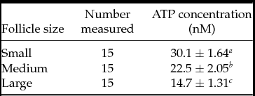
The ATP concentrations of pFF derived from each size of follicle are shown in Table 1. The ATP concentrations gradually decreased following increases in the diameter of the follicle. The ATP contents were significantly different (P < 0.05) among the three follicle sizes.
ATP concentration of the follicular fluid in each size

Values are presented as mean ± standard error (SE). Values in the same column (a, b, c) with different letters differ significantly (P < 0.05).
Nuclear maturation
Before maturation, all 72 oocytes collected from four replicates were at the germinal vesicle stage.
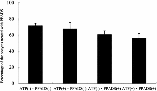
The nuclear maturation rates of the oocytes treated with PPADS are presented in Fig. 2. The nuclear maturation rates, collected from four replicates, up to the metaphase stage of the second meiotic division (MII stage) in all groups were almost the same, and no significant difference was observed regardless of the pre-treatment with PPADS and the addition of 20 nM ATP.
The nuclear maturation rates of the cumulus-cell-enclosed oocytes treated with pyridoxalphosphate-6-azophenyl-2′,4′-disulfonic acid (PPADS). Values are presented as mean ± standard error (SE). (–): non-addition or non-treatment, (+): addition or treatment. The total number of oocytes used for each treatment was: ATP(–)·PPADS(–), n = 117; ATP(+)·PPADS(–), n = 100; ATP(–)·PPADS(+), n = 104; ATP(+)·PPADS(+), n = 102; in four replicates.

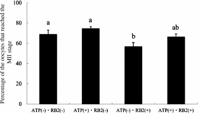
When the COs were treated with RB2, the nuclear maturation rates, collected from four replicates in both groups treated with RB2 were almost the same and no significant difference was observed. However, when these rates were compared with that in the control group [ATP(–) RB2(–)], the rate in the group treated with RB2 in the absence of ATP [ATP(–) RB2(+)] was significantly lower (P < 0.05) than that of the control group (Fig. 3).
The nuclear maturation of the cumulus-cell-enclosed oocytes treated with reactive blue 2 (RB2). Values are presented as mean ± standard error (SE). Values (a, b) with different letters differ significantly (P < 0.05). (–): non-addition or non-treatment, (+): addition or treatment. The total number of oocytes used for each treatment was: ATP(–)·RB2(–), n = 119; ATP(+)·RB2(–), n = 117; ATP(–)·RB2(+), n = 117; ATP(+)·RB2(+), n = 127; in four replicates.

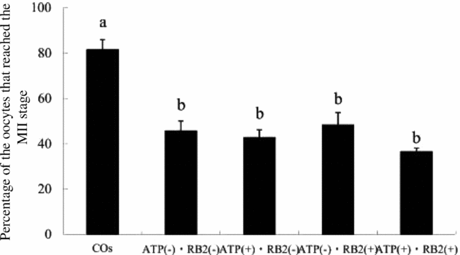
The effect of RB2 treatment on the nuclear maturation rates of the oocytes without cumulus cells, collected from five replicates is presented in Fig. 4. All DOs groups were significantly lower (P < 0.05) than the COs group without pre-treatment of RB2 in the absence of ATP. However, no significant differences were observed among the DOs groups regardless of RB2 treatment and the addition of ATP.
The nuclear maturation rates of the cumulus-cell-denuded oocytes treated with reactive blue 2 (RB2). Values are presented as mean ± standard error (SE). Values (a, b) with different letters differ significantly (P < 0.05). (–): non-addition or non-treatment, (+): addition or treatment. The total number of oocytes used for each treatment was: COs, n = 138; ATP(–)·RB2(–), n = 127; ATP(+)·RB2(–), n = 123; ATP(–)·RB2(+), n = 140; ATP(+)·RB2(+), n = 137; in five replicates.

The survivability of the oocytes
In total, 73.9% of 69 oocytes collected from four replicates before maturation survived as determined by FDA staining.
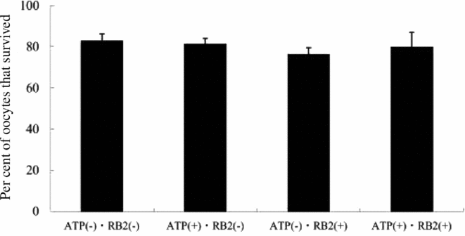
The survival rates in the oocytes with or without RB2 in the presence or absence of ATP collected from four replicates are shown in Fig. 5.
The survival rate of the oocytes treated with reactive blue 2 (RB2). Values are presented as mean ± standard error (SE). (–): non-addition or non-treatment, (+): addition or treatment. The total number of oocytes used for each treatment was: ATP(–)·RB2(–), n = 70; ATP(+)·RB2(–), n = 67; ATP(–)·RB2(+), n = 68; ATP(+)·RB2(+), n = 67; in four replicates.

The survival rates of the oocytes in each group were almost the same, and no significant differences were observed.
Development of the oocytes after activation
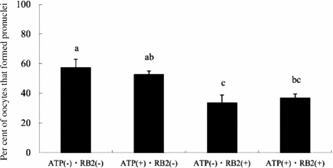
The pronuclear formation rates of the oocytes treated with RB2 collected from four replicates are presented in Fig. 6. When the rates of the groups matured with the ATP were compared with those of the groups matured without ATP, no significant differences were observed. However, when the rates of the RB2-treated or non-treated groups were compared, the rates of the both groups treated with RB2 [ATP(–)·RB2(+) and ATP(+)·RB2(+)] were significantly lower (P < 0.05) than that of the control [ATP(–)·RB2(–)] group.
Pronuclear formation rates in each group treated with reactive blue 2 (RB2). Values are presented as mean ± standard error (SE). Values (a, b) with different letters differ significantly (P < 0.05). (–): non-addition or non-treatment, (+): addition or treatment. The total number of oocytes used for each treatment was: ATP(–)·RB2(–), n = 76; ATP(+)·RB2(–), n = 69; ATP(–)·RB2(+), n = 74; ATP(+)·RB2(+), n = 76; in four replicates.

The cleavage rates from 2- to 16-cell stage cells at 2 days and the blastocyst formation rates on the 7th day after activation collected from four replicates are presented in Table 2.
Development rate of the oocytes up to the blastocyst stage

Values are presented by mean ± standard error (SE). Values collected from four replicates in the same column (a, b) with different letters differ significantly (P < 0.05). (–): non-addition or non-treatment, (+): addition or treatment.
As with the results of the pronuclear formation assays, both rates in the groups treated with RB2 [ATP(–) RB2(+) and ATP(+) RB2(+)]were significantly decreased (P < 0.05) compared with those of the control group.
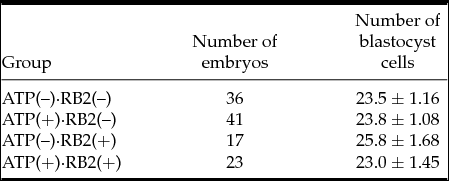
The cell numbers of the blastocysts (mean ± SE) among all groups were almost the same in each stage (Table 3), and no significant differences were found among the control group [ATP(–) RB2(–) group] and ATP-added or RB2-treated groups (Kruskal–Wallis rank sum test, K = 2.12, df = 3, P = 0.55).
Number of blastocyst cells in each group

Values are presented as mean ± standard error (SE). Values collected from four replicates in the same column (a, b) with different letters differ significantly (P < 0.05). (–): non-addition or non-treatment, (+): addition or treatment.
Discussion
In the present study, the ATP concentration of pFF was 11–31 nM, and along with follicle diameter increases, the ATP concentrations were significantly reduced. The plasma ATP concentration was reported to be 28 nM in human (Gorman et al., Reference Gorman, Feigl and Buffington2007) and 25–50 μM in dog (Farias et al., Reference Farias, Gorman, Savage and Feigl2005). Intracellular ATP concentration is in the range of 3–10 mM, whereas extracellular ATP level under normal physiological conditions is <100 nM (Trautmann, Reference Trautmann2009). Therefore, the ATP concentration in pFF obtained in this study was considered to be normal.
In addition, Fricke et al. (Reference Fricke, Ford, Reynolds and Redmer1996) indicated that the percentages of proliferating granulosa and thecal cells decreased throughout the final stages of antral follicular development in the pig. Extracellular ATP is involved in cell proliferation (Erlinge & Burnstock, Reference Erlinge and Burnstock2008; Burnstock & Ulrich, Reference Burnstock and Ulrich2011). Therefore, the decrease in the proliferation of granulosa cells may have induced the lower ATP concentration of the larger follicles compared with that in the smaller cells in this study.
RB2 treatment had a negative effect on the maturation rate up to the MII stage of the COs. In addition, RB2 treatment had no influence on the nuclear maturation of the oocytes without cumulus cells (DOs). Kamada et al. (Reference Kamada, Kubota, Hirata, Taguchi, Eguchi, Marumo and Aso1992, Reference Kamada, Blackmore, Oehninger, Gordon and Hodgen1994) reported that P2 receptors were present in human and porcine granulosa cells aspirated from follicles. Arellano et al. (Reference Arellano, Martínez-Torres and Garay2002) also reported that P2Y2, a member of the subfamily of the P2Y receptors, seemed to be present on the granulosa and cumulus cells in mice. Therefore, it is suggested that P2Y receptors may be present in cumulus cells and may regulate the nuclear maturation of porcine oocytes.
Furthermore, in the present study, we failed to find any effect on the nuclear maturation of oocytes treated with PPADS. It is indicated that the pharmacology of the recombinant P2X receptor subtypes expressed in oocytes or other cell types is often different from the pharmacology of P2X-mediated responses in naturally occurring sites (Gever et al., Reference Gever, Cockayne, Dillon, Burnstock and Ford2006; Burnstock, Reference Burnstock2007). Further study will be needed to clarify the presence or absence of P2X receptors on the surface of porcine oocytes using other antagonists such as TNP-ATP or Brilliant Blue Green and so on (Burnstock, Reference Burnstock2007; Erlinge & Burnstock, Reference Erlinge and Burnstock2008).
The nuclear maturation rate in the ATP(+) RB2(+) group, which was slightly higher than that in the ATP(–) RB2(+) group, was not significantly different compared with that in the ATP(+) RB2(–) and the control [ATP(–) RB2(–)] groups. In addition, no significant difference was observed between the pronuclear and blastocyst formation rates of the ATP(+) RB2(+) and ATP(+) RB2(–) groups, although significant differences were found between the ATP(–) RB2(+) and control [ATP(–) RB2(–)] groups. From these results, it is postulated that the addition of 20 nM ATP to the maturation medium seems to counter the negative effect of RB2 on the nuclear maturation of oocytes.
When ATP is released to the extracellular environment, it is diffused for about 300 μm2/s for up to 30 s to 3 min, then the ATP concentration drops from 1–10 mM intracellularly back down to 1 nM to 1 μM extracellularly (Amadio et al., Reference Amadio, Apolloni, D’Ambrosi and Volonté2011). It is well known that a higher concentration of ATP (at more than the micromolar level) has some influence on cell proliferation and differentiation, production of nitric oxide and relaxation, cell signalling, and so on (Dubyak & el-Moatassim, Reference Dubyak and El-Moatassim1993; Burnstock, Reference Burnstock2009; Burnstock & Ulrich, Reference Burnstock and Ulrich2011). However, the role(s) of extracellular ATP at lower concentrations (nM level) is still unclear. In the present study, follicular fluid contained nM level ATP (from 14.7 to 30.1 nM). These results let us infer that further study will be needed to clarify the role of ATP in follicular fluid.
In our preliminary study, we evaluated the nuclear maturation rates in the three groups: the COs pre-treated with 100 μM RB2 for 30 min, and matured without RB2; the COs non-pre-treated and matured with 100 μM RB2; and the control (non-pre-treated and non-matured with RB2) were compared. We found that the rate in the COs matured with, but not pre-treated with, RB2 was significantly lower (P < 0.05) than that of the COs pre-treated with RB2 and the control (43.0 ± 5.44, 59.3 ± 1.46 and 72.7 ± 6.04, mean ± SE collected from three replicates, these were matured without addition of ATP). In addition, both the maturation and pronuclear rates in the ATP(+) RB2(+) group were slightly increased compared with that of the ATP(–) RB2(+) group in this study. Based on these results, it is suggested that concentration of RB2 in the present study may not be enough to antagonize the receptors completely.
In the present study, the survivability of the oocytes treated with RB2 was almost the same as those of the oocytes without RB2 treatment, regardless of ATP addition to the maturation medium. This result indicates that both RB2 treatment and ATP addition to the maturation medium do not influence the membrane integrity of oocytes.
In this study, the nuclear maturation rates of the oocytes treated with RB2 were decreased compared with those of the non-RB2 treated oocytes. RB2 is a non-selective antagonist for the P2Y2, P2Y4, P2Y11 and P2X receptors, a subfamily of the P2 receptors, and the ligands for these receptors are ATP, ADP or UTP (Erlinge & Burnstock, Reference Erlinge and Burnstock2008; Tsuda et al., Reference Tsuda, Tozaki-Saitoh and Inoue2010). Webb et al. (Reference Webb, Bains, Cruttwell and Carroll2002) reported that both ATP and UTP could induce Ca2+ transients of mouse oocytes. Ca2+ transition is required not only for fertilization, but also for oocyte maturation, in vertebrates (Machaca, Reference Machaca2007). It is estimated that 0.44 mM uracil, which is a metabolite of UTP, is contained in pFF (Downs et al., Reference Downs, Coleman, Ward-Bailey and Eppig1985; Lecca & Ceruti, Reference Lecca and Ceruti2008). Therefore, RB2 treatment may block UTP action on the oocytes and inhibit the Ca2+ transition of pig oocytes, resulting in a decrease in their nuclear maturation up to the MII stage in this study.
The pronuclear formation, cleavage rates and blastocyst formation rates of the oocytes treated with RB2 after activation were significantly decreased compared with the control group. In parallel, the nuclear maturation rates of the group treated with RB2 were lower than those of cells not treated with RB2. Therefore, the lower development rates after activation may be induced by lower nuclear maturation following treatment with RB2 in this study.
However, the cell numbers in the blastocysts derived from the group treated with RB2 were the same as those of the non-treated groups with RB2. The cell number of blastocysts is one parameter of an oocyte's quality (Watson, Reference Watson2007; Catala et al., Reference Catala, Izquierdo and Rodriguez-Prado2012). The blastocyst cell number is an important indicator of embryo quality (Catala et al., Reference Catala, Izquierdo and Rodriguez-Prado2012). Therefore, it is postulated that RB2 treatment may not affect embryo quality.
In conclusion, P2Y receptors may play an important role in oocyte maturation via cumulus cells in the pig.
Acknowledgements
The authors would like to thank the staff of Miyakonojo Wellness Meat Co., Ltd., Miyakonojo City, Japan, for providing the porcine ovaries. This research was supported by University of Miyazaki, Miyazaki, Japan.