INTRODUCTION
The phylum Apicomplexa comprises a heterogeneous group of intracellular parasites, including species infectious for vertebrate and invertebrate organisms (Tenter et al. Reference Tenter, Barta, Beveridge, Duszynski, Mehlhorn, Morrison, Thompson and Conrad2002). Some members of Apicomplexa are relevant for humans due to their economic or public health impact, such as Plasmodium spp., the causative agent of malaria; Toxoplasma gondii, which causes toxoplasmosis; Cystoisospora belli, causing isosporiasis; and Cryptosporidium, responsible for cryptosporidiosis. Also, livestock business, mainly poultry and cattle, have to cope with severe losses by disease impact and prevention costs for Eimeria infections.
Like all eukaryotic cells, these protozoa are provided with a nucleus, endoplasmic reticulum, Golgi complex and mitochondrion (Joiner and Roos, Reference Joiner and Roos2002). Interestingly, most apicomplexans possess a single, ramified mitochondrion. They also possess some peculiar and exclusive structures, like the apical complex, formed by cytoskeletal components (conoid and apical rings) and secretory organelles (micronemes and rhoptries) that are essential for invasion of the host cell (Morrissette and Sibley, Reference Morrissette and Sibley2002; Dubremetz, Reference Dubremetz2007; Carruthers and Tomley, Reference Carruthers and Tomley2008). Other characteristic secretory organelles of Apicomplexa are the dense granules, which modify the parasitophorous vacuole surrounding the intracellular stages of these parasites (Cesbron-Delauw et al. Reference Cesbron-Delauw, Gendrin, Travier, Ruffiot and Mercier2008). In addition to the above features, apicomplexans also contain a unique organelle, the apicoplast. Though the presence of this organelle has been well documented for decades by electron microscopy studies, only in the late 1990s the same structure was identified as an essential organelle derived from a secondary endosymbiotic event (Köhler et al. Reference Köhler, Delwiche, Denny, Tilney, Webster, Wilson, Palmer and Roos1997). The apicoplast is a non-photosynthetic plastid essential for the Apicomplexa Type II fatty acid synthesis, isoprenoid biosynthesis and carbohydrate metabolism (Gleeson, Reference Gleeson2000; Mazumbar et al. Reference Mazumbar, Wilson, Masek, Hunter and Striepen2006; Fleige et al. Reference Fleige, Fischer, Ferguson, Gross and Bohne2007; Yeh and DeRisi, Reference Yeh and DeRisi2011). Interestingly, some Apicomplexa like Theileria and Cryptosporidium display further peculiarities: Theileria seems to lack fatty-acid biosynthesis enzymes, while Cryptosporidium appears to have lost the apicoplast during its evolution (Goodman and McFadden, Reference Goodman and McFadden2007).
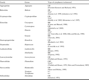
Some members of the Apicomplexa feature singular cytoplasmic inclusions sharing the same morphology but named with different terms, (Table 1) like the refractile body (RB) in Eimeria (de Venevelles et al. Reference de Venevelles, Chich, Faigle, Lombard, Loew, Péry and Labbé2006) and Lankesterella (Desser, Reference Desser and Kreler1993); and the crystalloid body (CB) in Cystoisospora and Plasmodium (Garnham et al. Reference Garnham, Bird, Baker, Desser and El-Nahal1969; Meis and Ponnudurai, Reference Meis and Ponnudurai1987; Gleeson, Reference Gleeson2000; Keithly et al. Reference Keithly, Langreth, Buttle and Mannella2005). In addition to RB and CB, virus-like particles (VLPs) have also been described in Apicomplexa (del Cacho et al. Reference del Cacho, Gallego, Montes, López-Bernad, Quílez and Sánchez-Acedo2001; Khramtsov et al. Reference Khramtsov, Woods, Nesterenko, Dykstra and Upton1997). Some eugregarines like Epicavus araeoceri present paracrystalline inclusions that can reach up to 4 μm in length within their endoplasmic reticulum (Schrével and Philippe, Reference Schrével, Philippe and Kreler1993). Babesia merozoites possess on their cytoplasm osmiophilic membrane bounded electron-dense organelles at their apical region, named spherical bodies (Kawai et al. Reference Kawai, Igarashi, Abgaandorjiin, Miyazawa, Ikadai, Nagasawa, Fujisaki, Mikami, Suzuki and Matsuda1999). Proteins secreted into the host cell cytoplasm by these organelles might have a role in the Babesia intracellular development and survival (Jasmer et al. Reference Jasmer, Reduker, Perryman and McGuire1992; Dowling et al. Reference Dowling, Perryman and Jasmer1996; Ruef et al. Reference Ruef, Dowling, Conley, Perryman, Brown, Jasmer and Rice-Ficht2000; Terkawi et al. Reference Terkawi, Seuseu, Eko-Wibowo, Huyen, Minoda, AbouLaila, Kawai, Yokoyama, Xuan and Igarashi2011). Curiously, some members of the Sarcocystidae family like Sarcocystis and Hammondia present crystalline inclusions in their cytoplasm like Sarcocystis and Hammondia (Speer and Dubey, Reference Speer and Dubey1989; Vlemmas et al. Reference Vlemmas, Kakanoudis, Tsangaris, Theodorides and Kaldrymidou1989), whereas no such structure has been observed in other members such as Toxoplasma and Neospora (Speer et al. Reference Speer, Dubey, McAllister and Blixt1999) (Table 1).
The RB, CB and VLP have been poorly characterized so far and still require further studies at both ultrastructural and functional levels, which will contribute significantly towards our understanding of the roles played by these structures in the biology of apicomplexa. Here, we present a short overview of the current knowledge about RB, CB, and VLPs.
THE REFRACTILE BODY
The refractile body (RB) has been described in members of the Eimeriidae family, like Eimeria, Goussia and Caryospora genera, as well as in members of the Lankesterellidae family, like Lankesterella and Schellackia genera (Fig. 1 and Table 1) (Upton and Barnard, Reference Upton and Barnard1988; Sundermann and Lindsay, Reference Sundermann and Lindsay1989; Paperna, Reference Paperna1992; Desser, Reference Desser and Kreler1993; Azevedo, Reference Azevedo2001). Generally, these protozoans feature 2 copies of this organelle located at different positions on the cytoplasm: a larger one that occupies most of the posterior part of the cell body; and a smaller one located at the apical part, between the nucleus and the apical complex (Danforth and Augustine, Reference Danforth and Augustine1989; Sundermann and Lindsay, Reference Sundermann and Lindsay1989; Jean et al. Reference Jean, Grosclaude, Labbé, Tomley and Péry2000). Interestingly, Goussia clupearum presents 4 RBs in its cytoplasm (Azevedo, Reference Azevedo2001).
Thin sections of refractile bodies of different apicomplexan sporozoites. (A) Eimeria acervulina (from Miranda et al. Reference Miranda, de Souza, Plattner, Hentschel, Kawazoe, Fang and Moreno2008; with permission). The refractile body (RB) occupies a large volume of the cytoplasm. Arrows point to acidocalcisomes. N, nucleus. Scale bar=500 nm. (B) Caryospora bigenetica (from Sundermann and Lindsay, Reference Sundermann and Lindsay1989; with permission). There are 2 copies of the refractile body: one close to the apical part (AR); and one at the posterior cell body (PR). N, nucleus; A, amylopectin granules; OC, caryocyst; PV, parasitophorous vacuole. Scale bar=1 μm. (C) Schellakia cf. agamae sporozoites (S) (from Paperna, Reference Paperna1992; with permission). The RB (R) occupies a large portion of the posterior part of the parasite cell body. W, mother cyst wall; M, mitochondrion; OR, oocyst residuum; H, intracellular matrix; db, dense bodies. Scale bar=2 μm. (D) Lankesterella minima intraerythrocytic sporozoite (from Desser, Reference Desser and Kreler1993; with permission). The refractile body (Rb) locates at the posterior part of the cell body. Pv, parasitophorous vacuole; Mn, micronemes. X 2100.

So far there has been no evidence for a membrane surrounding this organelle, although it can be positively labelled with the lipid fluorescent dye Nile Red, suggesting a lipid content within it (Greenspan et al. Reference Greenspan, Mayer and Fowler1985; de Venevelles et al. Reference de Venevelles, Chich, Faigle, Lombard, Loew, Péry and Labbé2006). The RB can also be stained green with Acridine Orange, indicating an acidic nature for this cytoplasmic inclusion (Miranda et al. Reference Miranda, de Souza, Plattner, Hentschel, Kawazoe, Fang and Moreno2008). Observing ultrathin sections of sporozoite forms of these protozoa, one can perceive that the RBs in Eimeria appear less electron dense than the ones from Caryospora or Schellackia (Fig. 1). Whether this is a matter of sample preparation or is linked to their physiological properties deserves further investigation. The RB is generally present in the sporozoite stage, fragmented during merogony and incorporated into the forming second-generation merozoites (Lal et al. Reference Lal, Bromley, Oakes, Prieto, Sanderson, Kurian, Hunt, Yates, Wastling, Sinden and Tomley2009).
Examples of apicomplexan presenting cytoplasmic inclusions

† CB, crystalloid body; RB, refractile body; SB, spherical body; VLP, virus-like particle.
n.c., not yet characterized.
* Still controversial whether Plasmodium presents CB and/or VLP.
The RB from Eimeria are the most studied, with several characterized proteins located at this structure characterized, including different enzymes, e.g. hydrolase, lactate dehydrogenase and an aspartyl protease (named eimepsin) (Vermeulen et al. Reference Vermeulen, Kok, van den Boogaart, Dijkema and Claessens1993; Jean et al. Reference Jean, Grosclaude, Labbé, Tomley and Péry2000; de Venevelles et al. Reference de Venevelles, Chich, Faigle, Loew, Labbé, Girard-Misguich and Péry2004, Reference de Venevelles, Chich, Faigle, Lombard, Loew, Péry and Labbé2006). Also, a protein called SO7′, the most abundant protein identified in Eimeria sporozoites, is located at the RB (de Venevelles et al. Reference de Venevelles, Chich, Faigle, Loew, Labbé, Girard-Misguich and Péry2004). In fact, some attempts have been made to use SO7′ to immunize chickens to prevent Eimeria infection (Kopko et al. Reference Kopko, Martin and Barta2000). Molecular analysis on different life-cycle stages demonstrated that transcripts of some RB proteins were not detected in merozoites (Abrahamsen et al. Reference Abrahamsen, Johnson, Jutila, Speer and White1994), while proteomic analysis showed that eimepsin and SO7′ are expressed only in oocyst and sporozoite stages and not detected in second-generation merozoites (Lal et al. Reference Lal, Bromley, Oakes, Prieto, Sanderson, Kurian, Hunt, Yates, Wastling, Sinden and Tomley2009). This is particularly interesting because RB is not present in the oocyst, which could indicate that these proteins could be localizing differently and might play an alternative role at this stage of the life cycle.
Some groups have proposed a role for eimepsin and SO7′ in the invasion process and intracellular development (Danforth and Augustine, Reference Danforth and Augustine1989; Augustine, Reference Augustine1999, Reference Augustine2001; Jean et al. Reference Jean, Grosclaude, Labbé, Tomley and Péry2000), suggesting a pathway for secretion of these proteins from the RB. Still, it remains to be determined whether Eimeria proteases and proteins are trafficked to the secretory organelles upon invasion or if, instead, the RB itself secretes its contents during this process. Indeed, a protease from Plasmodium – subtilisin 2 – is located within the micronemes and is essential for invasion of red blood cells by the merozoites forms (Uzureau et al. Reference Uzureau, Barale, Janse, Waters and Breton2004; Harris et al. Reference Harris, Yeoh, Dluzewski, O'Donnell, Withers-Martinez, Hackett, Bannister, Mitchell and Blackman2005), indicating that proteases play a role in invasion. Perhaps the smaller RB could supply essential mediators produced and/or exported at the apical complex that are required for the entrance into the host cell. Another functional interpretation is that re-localization from the RB to another organelle, e.g. micronemes, could be essential for the activation of these enzymes, by, for example, their exposure to variations in pH consequent to their relocation.
The Eimeria RB protein eimepsin is similar to the plasmepsins, proteases from Plasmodium that degrade heme in the food vacuole (Liu et al. Reference Liu, Gluzman, Drew and Goldberg2005). The presence of other enzymes, such as lactate dehydrogenase, indicates a role of the RB in energy metabolism in this parasite. In fact, the RB is in close contact with amylopectin granules (Scholtyseck and Ghaffar, Reference Scholtyseck and Ghaffar1981), suggesting a potential role for RB in the degradation of amylopectin inclusions during intracellular development (Michalski et al. Reference Michalski, Edgar and Prowse1992).
THE CRYSTALLOID BODY
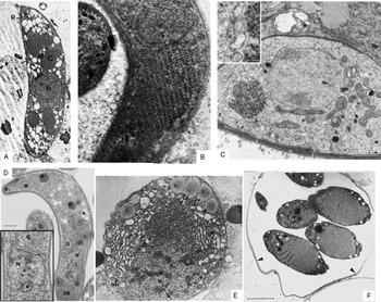
Members of the Aggregatidae family (Aggregata eberthi), Cryptosporidiidae family (Cryptosporidium), Haemogregarinidae (Hemolivia stellata), Hepatozidae family (Hepatozoon mocassini), Sarcocystidae family (Hammondia heydorni), archigregarines like Selenidium hollandei, the coccidian Coleotropha durchoni as well as the Eimeriidae member Cystoisospora, feature another unusual cytoplasmic inclusion called the crystalloid body (CB) (Table 1) (Porcht-Henneré, Reference Porchet-Henneré1971; Porchet-Henneré and Richard, Reference Porchet-Henneré and Richard1971; Schrével, Reference Schrével1971; Trefiak and Desser, Reference Trefiak and Desser1973; Mehlhorn and Markus, Reference Mehlhorn and Markus1976; Wong and Desser, Reference Wong and Desser1976; Beyer et al. Reference Beyer, Scholtyseck and Entzeroth1983; Speer and Dubey, Reference Speer and Dubey1989; Lowichik et al. Reference Lowichik, Lanners, Lowrie and Meiners1993; Smallridge and Paperna, Reference Smallridge and Paperna2000; Boulard et al. Reference Boulard, Paperna, Petit and Landau2001; Keithly et al. Reference Keithly, Langreth, Buttle and Mannella2005; Mitchell et al. Reference Mitchell, Zajac and Lindsay2009) (Fig. 2). This inclusion occupies a considerable volume of the cell cytoplasm reaching some micrometres in diameter. The CB may disappear during the differentiation process from one life stage to another, like in Cystoisospora, Hammondia and Hemolivia (Mehlhorn and Markus, Reference Mehlhorn and Markus1976; Smallridge and Paperna, Reference Smallridge and Paperna2000). Cryptosporidium is a unique member of the Apicomplexa phylum, with some distinctive characteristics, such as having lost the apicoplast during evolution. Because of that, one could also classify the Cryptosporidium CB separately from the other apicomplexan CBs. However, due to the current lack of data, there is no basis to argue about the nature and origin of CBs and potential differences from one another, and therefore we continue to consider Cryptosporidium CB as one example of apicomplexa CB.
Thin sections of crystalloid bodies of different apicomplexan protozoa. (A) The crystalloid (C) in a Hemolivia stellata sporokinete (from Boulard et al. Reference Boulard, Paperna, Petit and Landau2001; with permission). N, nucleus; A, amylopectin granules; n, nucleolus; R, rhoptries. X 12 000. (B) Leucocytozoon sporozoite form (from Trefiak and Desser, Reference Trefiak and Desser1973; with permission). X 42 500. (C) The crystalloid (CR) in a Plasmodium falciparum ookinete (from Meis and Ponnudurai, Reference Meis and Ponnudurai1987; with permission). M, mitochondrion; large arrow – pellicle; thin arrows – microtubules; double arrows – CR, individual particles spread in the cytoplasm. Scale bar=1 μm. Inset – Higher magnification of the CR. Arrow points to the particles that form the CR. Scale bar=100 nm. (D) Cryptosporidium parvum sporozoite (from Keithly et al. Reference Keithly, Langreth, Buttle and Mannella2005; with permission) with the crystalloid body (CB), the nucleus (N), Golgi complex (G), dense granules (D), micronemes (M), amylopectin granule (A) and rhoptry (R). The relict mitochondrion (asterisk) localizes near the nucleus. Scale bar=500 nm. (E) The crystalloid (Cst) in Aggregata eberthi sporoblast stage (from Porchet-Henneré and Richard, Reference Porchet-Henneré and Richard1971; with permission). Pg, paraglycogen granules; mit, mitochondrion; pn, nuclear pore; N, nucleus; E, endoplasmic reticulum; L, lipid inclusions. X 10 000. (F) Crystalloids (C) in Hepatozoon mocassini oocyst (from Lowichik et al. Reference Lowichik, Lanners, Lowrie and Meiners1993; with permission). Arrows – sporocyst wall. Scale bar=2 μm.

The CB is formed by electron-dense particles arranged in a tight honeycomb pattern. These particles display variable sizes depending on the genera in which they have been observed and characterized at the ultrastructural level. In the haemogregarine Karyolysus, these particles measure 37–50 nm and in Cystoisospora they measure 30 nm in diameter. Electron microscopy analyses of thin sections suggest that in both Cystoisospora and haemogregarines CB particles are not surrounded by a cell membrane (Beyer et al. Reference Beyer, Scholtyseck and Entzeroth1983; Mehlhorn and Markus, Reference Mehlhorn and Markus1976; Lindsay et al. Reference Lindsay, Dubey and Blagburn1997). However, Vlemmas et al. (Reference Vlemmas, Kakanoudis, Tsangaris, Theodorides and Kaldrymidou1989) through observation of sheep tissue samples processed by routine transmission electron microscopy showed a membrane surrounding the crystalloid in the cytoplasm of Sarcocystis tenella (Sarcocystis ovicanis).
In Aggregata, the particles that constitute the CB are scattered throughout the oocyst cytoplasm. During the budding of sporozoites, they aggregate, becoming crystalline and forming the CB in each sporozoite (Porchet-Henneré and Richard, Reference Porchet-Henneré and Richard1971). In the Leucocitozoon oocyst stage, the CB particles concentrate forming a massive core in the middle of the oocyst cytoplasm (Wong and Desser, Reference Wong and Desser1976). Part of this mass is carried to each budding sporozoite. While the oocyst sporozoite present a single CB at the posterior portion of the cell body, in the salivary gland sporozoite there are 2 CBs: one at the anterior portion of the cell body (close to the apical organelles); and one at the posterior portion.
In Cryptosporidium there is some divergence in the particle size. Keithly et al. (Reference Keithly, Langreth, Buttle and Mannella2005) determined sizes ranging from 16 to 25 nm. Tetley and co-workers (Reference Tetley, Brown, McDonald and Coombs1998) reported instead a diameter of 35 nm, while Petry and Harris (Reference Petry and Harris1999) described a size variation for these particles between 45 and 60 nm. Keithly et al. (Reference Keithly, Langreth, Buttle and Mannella2005) studied the ultrastructure of the CB by electron tomography – a high-resolution electron microscopy technique, and did not observe a membrane surrounding these particles. Analysing the CB, they determined that contrary to Cystoisospora or haemogregarines, the particles per se would be formed by densely stacked membrane vesicles in apparent continuity with each other. By cryopreparations followed by 3D reconstruction methods, Tetley et al. (Reference Tetley, Brown, McDonald and Coombs1998) were also unable to detect the presence of a cell membrane around the CB and, in addition, observed 1–2 CBs in the cytoplasm of a single parasite.
The functional role of CB in the physiology of these protozoa is still unknown. Ctrnacta et al. (Reference Ctrnacta, Ault, Stejskal and Keithly2006) localized a pyruvate:NADP+ oxidoreductase in both the cytoplasm and at the CB of Cryptosporidium. Although the CB is closely apposed to the relict mitochondrion of this parasite (Keithly et al. Reference Keithly, Langreth, Buttle and Mannella2005), no labelling for this enzyme was observed in the relict mitochondrion. This could indicate a potential role of the CB in Cryptosporidium metabolism. Another remarkable observation is the labelling of CB with Mitotracker Green FM, suggesting the presence of a membrane potential within this organelle (Ctrnacta et al. Reference Ctrnacta, Ault, Stejskal and Keithly2006). These data, as well as the juxtaposition of the CB with amylopectin inclusions, could suggest a role for them in the energy metabolism. It is tempting to infer that CB and RB could simply be two ways of naming the same organelle with the same functions, but additional evidence is necessary to corroborate or reject this hypothesis.
Plasmodium also features a CB in both the ookinete and the oocyst stages (Garnham et al. Reference Garnham, Bird and Baker1962, Reference Garnham, Bird, Baker, Desser and El-Nahal1969; Sinden and Strong, Reference Sinden and Strong1978; Meis and Ponnudurai, Reference Meis and Ponnudurai1987) (Fig. 4), though with a smaller size than other CBs detected in other Apicomplexa. The particles within the CB have a diameter of 42 nm and these, analogous to what has been observed in the previously described CBs from other parasites, do not posses a surrounding membrane. However, their structure is very similar to that of VLPs (discussed below), and this in turn could lead to novel implications on the role of CB in Plasmodium metabolism. More recently members of the LCCL family were shown to be directly connected to the formation of Plasmodium CB (Claudianos et al. Reference Claudianos, Dessens, Trueman, Arai, Mendoza, Butcher, Crompton and Sinden2002; Saeed et al. Reference Saeed, Carter, Tremp and Dessens2010; Dessens et al. Reference Dessens, Saeed, Tremp and Carter2011). The LCCL proteins are modular proteins with ER signal peptides and several adhesive domains involved in carbohydrate, protein and lipid binding, being essential for parasite development and transmission to the mosquito (Dessens et al. Reference Dessens, Sinde and Claudianos2004; Pradel et al. Reference Pradel, Hayton, Aravind, Iyer, Abrahamsen, Bonawitz, Mejia and Templeton2004; Carter et al. Reference Carter, Shimizu, Arai and Dessens2008). One of its members – PbSR – is synthesized in macrogametocytes and is trafficked to the CB, being important for CB formation (Carter et al. Reference Carter, Shimizu, Arai and Dessens2008). Some authors suggest that the CB could be an atypical protein trafficking route for LCCL members from the gametocyte to the oocyst (Dessens et al. Reference Dessens, Saeed, Tremp and Carter2011). This idea clearly deserves further investigation.
As to the function of such cytoplasmic inclusions, one could propose that the CB in Cryptosporidium and Cystoisospora would be working analogously to some cryo-protectants: binding water within the parasites, which leads to a rise in intracellular osmolality. In this way CB could stabilize cytoplasmic components by diminishing osmotic stress (Mazur, Reference Mazur1984). However, the small volume occupied by CB within the cytoplasm of such parasites seems to impair this possibility. This function would instead be more plausible in Eimeria, where the RB occupies a larger volume of the parasite cytoplasm. Further investigation is required to challenge this hypothesis.
VIRUS-LIKE PARTICLES
Virus-like particles (VLPs) have been detected in the cytoplasm of several protozoans, like the microaerophilic Trichomonas and Giardia (Benchimol et al. Reference Benchimol, Monteiro, Chang and Alderete2002; Ong and Tai, Reference Ong and Tai2000); the amoebas Entamoeba and Naegleria (Schuster, Reference Schuster1969; Mattern et al. Reference Mattern, Diamond and Daniel1972, Reference Mattern, Hruska and Diamond1974); the trypanosomatides Leishmania and Phytomonas (Molyneux, Reference Molyneux1974; Marche et al. Reference Marche, Roth, Manohar, Dollet and Baltz1993); and in the Apicomplexa Babesia, Cryptosporidium and Eimeria (Table 1) (Ellis and Revets, Reference Ellis and Revets1990; Johnston et al. Reference Johnston, Farias, Gonzales, Dewes, Masuda, Termignoni, Amako and Ozaki1991; del Cacho et al. Reference del Cacho, Gallego, Montes, López-Bernad, Quílez and Sánchez-Acedo2001). All the VLPs so far described are double-stranded RNA viruses (Wang et al. Reference Wang, Wang and Alderete1987; Marche et al. Reference Marche, Roth, Manohar, Dollet and Baltz1993; Roditi et al. Reference Roditi, Wyler, Smith and Braun1994; Ong and Tai, Reference Ong and Tai2000; del Cacho et al. Reference del Cacho, Gallego, Montes, López-Bernad, Quílez and Sánchez-Acedo2001; Benchimol et al. Reference Benchimol, Monteiro, Chang and Alderete2002). More recently, a giant double-stranded DNA giant virus, known as Mimivirus, was characterized in the opportunistic pathogen Acanthamoeba polyphaga (La Scola et al. Reference La Scola, Audic, Robert, Jungang, de Lamballerie, Drancourt, Birtles, Claverie and Raoult2003).
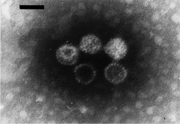
Like the VLPs described in other protozoans, the VLPs in Eimeria are particles ranging between 33 and 44 nm in diameter with the capsid presenting an icosahedral shape (Revets et al. Reference Revets, Dekegel, Deleersnijder, De Jonckheere, Peeters, Leysen and Hamers1989; Roditi et al. Reference Roditi, Wyler, Smith and Braun1994; del Cacho et al. Reference del Cacho, Gallego, Montes, López-Bernad, Quílez and Sánchez-Acedo2001) (Fig. 3). Interestingly, the RNAs sequences of VLPs from E. necatrix, E. acervulina and E. maxima are quite divergent from each other (Lee and Fernando, Reference Lee and Fernando2000). E. stiedae VLPs share sequence similarity with VLPs from Giardia (Revets et al. Reference Revets, Dekegel, Deleersnijder, De Jonckheere, Peeters, Leysen and Hamers1989), while in E. nieschulzi the VLPs feature significant homology to the VLPs from Leishmania (Roditi et al. Reference Roditi, Wyler, Smith and Braun1994). These data indicate a common origin for different VLPs that infect evolutionary divergent protozoa, which do not share the same hosts. The apicomplexan C. parvum also has VLPs (Khramtsov et al. Reference Khramtsov, Woods, Nesterenko, Dykstra and Upton1997). In this case, the VLPs also contain dsRNA and have a diameter of 31 nm, but they are not encapsulated, and are characterized by replication mechanisms similar to the viruses from the Partitiviridae family (Khramtsov and Upton, Reference Khramtsov and Upton2000) that have been so far characterized only in plants and fungi (Murphy et al. Reference Murphy, Fauquet, Bishop, Ghabrial, Jarvis, Martelli, Mayo and Summers1995).
Electron micrograph of isolated virus-like particles from Eimeria necatrix (from del Cacho et al. Reference del Cacho, Gallego, Montes, López-Bernad, Quílez and Sánchez-Acedo2001; with permission).

Although studied for more than 2 decades, the function of VLPs in Eimeria, C. parvum and Babesia is still unknown, as well as their possible role in the metabolism of the protozoa hosting them. In the case of Trichomonas, there is a direct connection between VLPs infecting the protozoa and the synthesis of a highly immunogenic protein located at the plasma membrane (Alderete et al. Reference Alderete, Demes, Gombosova, Valent, Yánoska, Fabusová, Kasmala, Garza and Metcalfe1987; Wang et al. Reference Wang, Wang and Alderete1987). VLPs are more frequently observed in Trichomonas under stressful conditions, e.g. by drug treatment (Benchimol et al. Reference Benchimol, Monteiro, Chang and Alderete2002). So far there is no direct evidence of a relationship between drug treatment and the increase in VLP presence in Apicomplexa or protein display by these VLPs.
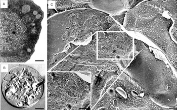
The presence of VLPs in Plasmodium is still controversial. Some authors suggested that the CB (commented on above) (Figs 2C and Fig 4A) observed in the cytoplasm of ookinetes near the nucleus is in fact an arrangement of VLPs (Garnham et al. Reference Garnham, Bird and Baker1962; Terzakis et al. Reference Terzakis, Vanderberg and Weiss1976; Meis and Ponnudurai, Reference Meis and Ponnudurai1987), while others suggested a different nature for plasmodial VLPs, proposing an energy storage function (Trefiak and Desser, Reference Trefiak and Desser1973). Since no VLPs are observed in erythrocytic or exo-erythrocytic stages, one could infer that the VLPs would infect Plasmodium in the midgut of the mosquito after the bloodmeal. However, the ookinetes shown in Fig. 4B and C were obtained in vitro from culture cells, as previously described by Raibaud et al. (Reference Raibaud, Lupetti, Paul, Mercati, Brey, Sinden, Heuser and Dallai2001), without the use of mosquitoes. Perhaps the mosquito midgut represents a stressful environment to Plasmodium that would increase the number of VLPs, forming the structure referred to as crystalloid body. Should this be the case, the VLPs might be present in the other stages of Plasmodium in such low numbers that they would escape detection at the electron microscopic level. Based on both particle size and morphology, we suggest here that the structure that is referred to as crystalloid body is in fact an aggregation of VLPs.
The crystalloid body (asterisk) from Plasmodium gallinaceum ookinetes observed by thin section (A), freeze-fracture (B) and quick-freeze/deep-etching techniques (C). Note that this cytoplasmic inclusion is similar to an aggregation of VLPs. Scale bars: A=200 nm; B and C=500 nm.

CONCLUSIONS
Although apicomplexan parasites have been extensively studied over the past decades, some basic questions remain unanswered on the very basic cell biology, and the physiology of these organisms remains wide open. As summarized here, little is known about the nature of the crystalloid and refractile bodies and their roles in the metabolism of the parasites. These structures are unique in Apicomplexa and, because of this, they could be the subjects for new exciting discoveries and could be promoted in the near future to suitable targets for the development of chemotherapeutical compounds with selective toxicity against those parasites endowed with such organelles. Like the structures reviewed here, the apicoplast and the secretory organelles were first recognized from morphological electron microscopy studies whereas their functions have been characterized much later. Thus we believe that more structural research can be performed on these structures, leading to a better understanding of their role on apicomplexan biology. The presence of VLPs per se also deserves additional studies. Should VLPs be essential for the metabolism of these protozoa, they too would become interesting targets for the development of new compounds selective against these deadly parasites.
ACKNOWLEDGEMENTS
We would like to thank Matthew Lewis (University of Heidelberg) and Friedrich Frischknecht (University of Heidelberg) for critically reading this manuscript. L.L. is supported by a Post-doctoral Fellowship from the University of Heidelberg Cluster of Excellence Cell Networks.