Introduction
Parasitic diseases in sheep, such as those infections caused by gastrointestinal nematodes (GINs), are responsible for causing substantial economic impacts in the sheep industry worldwide. Among the main GIN of small ruminants, Trichostrongylus colubriformis is considered the major parasite of the small intestine of sheep in Brazil (Cardia et al., Reference Cardia, Rocha-Oliveira, Tsunemi and Amarante2011; Amarante, Reference Amarante2014; Wilmsen et al., Reference Wilmsen, Silva, Bassetto and Amarante2014). Trichostrongylus colubriformis has a prepatent period of about three weeks, and its life cycle includes a free-living phase in the environment and a phase within the host where the animals become infected by ingesting grass contaminated with the infective larvae of the parasite (Zajac, Reference Zajac2006).
Environmental conditions have great influence on the distribution of different nematode species and on the risk of massive infections (Wilmsen et al., Reference Wilmsen, Silva, Bassetto and Amarante2014; Almeida et al., Reference Almeida, Albuquerque, Bassetto, Starling, Lins and Amarante2020). Unlike other countries where seasonality is observed in the epidemiology of intestinal parasites (De Chaneet & Dunsmore, Reference De Chaneet and Dunsmore1988; Makovcová et al., Reference Makovcová, Jankovská, Vadlejch, Langrová, Vejl and Lytvynets2009; McMahon et al., Reference McMahon, Gordon, Edgar, Hanna, Brennan and Fairweather2012), Brazil has favourable climatic conditions for the development and survival of nematode larvae in pastures throughout the year (Silva et al., Reference Silva, Amarante, Kadri, Carrijo-Mauad and Amarante2008; Rocha et al., Reference Rocha, Bricarello, Rocha and Amarante2014; Almeida et al., Reference Almeida, Albuquerque, Bassetto, Starling, Lins and Amarante2020). This fact contributes to high infection rates of animals, with cases of mortality, especially in young animals. Besides environmental conditions, several other factors influence the epidemiology of parasitic diseases; these include the presence of susceptible animals shedding large numbers of eggs in faeces, the stocking rate and the frequency and efficacy of anthelmintic treatments (Wilmsen et al., Reference Wilmsen, Silva, Bassetto and Amarante2014; Albuquerque et al., Reference Albuquerque, Bassetto, de Almeida and Amarante2017).
Trichostrongylus colubriformis infection causes a progressive reduction of feed intake in sheep, which can begin earlier than patency, and the severity of the clinical signs may vary according to several factors such as the age, physiological and nutritional status, and breed of the host, and also according to the larval challenge, time of exposure and establishment rate of the parasite (Dias-Silva et al., Reference Dias-Silva, Louvandini and Do Amarante2021). In heavy infections, T. colubriformis may cause severe enteritis due to tissue damage and inflammation in the intestinal mucosa (Andronicos et al., Reference Andronicos, Hunt and Windon2010). The formation of tunnels by T. colubriformis in epithelium mucosa affects tissue integrity, provoking hyperplasia and crypt hypertrophy, atrophy of microvillus and epithelium erosion (Cardia et al., Reference Cardia, Rocha-Oliveira, Tsunemi and Amarante2011; Dimitrijević et al., Reference Dimitrijević, Borozan, Katić-Radivojević and Stojanović2012), and thus compromising retention time of digesta, and the digestibility and absorption of nutrients (Cardia et al., Reference Cardia, Rocha-Oliveira, Tsunemi and Amarante2011; Dias-Silva et al., Reference Dias-Silva, Ventoso Bompadre and Danasekaran2019, Reference Dias-Silva, Filho, Katiki, Do Amarante, Abdalla and Louvandini2020). Furthermore, it may lead to decreased feed intake due to inappetence (Kyriazakis et al., Reference Kyriazakis, Anderson, Oldham, Coop and Jackson1996; Coop & Kyriazakis, Reference Coop and Kyriazakis1999; Dias-Silva et al., Reference Dias-Silva, Louvandini and Do Amarante2021).
As a consequence of the uneven development of the immune response, parasite distribution is binomial negative, in which most animals present a low parasite burden, while a few susceptible animals present a high parasite burden (Barger, Reference Barger1985). Susceptible animals, with a high parasite infection intensity, may present clinical signs of parasitism (Albuquerque et al., Reference Albuquerque, Bassetto, de Almeida and Amarante2017). The treatment of T. colubriformis infection is heavily based on the administration of anthelmintics; however, their suppressive use has led to the appearance of multidrug resistance (Thomaz-Soccol et al., Reference Thomaz-Soccol, De Souza, Sotomaior, Castro, Milczewski, Mocelin and Do Carmo Pessoa E Silva2004; Almeida et al., Reference Almeida, Garcia, Torgerson and Amarante2010; Cintra et al., Reference Cintra, Teixeira, Nascimento and Sotomaior2016). In targeted selective treatment (TST), animals receive treatment based on indicators of parasitism. The most common indicators are body weight gain, body score, scouring, packed cell volume (PCV), ocular conjunctiva colour (FAMACHA©) and faecal egg count (FEC) (Amarante, Reference Amarante2014; Medina-Pérez et al., Reference Medina-Pérez, Ojeda-Robertos, Reyes-García, Cámara-Sarmiento and Torres-Acosta2015; Valcárcel et al., Reference Valcárcel, Aguilar and Sánchez2015).
According to the TST precepts, only animals suffering the adverse effects of the infection should be treated, thus preserving a refugia population. This refugia population of parasites without exposure to the anthelmintic compounds contributes to the dilution of the resistance genes for the next parasite generations (van Wyk, Reference van Wyk2001). Famacha® is the most commonly used TST method in Brazil due to its easy application by sheep farmers. The treatment is recommended when animals present anaemia based on the evaluation of the ocular conjunctiva colour (van Wyk et al., Reference van Wyk, Malan and Bath1997). Nevertheless, even when TST was being applied, the quick emergence of anthelmintic resistance in GIN populations was recorded in some situations (Mederos et al., Reference Mederos, Ramos and Banchero2014; Cintra et al., Reference Cintra, Teixeira, Nascimento and Sotomaior2016; Albuquerque et al., Reference Albuquerque, Bassetto, de Almeida and Amarante2017).
Besides the use of anthelmintics, another alternative for GIN control is the use of a resistant breed, with a more efficient immune response against parasites (Amarante et al., Reference Amarante, Bricarello, Rocha and Gennari2004; Albuquerque et al., Reference Albuquerque, Bassetto, Almeida, Hildersley, McNeilly, Britton and Amarante2019). The development of the immune response to GIN varies among breeds and hosts of the same breed (Stear & Murray, Reference Stear and Murray1994). Resistant animals demonstrate the capability of developing a rapid and strong immune response, limiting the establishment of infective larvae, decreasing the fecundity of the females and also promoting the expulsion of parasites (Mowen & Glimcher, Reference Mowen and Glimcher2004; Amarante et al., Reference Amarante, Susin, Rocha, Silva, Mendes and Pires2009; Shakya et al., Reference Shakya, Miller, Lomax and Burnett2011; de la Chevrotière et al., Reference de la Chevrotière, Bambou, Arquet, Jacquiet and Mandonnet2012). Several studies carried out in Brazil have shown that the Brazilian hair sheep breed, Santa Ines, is more resistant to Haemonchus contortus parasitism in comparison with wool sheep (Amarante et al., Reference Amarante, Bricarello, Rocha and Gennari2004, Reference Amarante, Susin, Rocha, Silva, Mendes and Pires2009; Bricarello et al., Reference Bricarello, Amarante, Rocha, Cabral Filho, Huntley, Houdijk, Abdalla and Gennari2005; Costa et al., Reference Costa, Bueno, Veríssimo, Cunha, Santos, Oliveira, Spósito Filha and Otsuk2007; Rocha et al., Reference Rocha, Bricarello, Silva, Houdijk, Almeida, Cardia and Amarante2011). However, this breed did not show the same resistance to parasitism by T. colubriformis (Amarante et al., Reference Amarante, Susin, Rocha, Silva, Mendes and Pires2009; Cardia et al., Reference Cardia, Rocha-Oliveira, Tsunemi and Amarante2011).
GINs induce a T helper 2 (Th2)-type response that entails the activation of innate and adaptive cells of the immune system, signalled by specific cytokines such as interleukin 4 (IL-4), IL-5 and IL-13 (Inclan-Rico & Siracusa, Reference Inclan-Rico and Siracusa2018; Sorobetea et al., Reference Sorobetea, Svensson-Frej and Grencis2018). Mucosa mast cells (MCs), globule leukocytes (GL) and eosinophils are important effector cells involved in parasite clearance (McClure et al., Reference McClure, Emery, Wagland and Jones1992; Pernthaner et al., Reference Pernthaner, Cole, Morrison, Green, Shaw and Hein2006; Amarante et al., Reference Amarante, Rocha and Bricarello2007; Beasley et al., Reference Beasley, Kahn and Windon2010). In the immune-mediated host protection, goblet cells and their secreted products, mucin and mucus-associated proteins, also have key roles in altering the intestinal niche, thus contributing, in combination with parasite-specific immunoglobulins (IgA, IgG1 and IgE), to enhancing parasite expulsion (Harrison et al., Reference Harrison, Pulford, Hein, Barber, Shaw, McNeill, Wakefield and Shoemaker2003; Sharpe et al., Reference Sharpe, Thornton and Grencis2018).
Animals resistant to GIN infection develop an earlier and more pronounced Th2-type response, which might impair parasite establishment and development, resulting in a low worm burden (Pernthaner et al., Reference Pernthaner, Cole, Morrison, Green, Shaw and Hein2006; McRae et al., Reference McRae, Stear, Good and Keane2015; Sorobetea et al., Reference Sorobetea, Svensson-Frej and Grencis2018).
The current status of anthelmintic resistance has led to the development of alternative GIN control strategies such as the selection of resistant animals according to their natural immune response (Amarante et al., Reference Amarante, Bassetto, Neves and Amarante2014; Zvinorova et al., Reference Zvinorova, Halimani, Muchadeyi, Matika, Riggio and Dzama2016). In this context, we evaluated and compared the dynamics of natural intestinal nematode infections and immunological response of hair and wool lambs, under suppressive or TST with anthelmintics.
Material and methods
Experimental design
The experiment was carried out at the São Paulo State University (UNESP), Brazil. The experimental design was described previously (Albuquerque et al., Reference Albuquerque, Bassetto, de Almeida and Amarante2017). Briefly, 38 three-month-old uncastrated male lambs were purchased from commercial farms, where they had been raised indoors. The animals were distributed into four groups by stratified randomization, balanced as far as possible according to their nematode FECs and body weight measurements on arrival at the experimental area. The Santa Ines hair (SI) and Ile de France wool (IF) lambs were allocated to two anthelmintic treatment groups: nine SI and ten IF under suppressive treatment, in which lambs were drenched every two weeks with monepantel (2.5 mg/kg, Zolvix®, Elanco, Dundee, Scotland, United Kingdom) in order to keep the grazing lambs and their pastures as free of worms as possible; and ten SI and nine IF under TST, in which lambs were drenched only when they had shown PCV ≤ 20%, corresponding to category 3 of the FAMACHA method (van Wyk et al., Reference van Wyk, Hoste, Kaplan and Besier2006), in order to prevent the occurrence of mortality and/or severe haemonchosis. According to FEC performed on arrival at experimental facilities, all the animals presented gastrointestinal infections, with IF showing an average of 300 eggs per gram of faeces (EPG) (range 0–1100 EPG) and SI with an average of 950 EPG (range 0–7300 EPG) (Albuquerque et al., Reference Albuquerque, Bassetto, de Almeida and Amarante2017). For this reason, seven days prior to the start of the experiment, lambs under suppressive treatment were treated with the following combination of anthelmintics: albendazole (10 mg/kg, Valbazen® 10% Cobalto, Zoetis, Campinas, São Paulo, Brazil), levamisole (9.4 mg/kg, Ripercol® L 150 F, Zoetis, Campinas, São Paulo, Brazil) and monepantel (2.5 mg/kg, Zolvix®, Elanco, Dundee, Scotland, United Kingdom), which resulted in no nematode eggs detected in a sequence of faecal examinations performed. In contrast, the lambs from the TST group were not drenched on arrival and were introduced to the paddocks harbouring natural parasite infections acquired at their origin farm.
Some lambs under suppressive treatment were shedding Strongyloides spp. eggs (mean 42, range 0–200 EPG) in the seventh week, and, for that reason, those animals were dewormed with a single additional moxidectin dose (0.4 mg/kg, Cydectin®, Zoetis, Campinas, São Paulo, Brazil) in an attempt to clear the infection. In the 14th week, strongyle (379, 0–1000 EPG) and Strongyloides spp. (874, 0–5300 EPG) FEC increased as a consequence of the reduction in monepantel efficacy (Albuquerque et al., Reference Albuquerque, Bassetto, de Almeida and Amarante2017). The final monepantel drench for lambs under suppressive treatment was administered on the 18th week, and they were slaughtered on the 21st week (supplementary table S1).
The animals were allocated to clean pastures of Cynodon spp. and Urochloa decumbens at the beginning of the experiment, where the treatment groups grazed in separate paddocks until the end of the experiment. To confirm the pasture clean status, the herbage collection and processing method was performed 14 days before the animals entered the paddocks according to Taylor (Reference Taylor1939) and Niezen et al. (Reference Niezen, Miller, Robertson, Wilson and Mackay1998). The lambs had free access to tap water and mineral salt (Presencefós®, Presence Animal Nutrition, Paulínia, São Paulo, Brazil) during the whole experiment. In addition, they received a daily dietary supplement with a commercial concentrate containing 16% of crude protein (Suplementa Ovinos®, Presence Animal Nutrition, Paulínia, São Paulo, Brazil).
Blood collection
Blood samples (5 mL) were collected weekly by jugular vein puncture into Vacutainer® tubes containing anticoagulant (EDTA). The blood was centrifuged for 15 min at 4°C at 590×g (Universal 320 R, Hettich Zentrifugen, Tuttlingen, Germany) to allow plasma separation, and aliquots of plasma samples were stored at −20°C prior to enzyme-linked immunosorbent assay (ELISA).
Faecal examination
Faecal samples were collected when the animals arrived and then weekly, directly from the rectum of each animal in polyethylene bags previously labelled and kept refrigerated until processing. The modified McMaster technique (Ueno & Gonçalves, Reference Ueno and Gonçalves1998) was used for FEC, in which each nematode egg counted represented 100 EPG. On the same sampling dates, faecal cultures were prepared separately for each group for the production of GIN infective third-stage larvae (L3) that were later morphologically identified according to Ueno & Gonçalves (Reference Ueno and Gonçalves1998).
Histology
All lambs were slaughtered on the 21st week, and the gastrointestinal tract was removed. Subsequently, intestinal tissue samples were taken from the duodenum and fixed in 4% buffered formaldehyde for 48 h, then moved to 70% ethanol and paraffin-embedded. Tissue sections were cut to 5 μm thick and mounted on glass slides. Eosinophils and GL were counted on haematoxylin and eosin (H&E)-stained sections and MCs were counted on 1% toluidine blue-stained sections. All cells were counted in 60 randomly selected fields of view per animal in a 0.01 mm2 area at ×1000 magnification (adapted from Balic et al., Reference Balic, Bowles and Meeusen2002). The counts were expressed as cells/mm2 tissue surface.
Intestinal worm burden
After collecting the tissue samples, each small intestine was opened and had the total intestinal contents collected for recovering all developmental nematode stages. Afterwards, intestines were suspended in a 2 L graduated cylinder with saline solution for 4 h at 37°C to recover nematodes present in the mucosa (Ueno & Gonçalves, Reference Ueno and Gonçalves1998). Both contents were placed in graduated buckets and aliquots of 10% were collected individually, stored in plastic flasks and preserved at −20°C. All nematodes present in the aliquots were identified and quantified, according to their developmental stage (Ueno & Gonçalves, Reference Ueno and Gonçalves1998; Almeida et al., Reference Almeida, Bassetto, Amarante, Albuquerque, Starling and Amarante2018).
ELISA
Trichostrongylus colubriformis-specific IgG
Plasma samples collected at 11-time points were used to determine IgG levels against L3-soluble antigen extract of T. colubriformis. The L3 extract preparation and the protocol to determine the parasite-specific plasma IgG levels were previously described by Amarante et al. (Reference Amarante, Susin, Rocha, Silva, Mendes and Pires2009) and Silva et al. (Reference Silva, Bassetto and Amarante2012), but with some modifications: the plates were coated with 2 μg of antigen/mL; each wash was performed three times, rotating through 180° and re-washing three more times; the negative control sample used was from a worm-free animal, as previously described (Santos et al., Reference Santos, Xavier, Amarante, Bassetto and Amarante2014); the plasma positive control (PC) sample used was from a sheep artificially infected with both H. contortus and T. colubriformis every three days for 84 days. Results were expressed as the percentage of optical density (OD) value of the PC plasma sample (Kanobana et al., Reference Kanobana, Vervelde, van der Veer, Eysker and Ploeger2001).
Trichostrongylus colubriformis-specific IgA
A 5 cm tissue piece from the duodenum of each animal was sampled and stored at −20°C until processing for the mucus extraction. Tissues were thawed and mucus was scraped off with a glass slide and stored in a falcon tube on ice, followed by the addition of phosphate-buffered saline (PBS) supplemented with protease inhibitors (Complete Mini Solution1®, Roche, Indianapolis, USA) to each sample in a proportion of 4:1 (4 mL PBS+1 mL of mucus). The samples were shaken for 1 h at 4°C and centrifuged for 30 min at 4°C and 3000×g. The supernatant was collected and centrifuged again for 30 min at 4°C and 15,000×g.
ELISA assays for parasite-specific IgA in the intestinal mucus were carried out with anti-T. colubriformis L3 and adult extract, as previously described (Silva et al., Reference Silva, Bassetto and Amarante2012), with a few differences: plates were coated with 5 μg of antigen/mL; each wash was performed three times, rotating through 180° and re-washing three more times; mucus samples were diluted (1:20) in PBS-GT (0.1% Gelatin and 0.05% Tween 20 in PBS) and rabbit anti-sheep IgA peroxidase-conjugated antibody was diluted at 1:20,000. The results were expressed as OD value minus OD-blank sample (Kanobana et al., Reference Kanobana, Vervelde, van der Veer, Eysker and Ploeger2001).
Statistical analysis
All data were submitted to the normality test (Shapiro–Wilk test). Cell counts, worm burden and immunoglobulin levels were transformed using log10 (x + 1) prior to analysis. Data with single measures and data for repeated measures at several time points were analysed by analysis of variance using the General Linear Model to assess the breed and treatment effects, and the interaction between them. Group means were compared by Tukey's test using the Statistical Analysis System, version 9.2 (SAS Institute, Inc., Cary, North Carolina, USA). Values of P < 0.05 were considered statistically significant.
Results
Parasitology
Three species of small intestine nematodes were identified in the experimental animals: T. colubriformis, Cooperia curticei and Strongyloides papillosus.
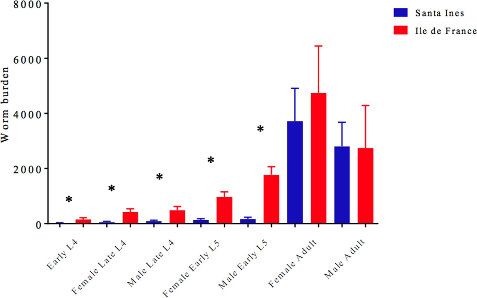
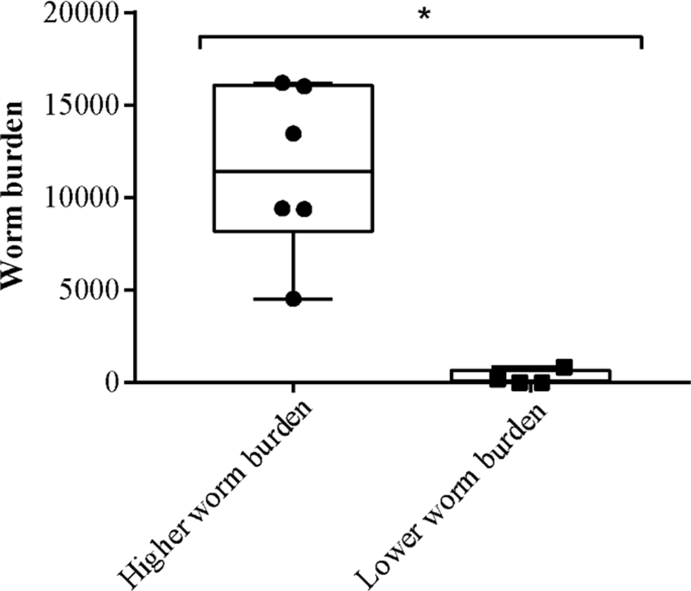
Overall, Santa Ines lambs had a lower total worm burden compared with Ile de France lambs regarding all small intestine species recovered. However, statistical difference was only observed for the C. curticei burden (supplementary table S2). Among the Santa Ines lambs under TST there were great variations in worm burden of T. colubriformis (fig. 1 and supplementary table S3) and S. papillosus (supplementary table S4) species, in which four lambs had a low T. colubriformis burden ranging from 0 to 850 worms (lower worm burden), and the other six presented high infection intensities, with worm burdens ranging from 4330 to 16,190 specimens (higher worm burden) (fig. 2). Additionally, there was a statistical difference between individuals with low and high worm burdens (P < 0.05). The same trend was observed with regards to S. papillosus counts, in which six Santa Ines lambs had a low number of parasites, ranging from 80 to 880 parasites, whereas the other four had a high worm burden ranging from 1430 to 36,460 (supplementary table S4). Regarding the Ile de France lambs, most of them had counts ranging from 4010 to 10,090 T. colubriformis worms, and one lamb showed the highest worm burden with 28,890 specimens. Nearly all of Ile de France lambs had high S. papillosus counts, ranging from 2960 to 79,260 worms, with the exception of one Ile de France lamb which had only 790 specimens.
Trichostrongylus colubriformis averages (± standard error) of Ile de France and Santa Ines lambs under TST. *Statistically different (P < 0.05).

Trichostrongylus colubriformis burden of Santa Ines lambs under TST, presenting lower and higher worm burden. The ends of the box are the upper and lower quartiles; the median is marked by a horizontal line inside the box; and the two lines outside the box extend to the highest and lowest observations. *Values significantly different by Tukey test (P < 0.05).

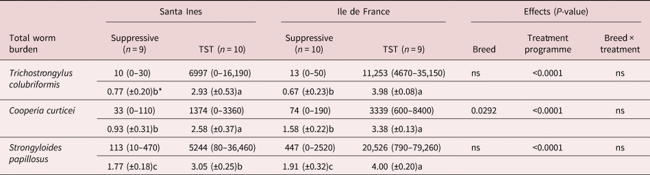
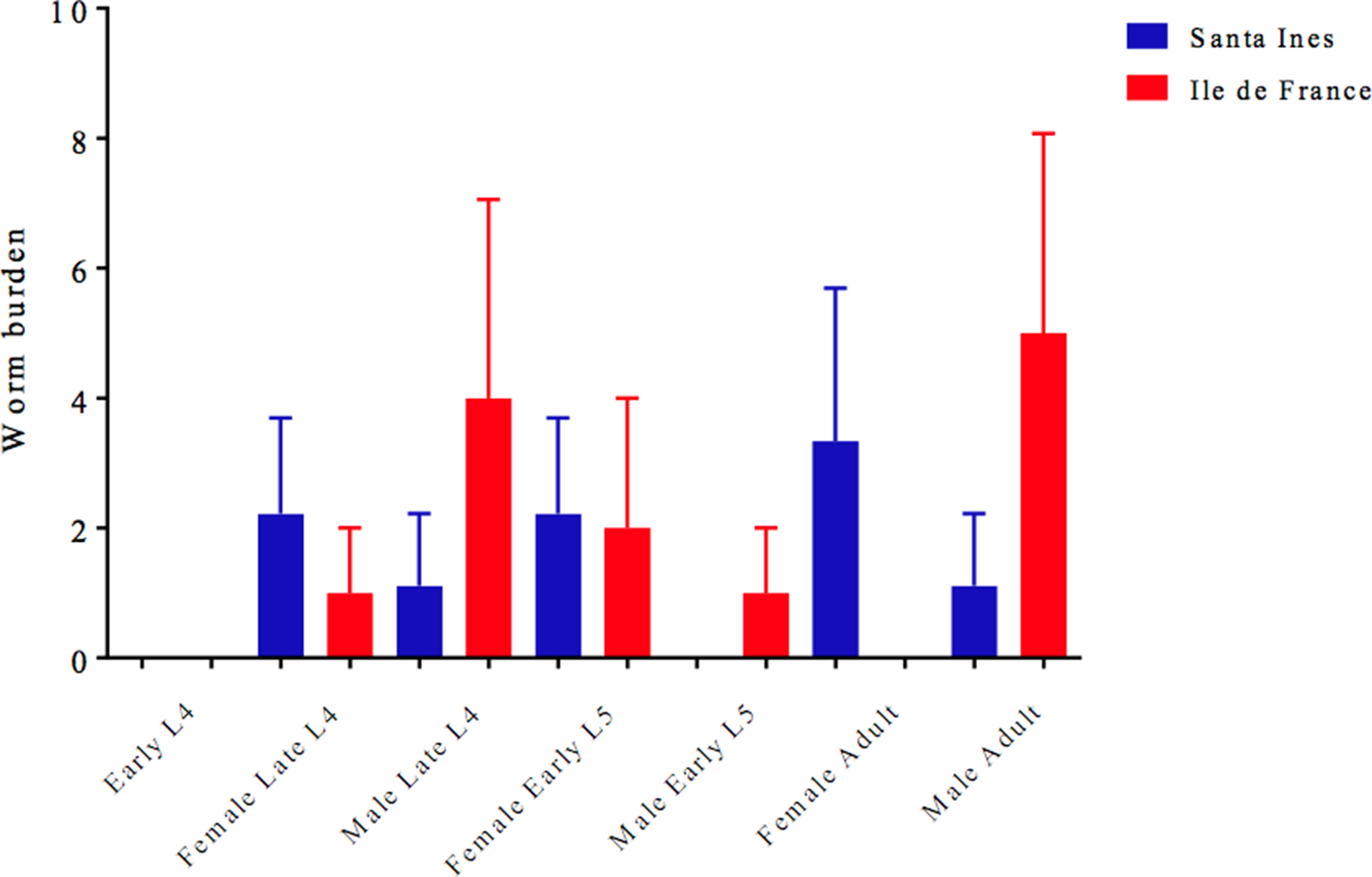
The suppressive treatment program had the aim to maintain the animals as free as possible of nematode infection (worm-free status) under field conditions, and reaching the goal, the suppressive treatment program had high efficacy against all small intestine species (table 1), reflecting in low T. colubriformis burden (fig. 3). In contrast, TST animals had higher counts of these nematodes to all developmental stages (table 1 and supplementary tables S2–S4), with Ile de France lambs showing the highest counts to immature developmental stages of all intestinal parasites, also to C. curticei burden (supplementary table S2), with a significant breed effect (P < 0.05).
Mean (minimum–maximum) values of small intestinal nematodes in Santa Ines and Ile de France lambs under suppressive or targeted selective treatment (TST) programmes with anthelmintics.

* Means (± standard error) of log transformed values followed by different letter in the same line differ from each other by Tukey's test (P < 0.05). ns, not significant (P > 0.05).
Trichostrongylus colubriformis averages (± standard error) of Ile de France and Santa Ines lambs under suppressive treatment. There was no statistical difference (P > 0.05).

IgG plasma levels against T. colubriformis L3
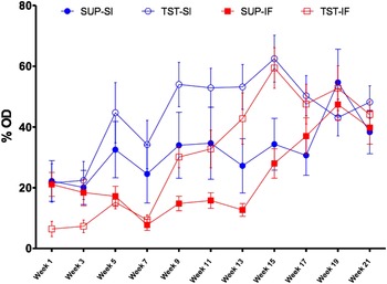
A significant breed effect on the mean levels of anti-T. colubriformis L3 IgG in plasma occurred early in the trial, on the fifth (P = 0.0187), seventh (P = 0.0453) and ninth (P = 0.0187) weeks, with Santa Ines lambs showing the highest T. colubriformis L3-specific IgG levels (fig. 4). IgG levels were similar among all the groups in the final two samplings. The treatment program had an effect on IgG levels at five time points, between the ninth and 17th weeks (P < 0.05) when animals under TST presented higher means than the suppressive treatment groups.
Mean levels of anti-Trichostrongylus colubriformis L3 IgG (% OD) in plasma of Santa Ines (SI) and Ile de France (IF) lambs under suppressive (SUP) or TST with anthelmintics. Bars represent standard error.

Small intestine mucus IgA
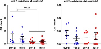
There was a significant breed effect (P = 0.0185) on T. colubriformis L3-specific IgA levels in the intestinal mucus (fig. 5), with Santa Ines lambs (suppressive 0.265 ± 0.12; TST 0.220 ± 0.06) presenting higher levels in comparison with Ile de France lambs (suppressive 0.082 ± 0.02; TST 0.090 ± 0.03). Values of IgA against antigens of adult T. colubriformis were relatively low and similar among groups (fig. 5).
Values of anti-Trichostrongylus colubriformis L3- and adult-specific IgA optical density (OD – blank) in the intestinal mucus collected post-mortem from Santa Ines (SI) and Ile de France (IF) lambs under SUP or TST with anthelmintics. Vertical bars represent mean ± standard error.

Intestinal cellular immune responses
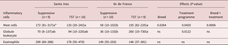
There was a significant breed × treatment interaction (P < 0.05) on MC counts and a significant effect of treatment on both MC and globular leukocytes (P < 0.05), with higher MC and globular leukocyte counts in lambs undergoing TST, although Santa Ines lambs under suppressive treatment presented the highest MC counts. The numbers of eosinophils were similar among groups, without any breed or treatment effect (P > 0.05) (table 2).
Means (minimum–maximum values) of eosinophils, mast cells and globule leukocytes (cells/mm2) in the intestinal mucosa of the Santa Ines and Ile de France under suppressive or TST programme with anthelmintic.

* Means followed by different letter in the same line differ from each other by Tukey's test (P < 0.05). ns, not significant (P > 0.05).
Even though there was a large difference in T. colubriformis counts among the Santa Ines lambs (fig. 2), no statistical difference was observed for IgA (against L3 and adult), IgG and cell count values (P > 0.05) regarding the animals that had low and high parasite burden (supplementary figs S1 and S2).
Discussion
The mechanism and robustness of nematode expulsion appear to be parasite-specific and vary among individuals. The Santa Ines is well known as a breed that has a high resistance to H. contortus infection (Amarante et al., Reference Amarante, Bricarello, Rocha and Gennari2004; Silva et al., Reference Silva, Bassetto and Amarante2012; Albuquerque et al., Reference Albuquerque, Bassetto, Almeida, Hildersley, McNeilly, Britton and Amarante2019); however, this strong resistance against H. contortus apparently was not able to induce greater resistance to T. colubriformis infection, in agreement with other studies (Amarante et al., Reference Amarante, Bricarello, Rocha and Gennari2004; Cardia et al., Reference Cardia, Rocha-Oliveira, Tsunemi and Amarante2011). The Santa Ines lambs presented a large variation on T. colubriformis burden among animals, and despite the less consistent differences between breeds, few Santa Ines lambs showed the ability to limit the small intestinal infection by T. colubriformis and S. papillosus – a fact that was not observed in Ile de France lambs. Sixty percent of Santa Ines lambs displayed relatively susceptibility to T. colubriformis infection; this trait does not seem to be an exclusive characteristic of the Santa Ines breed, as other native tropical breeds are also known as resistant to H. contortus infection, such as the Gulf Coast (Bahirathan et al., Reference Bahirathan, Miller, Barras and Kearney1996) and Florida Native (Amarante & Barbosa, Reference Amarante and Barbosa1998), which also harboured Trichostrongylus in numbers similar to those of breeds considered susceptible to Haemonchus infection. The establishment rate of T. colubriformis (33.08%) was higher than H. contortus (3.75%) following simultaneous experimental infections in Nigerian West African Dwarf sheep (Idika et al., Reference Idika, Chiejina, Mhomga, Ngongeh and Nnadi2012). Taken together, all these results suggest that T. colubriformis has a greater ability to evade the host's immune response. The transcriptomic analyses of T. colubriformis identified serine, metalloproteases and proteases inhibitors in the excretory/secretory products related to the promotion of the establishment of the nematode and immunological evasion by lysis of immune cells, antibody digestion, cleavage of surface receptors of cytokines and immunological cells proteases (Cantacessi et al., Reference Cantacessi, Mitreva, Campbell, Hall, Young, Jex, Ranganathan and Gasser2010).
The breed difference in immature T. colubriformis worm counts weeks before slaughtering may indicate that Santa Ines lambs have already acquired some degree of protection against incoming larvae of T. colubriformis in comparison with Ile de France lambs. This observation was confirmed by the higher levels of anti-L3 IgA in the intestinal mucus of the Santa Ines lambs. According to Harrison et al. (Reference Harrison, Pulford, Hein, Barber, Shaw, McNeill, Wakefield and Shoemaker2003), IgG and IgA are responsible for larvae aggregate, impairing their motility and causing an 80% reduction in larvae establishment, as well the antibodies remain in mucus for many days after the challenge. In the present study, IgG was tested at 11 time points in plasma, while IgA was measured in mucus at post-mortem. The Santa Ines lambs were able to respond earlier and produce higher levels of anti-T. colubriformis L3-specific IgG, as observed in other resistant sheep (Pernthaner et al., Reference Pernthaner, Cole, Morrison, Green, Shaw and Hein2006), although Ile de France lambs were able to reach the same IgG levels as Santa Ines over the time. Furthermore, Santa Ines lambs had significantly higher IgA levels than Ile de France regardless of the treatment performed.
In resistant sheep, most of the larvae are rejected within the first day after infection, and this early rejection is associated with increased concentration of T. colubriformis-specific IgA and IgG2 in intestinal mucus, and also with the local appearance of globule leukocytes, T-cell infiltration, activation and differentiation, and epithelial necrosis (McClure et al., Reference McClure, Emery, Wagland and Jones1992). In this study, adult parasites corresponded to the largest proportion of the T. colubriformis population in our lambs; however, there were no differences between breeds in the counts of these mature worms. Apparently, at this stage, T. colubriformis is less affected by the immune response, probably due to proteinases inhibitors production related to immune response evasion (Cantacessi et al., Reference Cantacessi, Mitreva, Campbell, Hall, Young, Jex, Ranganathan and Gasser2010). In addition, the efficiency of the immune response against T. colubriformis was not uniform within the same breed. An aggregated parasite distribution was observed amongst Santa Ines lambs, whereby 40% showed low parasitic load (0–850), indicating a satisfactory capacity to prevent T. colubriformis parasitism, while 60% had large worm burdens (4330–16,190). Cardia et al. (Reference Cardia, Rocha-Oliveira, Tsunemi and Amarante2011) also observed an aggregated parasite distribution in Santa Ines lambs serially infected with T. colubriformis. However, in that trial, most of the animals (60%) showed a low worm burden (ranging from 13 to 1540), and 40% had a high worm burden (ranging from 6310 to 26,830).
The inflammatory cells such as eosinophils, MC and GL are related to an effective local immune response against GIN (Douch et al., Reference Douch, Harriston, Elliott, Buchanan and Greer1986; McClure et al., Reference McClure, Emery, Wagland and Jones1992). These cells are abundant in the intestinal tract and, with the mucus barrier, are associated with parasite clearance (Huntley et al., Reference Huntley, Nwelands, Jackson and Miller1992; Sorobetea et al., Reference Sorobetea, Svensson-Frej and Grencis2018). The MC is an important component of immunological response against parasite infection, one that is responsible for linking the innate and adaptive immune response and also acts as an effector cell on the nematode, limiting their development (McRae et al., Reference McRae, Stear, Good and Keane2015). The highest MC count in Santa Ines lambs showed the great ability of this breed to assemble an efficient immune response against GIN, which culminates in lower worm burden in most of the animals. The marked difference in MC and GL counts between treatment groups presumably occurred as a result of a longer and greater parasite challenge in lambs under TST in comparison to those under suppressive treatment, which remained virtually nil. Huntley et al. (Reference Huntley, Nwelands, Jackson and Miller1992) suggested that a continuous nematode challenge is necessary for the occurrence of MC hyperplasia. However, in a T. colubriformis infection, Douch et al. (Reference Douch, Harriston, Elliott, Buchanan and Greer1986) did not find a correlation between MC and resistance, but observed a strong association between GL and larval migration in an inhibitory assay. It is important to emphasize that GLs derive from MCs (Huntley et al., Reference Huntley, Nwelands, Jackson and Miller1992).
Finally, in comparison with Ile de France sheep, Santa Ines lambs showed a stronger local immune response against intestinal nematodes and, consequently, the lowest worm burdens. However, this response was only more efficient in limiting parasitism by C. curticei, whereas for the outcome of T. colubriformis and S. papillosus infection, the efficiency of the immune response appears to be individual, regardless of breed.
Supplementary material
To view supplementary material for this article, please visit https://doi.org/10.1017/S0022149X21000821
Financial support
This work was supported by the Brazilian Federal Agency for Support and Evaluation of Graduate Education – CAPES–PDSE (A.C.A.A., 8881.188861/2018-01); the São Paulo Research Foundation (F.A.A., FAPESP 2015/25718-7); FAPESP (C.C.B., 2015/00221-2); a scholarship from CAPES (J.G.G.L.); and the National Council for Scientific and Technological Development (CNPq) (A.F.T.A., 303712/2013-9).
Conflicts of interest
None.
Ethical approval
The authors assert that all animal procedures contributing to this work comply with the ethical standards of the relevant national and institutional guides on the care and use of animals and were approved by the Animal Use Ethics Committee of FMVZ/UNESP (protocol number 47/2016).
Author contributions
A.C.A.A.: resources, methodology, investigation, project administration, software, data curation, visualization, writing – original draft preparation; F.A.A.: investigation, validation, writing – review and editing; C.C.B.: investigation, validation, writing – review and editing; J.G.G.L.: software, formal analysis, writing – review and editing; A.F.T.A.: conceptualization, methodology, supervision, funding acquisition, software, formal analysis, data curation, writing – review and editing.