Introduction
The genetically altered mouse has become a valuable model system in biomedical research to study the genetics and pathogenesis of human diseases. Hundreds, even thousands, of newly derived genetically altered mouse lines are generated each year, outpacing the time and resources available to adequately study them. As this increasing number of mouse lines is stretching the nation's animal housing and care infrastructure to the breaking point, significant efforts are underway to find simple, effective, and efficient means for preserving mouse lines amenable for long-term inanimate storage.
By preserving the diploid genome, embryo cryopreservation is probably the ‘gold standard’ for effective, yet complex, slow, and costly, management of mutant mice. On the other hand, sperm cryopreservation, although only preserving the haploid genome, has been shown to be substantially more efficient and cost effective (Mazur et al., Reference Mazur, Leibo and Seidel2008). As male mice do not require exogenously administered gonadotrophins, sperm collection from the epididymis is quick and easy, and only one or two males are needed to ensure an adequately sized archive (Marschall et al., Reference Marschall and Hrabé de Angelis1999; Critser & Mobraaten, Reference Critser and Mobraaten2000). Since the first reports of mouse sperm cryopreservation in 1990 (Okuyama et al., Reference Okuyama, Isogai, Saga, Hamada and Ogawa1990; Tada et al., Reference Tada, Sato, Yamonoi, Mizorgi, Kasai and Ogawa1990; Yokoyama et al., Reference Yokoyama, Akiba, Katsuki and Nomura1990), a number of newer and improved techniques have emerged, including the popular ‘Nakagata’ protocol which uses raffinose and skim milk as cryoprotectants (Nakagata, Reference Nakagata2000). Nevertheless, as with embryos, sperm must be cryopreserved, stored, and even transported at liquid nitrogen temperatures (Mazur et al., Reference Mazur, Leibo and Seidel2008), imposing higher costs for necessary storage tanks and a suitable environment, space, and monitoring, to store them. Further, cryo-recovered sperm exhibits substantial strain to strain variability when used for in vitro fertilization (IVF), and is difficult when applied to the preservation of certain mouse strains, including C57BL/6, BALB/c, and 129S3/SvIm, etc. (Critser & Mobraaten, Reference Critser and Mobraaten2000; Sztein et al., Reference Sztein, Farley and Mobraaten2000). During the preservation process itself, normal sperm can be damaged by oxidative stress (Twigg et al., Reference Twigg, Irvine, Houston, Fulton, Michael and Aitken1998; Chatterjee & Gagnon, Reference Chatterjee and Gagnon2001), activated endonuclease (Maione et al., Reference Maione, Pittoggi, Achene, Lorenzini and Spadafora1997; Aitken & De Iuliis, Reference Aitken and De Iuliis2007) or cryodamage (Peris et al., Reference Peris, Morrier, Dufour and Bailey2004; Li et al., Reference Li, Meyers, Tollner and Overstreet2007a; Yildiz et al., Reference Yildiz, Ottaviani, Law, Ayearst, Liu and McKerlie2007). Because of these and other reasons, more convenient and cost-effective means of sperm preservation are needed to fully exploit the potential of sperm as a suitable and appropriate format for the preservation, storage, and sharing of genetically altered mice.
In recent years, however, alternative approaches to traditional cryopreservation have been developed to facilitate, simplify, and reduce the expense of preserving and storing mouse sperm. For example, freeze drying, a three-step process involving freezing, primary drying (water sublimation), and secondary drying (removal of bound water), was first demonstrated for mouse sperm in 1998 (Wakayama & Yanagimachi, Reference Wakayama and Yanagimachi1998). In 2001, Kusakabe et al. (2001) introduced a new medium consisting of EGTA–Tris-buffered solution (ETBS) that extended and improved freeze drying of sperm from mouse (Ward et al., Reference Ward, Kaneko, Kusakabe, Biggers, Whittingham and Yanagimachi2003; Kaneko & Nakagata, Reference Kaneko and Nakagata2005; Kawase et al., Reference Kawase, Araya, Kamada, Jishage and Suzuki2005, Reference Kawase, Hani, Kamada, Jishage and Suzuki2007), ox (Keskintepe et al., Reference Keskintepe, Pacholczyk, Machnicka, Norris, Curuk, Khan and Brackett2002; Martins et al., Reference Martins, Báo, Dode, Correa and Rumpf2007), rabbit (Liu et al., Reference Liu, Kusakabe, Chang, Suzuki, Schmidt, Julian, Pfeffer, Bormann, Tian, Yanagimachi and Yang2004), rat (Hirabayashi et al., Reference Hirabayashi, Kato, Ito and Hochi2005), boar (Kwon et al., Reference Kwon, Park and Niwa2004; Nakai et al., Reference Nakai, Kashiwazaki, Takizawa, Maedomari, Ozawa, Noguchi, Kaneko, Shino and Kikuchi2007) and human (Kusakabe et al., Reference Kusakabe, Yanagimachi and Kamiguchi2008). However, despite the presence of 50 mM EGTA as a Ca2+ chelating agent, freeze drying has been shown to cause chromosome damage in 20–40% of mouse sperm (Ward et al., Reference Ward, Kaneko, Kusakabe, Biggers, Whittingham and Yanagimachi2003; Kusakabe et al., Reference Kusakabe, Yanagimachi and Kamiguchi2008). Further, freeze-dried sperm requires frozen storage temperatures to prevent slow deterioration (Kaneko & Nakagata, Reference Kaneko and Nakagata2005; Kawase et al., Reference Kawase, Araya, Kamada, Jishage and Suzuki2005).
Upon rehydration, freeze-dried sperm is amotile, which excludes the use of IVF and other artificial reproductive technologies that require sperm to retain motility. On the other hand, the mechanical injection of oocytes by ICSI eliminates this requirement. Although live animals have been derived by ICSI using freeze-dried sperm from the mouse (Kaneko et al., Reference Kaneko, Whittingham, Overstreet and Yanagimachi2003), rat (Hirabayashi et al., Reference Hirabayashi, Kato, Ito and Hochi2005; Hochi et al., Reference Hochi, Watanabe, Kato and Hirabayashi2008) and rabbit (Liu et al., Reference Liu, Kusakabe, Chang, Suzuki, Schmidt, Julian, Pfeffer, Bormann, Tian, Yanagimachi and Yang2004), a comprehensive analysis of the health and genetics of derived mice has not. Such analysis is necessary, because since ICSI bypasses all natural selection barriers to fertilization (cumulus layer, zona pellucida, and oolemma) (Li & Lloyd, Reference Li, Lloyd, Pease and Lois2006), and genetic defects and/or damaged DNA in sperm can be passed on to progeny and their subsequent generations. For example, ICSI enables infertile men with genetically compromised sperm to successfully fertilize oocytes (Liu et al., Reference Liu, Tsao, Cheng, Wu, Huang, Chen, Lin and Lee2004; Clementini et al., Reference Clementini, Palka, Iezzi, Stuppia, Guanciali-Franchi and Tiboni2005; Hansen et al., Reference Hansen, Bower, Milne, de Klerk and Kurinczuk2005; Pang et al., Reference Pang, Kim, Lee and Kim2005). Further, although most appear normal and healthy (Yanagimachi, Reference Yanagimachi2005; Agarwal, Reference Agarwal2007), ICSI-conceived children are at greater risk of birth defects compared with spontaneous conceptions (Hansen et al., Reference Hansen, Bower, Milne, de Klerk and Kurinczuk2005). Of most concern since the birth of the first successful ICSI-conceived child in 1992 (Palermo et al., Reference Palermo, Joris, Devroey and Van Steirteghem1992), is that the inability to assess carefully sufficient numbers and generations of children has meant that the long-term safety and efficacy of ICSI across multiple generations remain largely unknown.
Therefore, in order to further assess ICSI and freeze drying as a means to preserve valuable genetically altered mice, it is necessary to demonstrate that both ICSI and freeze drying are safe, reliable, and reproducible techniques by the demonstration that mice that have been derived by ICSI using freeze-dried sperm are viable, healthy, and fertile. The present study was designed systematically and comprehensively to study the health and genetics of mice derived by ICSI using freeze-dried sperm and their progeny. Specifically, we examined in vitro and in vivo embryo development, birth rate, litter size, sex ratio, weaning rate, fertility, pathology, and genomic stability of two strains (C57BL/6J and B6D2F1/J) of wild-type mice across three generations.
Materials and Methods
Animals
All B6D2F1/J and C57BL/6J (B6) mice were purchased from the Jackson Laboratory. Mice were housed in individually ventilated plastic cages (BioZone Inc.) with bedding made from reclaimed wood pulp (Absorption Corporation) in a specific pathogen-free barrier facility with light cycle of 14 h light and 10 h dark, according to standard operating procedures of the Center for Laboratory Animal Science at the University of California, Davis. Mice were fed ad libitum PicoLab Mouse Diet 20 from Purina Mills (St. Louis, MO, USA). Mice had free access to deionized, autoclaved drinking water. Mouse euthanasia was carried out by a combination of CO2 asphyxiation and cervical dislocation. The care, use and disposition of all mice used in this study were reviewed and approved by the Institutional Animal Care and Use Committee of the University of California, Davis.
Chemicals and media
Non-essential and essential amino acids (NEAA and EAA) required for KSOM medium (Biggers et al., Reference Biggers, McGinnis and Raffin2000) were purchased from GIBCO BRL. PVP (polyvinyl pyrrolidone) was purchased from Irvine Scientific, mercury was purchased from Fisher Scientific, and equine chorionic gonadotropin (eCG), human chorionic gonadotrophin (hCG) and all other chemicals were purchased from Sigma Chemical Co. unless otherwise stated. Microsatellite oligos were purchased from MWG-Biotech.
Experimental design
Experiments were conducted to compare results between two different strains (B6D2F1/J and C57BL/6J) and among three different treatment groups based on how embryos were initially fertilized (natural mating group, fresh sperm ICSI group, and freeze-dried sperm ICSI group). All experiments were begun using four male mice (parental generation) at 8 weeks of age that were chosen randomly from a breeding colony for each of the two strains (B6D2F1/J and C57BL/6J) as founder sires for natural mating with females (natural mating group). After obtaining one litter from each sire by natural mating, the male was euthanatized and sperm from the caudal epididymidis were collected into prewarmed ETBS medium. The freshly harvested sperm were split into two samples, one of which was used immediately for ICSI into freshly harvested ova to produce fertilized embryos (fresh sperm ICSI group, motile sperm with normal morphology were randomly selected for ICSI), while the other was freeze dried and subsequently stored at 4 °C for 1–2 months, after which time it was rehydrated and used for ICSI into freshly harvested ova to produce fertilized embryos (freeze-dried sperm ICSI group, sperm with head and tail together and with normal morphology were randomly selected for ICSI). The 2-cell embryos obtained by overnight culture after ICSI were blind coded and transferred into the oviducts of pseudopregnant female CD-1 recipient mice 0.5 days post coitus (dpc) with vasectomized stud male CD-1 mice. All pregnant recipients were allowed to go to term and give birth (first generation). To avoid variation among experiments, all ICSI and embryo transfer procedures were performed by the same injectionist and surgeon, respectively. After birth, pups from all three treatment groups were weaned at 21 days of age. When the young were 8 weeks old, one male and one female mouse from each litter within each treatment group were chosen randomly as breeding pairs to generate the second generation offspring. After birth and weaning of these offspring, one male and one female mouse at 8 weeks of age from each litter within each treatment group were chosen randomly as breeding pairs to generate the third generation offspring which were assessed after birth until weaning.
Sperm collection and freeze drying (parental generation)
Male mice (10–11 weeks old) were sacrificed, and then the two caudal epididymides were cut out and placed in an empty Petri dish. The caudal epidydimides were punctured gently with 30-G needles under a dissecting microscope, transferred with sterile forceps into 1 ml prewarmed (37 °C) ETBS containing 10 mM Tris–HCl, pH 8.2, 50 mM EGTA and 50 mM NaCl in a 1.5 ml microcentrifuge tube, and then incubated at 37 °C for 10 min. The top 0.8 ml sperm suspension was carefully aspirated out and split into two parts: one part was used immediately for ICSI (fresh sperm group), and the other part was used for freeze drying.
Freeze drying of sperm was performed using a modification of the Kusakabe protocol (Kusakable et al., Reference Kusakable, Szczygiel, Whittingham and Yanagimach2001). Briefly, 0.1 ml sperm suspensions were dispensed into dry and autoclaved 1 ml Wheaton longneck glass ampules, which subsequently were plunged into liquid nitrogen for 1 min to freeze the sperm samples. Each ampule then was immediately connected to a manifold dryer of a Freezone® 4.5 freeze drying machine. The sperm contained within the ampule was freeze dried for 4 h under single vacuum pressure at 0.03–0.04 mBar. After freeze-drying, ampules were flame-sealed under vacuum (0.03–0.04 mBar), and then stored at 4 °C until rehydrated and used for ICSI.
ICSI and embryo transfer
Six- to 8-week-old B6D2F1/J and C57BL/6J female mice were superovulated by an intraperitoneal (i.p.) injection of 5 IU eCG followed by injection of 5 IU hCG (i.p.) 47–48 h later. Mice were sacrificed by a combination of CO2 asphyxiation and cervical dislocation 14–15 h after hCG injection, the abdomen surgically opened, and cumulus–oocyte complexes (COCs) collected from oviducts into warm HEPES–CZB medium (Chatot et al., Reference Chatot, Lewis, Torres and Ziomek1990) supplemented with 5.56 mM d-glucose. Cumulus cells were dissociated from eggs by treatment with 300 U/ml bovine testis hyaluronidase in HEPES–CZB medium at 37 °C, and then oocytes were cultured in pre-equilibrated KSOM medium at 37 °C in humidified 5% CO2 and 95% air prior to ICSI.
Fresh sperm ICSI was performed immediately after sperm collection. For ICSI using freeze-dried sperm, an ampule was opened and 100 μl of culture grade distilled water was added to rehydrate the sperm. The ICSI procedure was performed using piezo-assistance on a Nikon TE300 inverted microscope with Narishige micromanipulators at room temperature as previously described (Li & Lloyd, Reference Li, Lloyd, Pease and Lois2006). Briefly, sperm heads were separated from sperm tails in 10% PVP (w/v in BSA-free HEPES–CZB) by applying a few piezo pulses (intensity 3–4, speed 3, PMM controller, Prime Tech), to the sperm neck region. After washing in another 10% PVP drop, a group of about 10 sperm heads was loaded into the ICSI pipette with appropriate intervals between sperm heads. ICSI into oocytes was performed in a drop of BSA-free HEPES–CZB medium containing 0.01% (w/v) PVA (polyvinyl alcohol, cold water soluble, molecular weight 30–70 kDa). The zona was penetrated by applying a few piezo pulses (intensity 2–3 and speed 3), and then the zona piece was expelled into the perivitelline space before the ICSI needle was pushed against the oolemma to the opposite side of the oocyte where the oolemma was broken by one weak piezo pulse (intensity 1, speed 1). One sperm head was released into the ooplasma with minimal accompanying medium. Injected oocytes were washed and incubated in equilibrated KSOM medium (50 μl drops under mineral oil) at 37 °C in humidified 5% CO2 and 95% air overnight until 2-cell embryo transfer, or for 96 h after ICSI until blastocyst stage. Embryos were scored every 24 h.
Embryos at the 2-cell stage were transferred into the oviducts (maximum of 12 per oviduct) of 0.5 d.p.c. pseudopregnant CD-1 recipient female mice anesthetized with 2.5% Avertin i.p. Recipients were kept warm on a heating pad until fully recovered from anesthesia. All pregnant recipients were allowed to go to term and give birth to live offspring for analysis and subsequent breeding.
Necropsy and pathology examination
To determine if the ICSI procedure and/or sperm freeze drying caused any pathological defects in liveborn offspring in either the B6D2F1/J or C57BL/6J strains studied, two to four mice (both male and female) at 12–13 weeks of age in the first and second generations or at 3–4 weeks of age in the third generation from each of the four representative litters in the three experimental groups (natural mating group, fresh sperm ICSI group, and freeze-dried sperm ICSI group) were randomly selected for gross necropsy and histological pathology examinations. Organs for histological pathology examination included liver, kidney, spleen, mesenteric lymph node, pancreas, heart, lung, thymus, brain, urinary bladder, stomach, cecum, small intestine, colon, reproductive system (ovaries, oviducts, uterus, vagina, testes, seminal vesicles, prostates) and endocrine system (pituitary, thyroid, parathyroid, adrenal, pancreas). Five μm sections were stained with hematoxylin and eosin. All analyses (gross exams and histology exams) were performed on blinded samples that had been assigned coded, random numbers.
Microsatellite analysis
To determine if ICSI and/or freeze drying induces genomic instability in sperm from either the B6D2F1/J or C57BL/6J strains, all mice (parental, first, second, and third generations) from each treatment group (natural mating group, fresh sperm ICSI group, and freeze-dried sperm ICSI group) were sampled for microsatellite instability (MSI) analysis. Briefly, approximately 1 cm long tail snips taken from mice at 10–12 days of age were digested in 0.5 ml of 0.2 mg/ml Proteinase K (Roche Diagnostics) in 50 mM KCl, 10 mM Tris (pH 8.0), 1.0 M EDTA, and 0.1% (v/v) Triton X-100 at 55 °C for 12 h or until complete digestion. Samples were then heated at 95 °C for 10 min to inactivate the protease before being diluted (1:2) with TE buffer (10 mM Tris and 1 mM EDTA, pH 8.0) and stored at −20 °C.
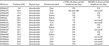
Multiplex PCR was conducted on individual DNA samples using pools of fluorescent 5′-labelled (FAM, TAMRA, Cy3 and HEX) microsatellite primer sets incorporating one 5′-labelled primer per primer pair. Of the 16 microsatellite markers tested, four were included as potentially unstable markers since they had shown polymorphisms that may have been indicative of genetic instability in a previous long-term backcrossing experiment (unpublished data). Both stable and suspected unstable markers were used to enhance identification of high and low frequency MSI, respectively (Table 1).
Microsatellite (MS) loci utilized for C57BL6/J and B6D2F1/J MSI analysis.

Hot start PCR amplification was conducted in a 18 μl reaction mixture consisting of approximately 50 ng DNA, 0.4 μM for each multiplexed primer, 0.175 mM dNTP, 1% (w/v) bovine serum albumin, 1.2 units Taq polymerase (Promega), 1 mM EDTA, 1.5 mM MgCl2, and 10 mM Tris, pH 8.0, overlaid with light mineral oil. Reactions were amplified on a PTC-100 (Bio-Rad) using the following PCR amplification cycles: 95 °C for 3 min (DNA and primers only), hold at 81 °C while reaction cocktail was added, followed by 10 cycles of 94 °C for 50 s, 57 °C for 50 s, 72 °C for 1 min, 21 cycles of 92 °C for 1 min, 57 °C for 50 s, 72 °C for 1 min, final extension of 72 °C for 8 min, and hold at 4 °C. When initial PCR amplification failed, MgCl2 concentration of the final cocktail was increased by 20% to enable amplification.
For electrophoresis separation, approximately 15 μl of gel loading buffer consisting of 10 mM NaOH, 3 mM EDTA, 0.02 % (w/v) bromophenol blue, in 98% (v/v) formamide were mixed with each microsatellite reaction mixture, denatured at 95 °C for 15 min, and then 3 μl of prepared mix was loaded using a multichannel syringe (Hamiliton) onto a long (24 cm) 7% denaturing polyacrylamide gel in 0.75× TBE buffer (final 67.5 mM Tris–borate and 10 mM EDTA, pH 8.0). Amplicons were separated at 2000 volts for approximately 1.5 h on a nucleic acid sequencer (CBS Scientific) or a Genomix LR (Beckman Analytical). Gels were imaged on a multi-channel FMBIO2 laser scanner (Hitachi) which allowed for spectral separation of FAM labelled microsatellite amplicons from TAMRA, Cy3, and HEX-labelled microsatellite amplicons. A 60–400 bp CXR fluorescent ladder (Promega, Madison, WI, USA) was used as size standard.
For comparison, the tail samples collected from each generation in all experimental groups (natural mating group, fresh sperm ICSI group, and freeze-dried sperm ICSI group) were stored at −20 °C in the same freezer for almost the same length of time before DNA extraction, and then all extracted DNA samples were stored at −20 °C in the same freezer for almost the same length of time before being used as genomic templates in subsequent PCR genotyping reactions. All PCR amplified samples were stored at −20 °C for almost the same length of time before being used for electrophoresis separations. Although samples were not blind coded, all microsatellite analyses were performed by the same experienced, unbiased and removed technician. Further, microsatellite analysis was performed using the same protocol throughout the entire project, PCR primers, and chemicals, buffers, enzymes, test tubes, etc., were purchased from the same vendors, and the same thermal cyclers, electrophoresis equipment and gel imaging system were used for all samples.
Statistical analysis
Statistical software at http://www.swogstat.org/statoolsout.html (Southwest Oncology Group) was used for the 95% binomial distributions and binomial probabilities for MSI analysis, and GraphPad Prism 5 software (GraphPad Software, Inc.) was used for statistical analysis of other endpoints. The sperm developmental potential data, including fertilization rate, blastocyst formation rate, birth rate and weaning rate, and the percentage of males (sex ratio) were analyzed by Fisher's Exact probability test. Litter size data were analyzed by one-way ANOVA. p < 0.05 was chosen as an indication of statistical significance. In order to minimize male-to-male variability, all of the sperm developmental potential data, litter sizes and pathology lesions were tracked on a per male basis, and no ‘sire effects’ in the experiments of this study were detected by nested ANOVA tests.
Results
Effects of sperm freeze drying on fertilization and blastocyst formation
As shown in Tables 2 and 3, freeze-dried sperm from both C57BL/6J and B6D2F1/J strains were able to fertilize eggs normally, and there was no difference in fertilization rate (as indicated by percentage of 2-cell embryos) between fresh and freeze-dried sperm. However, the blastocyst formation rate of embryos derived from freeze-dried sperm was significantly lower than that from fresh sperm in both C57BL/6J (54% vs. 69%) and B6D2F1/J (57% vs. 73%) mouse strains (p < 0.05).
In vitro development of embryos derived by ICSI from fresh or freeze-dried (FD) sperm in B6D2F1/J and C57BL/6J mouse strains. Freeze-dried sperm were stored at 4 °C for 1 week.

a ,b Values with different superscripts within each column are significantly different (p < 0.05). Four replicates using four different male mice were performed for each group. In each strain, the same four males were used as sperm donors for fresh and freeze-dried sperm, respectively.
In vitro and in vivo development (First Generation) of embryos derived from fresh and freeze-dried (FD) sperm ICSI in B6D2F1/J and C57BL/6J strains.

Within each strain, the same four males were used as sperm donors for natural mating, fresh sperm and freeze-dried sperm, respectively. Freeze-dried B6D2F1/J and C57BL/6J sperm were stored at 4 °C for 43–46 and 34–40 days, respectively before ICSI.
a ,b Values with different superscripts within each column and the same mouse strain are significantly different (p < 0.05). Four different male mice were used for each group.
Effects of sperm freeze drying on birth and weaning rates of the first generation
As shown in Table 3, the birth rate (number of pups born/number of 2-cell embryos transferred) of 2-cell embryos derived from freeze-dried sperm was significantly lower than that derived from freshly harvested sperm in B6D2F1/J strain (14% vs. 33%, p < 0.05). The birth rate of the embryos derived from freeze-dried C57BL/6J sperm was marginally lower than that from freshly harvested sperm (Table 3) (17% vs. 29%, p = 0.06). Of the mice born from freeze-dried sperm in both B6D2F1/J and C57BL/6J strains, essentially all were successfully weaned by day 21 after birth, and no significant differences were found in weaning rates (number of pups weaned/number of pups born) of mice derived from fresh or freeze-dried sperm from either of the 2 strains.
Neither ICSI nor freeze drying of sperm significantly affected the sex ratio of progeny (Table 3). Within C57BL/6J, the sex ratio (percentage of male mice) of pups derived from freeze-dried sperm (59%) by ICSI was not significantly different from the sex ratio of pups derived from either fresh sperm (54%) by ICSI or derived by natural mating (54%). Within B6D2F1/J, the percentage of B6D2F1/J male pups derived from freeze-dried sperm (58%) by ICSI was also not significantly different from the pups from natural mating (51%), but was just significantly greater than those derived from fresh sperm (35%).
Effects of sperm freeze drying on litter size and sex ratio of the second and third generations
To determine the extent to which mice derived by ICSI using freeze-dried sperm were reproductively sound, one male and one female at 8 weeks of age from each of the four or five litters in the first generation from each treatment group (natural mating group, fresh sperm ICSI group, or freeze-dried sperm ICSI group) in both C57BL/6J and B6D2F1/J strains were randomly chosen as breeding pairs to generate the second generation. As shown in Table 4, all breeding pairs in both C57BL/6J and B6D2F1/J strains successfully mated and produced offspring (second generation), then from which mice were successfully mated to produce offspring (third generation). Moreover, there were no significant differences in litter sizes among the three treatment groups in both the second and third generations. Weaning rates for all treatment groups in both the second and third generations were 100%.
Reproductive performance of second (F2) and third (F3) Generations of B6D2F1/J and C57BL/6J strain mice derived by natural mating, fresh sperm ICSI or freeze-dried (FD) sperm ICSI.

Means ± SEM, n = 4 or 5 breeding pairs.
a ,b Values with different superscripts within each column and the same mouse strain are significantly different (p < 0.05).
Neither ICSI nor freeze drying affect the sex ratio of C57BL/6J mice in either generation (Table 4), and there was no difference in the sex ratio among the natural mating, fresh sperm and freeze-dried sperm groups. There was also no significant difference in sex ratio of B6D2F1/J mice in either generation between the freeze-dried sperm ICSI group and the natural mating group. There was a statistically significant difference in the percentage of B6D2F1/J males derived by ICSI from freeze-dried sperm versus fresh sperm in both the second generation (59% vs. 38%, respectively) and the third generation (53% vs. 35%, respectively), although the origin of these differences was not investigated.
Effects of sperm freeze drying on the incidence of non-lethal pathologic changes
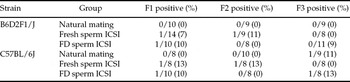
To determine if ICSI and/or freeze drying caused any pathological abnormalities, gross and microscopic examination of tissues from mice across three progeny generations was performed (Table 5). The age of mice examined for pathologic changes was 12–13 weeks old for the first and second generations and 3–4 weeks old for the third generation in all experimental groups. No significant differences in the number of mice with gross and or histological pathology lesions, within any generation of either mouse strain, were observed between any of the three treatment groups (natural mating group, fresh sperm ICSI group, and freeze-dried sperm ICSI group). The lesions found in B6D2F1/J mice included unilateral oligospermia of unknown etiology (n = 1), chronic bilateral bacterial nephritis (n = 1), and mild chronic lymphadenitis (n = 1). The lesions found in C57BL/6J strain mice included mild multifocal hepatitis (n = 1), mild chronic focal pancreatitis (n = 1) focal pulmonary perivascular lymphocyte aggregate (n = 1), renal tubular amyloidosis (n = 1), and unilateral aplasia of the eye (n = 1).
Number and percentage of the mice with lesions in the first (F1), second (F2) and third (F3) generations of mice.

Effects of sperm freeze drying on genomic stability
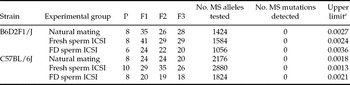
To determine if ICSI and/or freeze drying caused genomic instability, microsatellite alleles were amplified from the original founder males (parental generation) of both strains (B6D2F1/J and C57BL/6J) in all three experimental groups and from mice across three generations (first, second, and third) of progeny compared with the parental generation. As shown in Table 6, a total of 10 994 microsatellite alleles were evaluated for polymorphic mobility shifts at 16 loci in C57BL/6J and at eight loci in B6D2F1/J (Table 1). One C57BL6/J foundation male was found to exhibit a pre-existing polymorphism at D2Mit271 which was transmitted in a manner consistent with normal Mendelian inheritance and this locus was excluded as evidence of MSI induced by either treatment group (Table 1). There was no evidence that either sperm freeze drying or the ICSI procedure elicited any microsatellite instability in the derived mice in each of the treatment groups or across three generations of progeny.
Number of mice in the parental founder lines (P) and three subsequent generations (F1, F2, F3) from which DNA was extracted from tail tips and used for analysis of 16 microsatellite markers in C57BL/6J strain and eight microsatellite markers in B6D2F1/J strain.

a Only upper limit shown for 95% binomial confidence interval; all lower limits are zero. Estimated proportions based on F1–F3 only for each strain per treatment group.
FD, freeze-dried. MS, microsatellite. MPMR, microsatellite polymorphic mutation rate.
Discussion
The present study has shown that ICSI using freeze-dried sperm from mice on either an inbred (C57BL/6J) or hybrid (B6D2F1/J) genetic background and stored short term at refrigerator temperature can be used to produce at least three generations of mice that are both genotypically stable and phenotypically normal. Fertilization rate (as measured by percentage of 2-cell embryos) achieved after ICSI using freeze-dried sperm from either strain was comparable with that using fresh sperm, indicating that mouse sperm factors required for egg activation and fertilization are not adversely affected by freeze drying. Thereafter, the blastocyst formation rate and subsequent birth rate of transferred embryos derived from freeze-dried sperm were significantly (p < 0.05) to marginally (p = 0.06) lower than that from fresh sperm. Nevertheless, once born, mice derived by ICSI using freeze-dried sperm were healthy and reproductively sound, and no major differences in litter size, weaning rate, and sex ratio were noted in two generations of progeny (F2 and F3) from naturally mated ICSI-derived offspring compared with the natural mating group. Further, there was no evidence that either ICSI or freeze drying-induced genomic instability as determined by microsatellite analysis of the derived mice and subsequent generations in comparison to both parental genotypes, nor any differences in the number or types of pathological changes in any of the three generations of progeny. To our knowledge, this report is the first to study the transgenerational production of mice generated from the breeding of progeny derived by ICSI using freeze-dried sperm.
The lower blastocyst formation rate and birth rate of the embryos derived by ICSI using freeze-dried sperm are consistent with our previously published results found by ICSI using evaporatively dried mouse sperm (Li et al., Reference Li, Meyers, Tollner and Overstreet2007a, Reference Li, Biggers, Elmoazzen, Toner and Lloydb). While a precise determination of the cause of these phenomena was outside the scope of the present study, observations by others (Ward et al., Reference Ward, Kaneko, Kusakabe, Biggers, Whittingham and Yanagimachi2003; Kawase et al., Reference Kawase, Araya, Kamada, Jishage and Suzuki2005; Kusakabe et al., Reference Kusakabe, Yanagimachi and Kamiguchi2008) suggest that the freeze-drying process probably damages sperm DNA and/or chromosomes. The freeze-drying procedure was reported to cause chromosome damage in about 20–40% of sperm (Ward et al., Reference Ward, Kaneko, Kusakabe, Biggers, Whittingham and Yanagimachi2003; Kusakabe et al., Reference Kusakabe, Yanagimachi and Kamiguchi2008), and subsequent storage of freeze-dried sperm at 4 °C also causes sperm DNA damage (Kawase et al., Reference Kawase, Araya, Kamada, Jishage and Suzuki2005). In other studies, mouse sperm with DNA damaged by gamma radiation could fertilize oocytes in vitro normally, but the blastocyst formation rate decreased significantly (Ahmadi & Ng Reference Ahmadi and Ng1999). Using a bovine model, Fatehi et al. (Reference Fatehi, Bevers, Schoevers, Roelen, Colenbrander and Gadella2006) also demonstrated that sperm DNA damage caused by X-ray radiation does not affect fertilization and embryo development until after the first cleavage, when embryonic gene expression begins.
Previous studies in mice have found that paternal chromosome-type aberrations due to double-stranded breaks (DSBs) can be repaired completely or partially, depending on the level of damage, via maternal DSB repair genes in the early zygote (Marchetti et al., Reference Marchetti, Essers, Kanaar and Wyrobek2007; Derijck et al., Reference Derijck, Van Der Heijden, Giele, Philippens and de Boer2008). Nevertheless, embryos with incompletely repaired DSBs are still capable of developing to birth, and may elicit transgenerational genomic instability, as evidenced by point mutations, small deletions, amplifications, and microsatellite instability (MSI). In addition to DNA-repair mechanisms, epigenetic mechanisms may be altered in zygotes derived with damaged sperm (Fernández-Gonzalez et al., Reference Fernández-Gonzalez, Moreira, Pérez-Crespo, Sánchez-Martín, Ramirez, Pericuesta, Bilbao, Bermejo-Alvarez, Hourcade Jde, Fonseca and Gutiérrez-Adán2008), resulting in aberrant DNA methylation and histone modifications (Barber & Dubrova, Reference Barber and Dubrova2006).
For our study, we chose to assess microsatellite instability (MSI) in an effort to detect transgenerational genomic instability induced by sperm freeze drying. MSI is considered a hallmark of genomic instability and is characterized by small deletions or insertions from unrepaired replication slippage due to defects in DNA-repair pathways (Gafà et al., Reference Gafà, Maestri, Matteuzzi, Santini, Ferretti, Cavazzini and Lanza2000; González-García et al., Reference González-García, Moreno, Navarro, Martí-Ragué, Marcuello, Benasco, Campos, Capellà and Peinado2000; van Gent et al., Reference van Gent, Hoeijmakers and Kanaar2001). Microsatellites are tandem arrays of simple sequence repeats (SSRs) dispersed throughout the genome (Pabst et al., Reference Pabst, Schwaller, Bellomo, Oestreicher, Mühlematter, Tichelli, Tobler and Fey1996). Genomic instability of a microsatellite region is a function of the number of SSRs present: SSRs with <10 repeats are unlikely to reveal polymorphism whereas SSRs > 15 offers maximal probability of finding polymorphism (Silver, Reference Silver1995). Microsatellite variability is heritable, and insertions or deletions of single or more repeats can readily be identified by PCR. We selected microsatellite regions with SSRs ≥ 15 in order to detect non-repaired replication slippage due to MSI. In a previous study analyzing colorectal cancer cells, a minimum of five microsatellite markers were sufficient to detect and distinguish between both high and low frequency mitotic MSI in somatic tissues (Boland et al., Reference Boland, Thibodeau, Hamilton, Sidransky, Eshleman, Burt, Meltzer, Rodriguez-Bigas, Fodde, Ranzani and Srivastava1998). However, MSI is highly variable and is dependent on species, strain, microsatellite repeat size and repeat number, which DNA-repair genes are affected, and the type and extent of damage inducing MSI. We thus selected a total of 16 and 8 markers to analyze the C57BL/6J and B6D2F1/J strains, respectively, to increase the probability of detecting meiotic genomic instability ranging from a conservative six-fold increase in MSI to a >1000 fold increase in MSI above the natural microsatellite mutation rate. Assuming a previously predicted (Marchetti et al., Reference Marchetti, Essers, Kanaar and Wyrobek2007) and empirically documented average natural mutation rate of 1.9 × 10⁻⁴ mutations/generation/locus for dinucleotide SSRs (van Gent et al., Reference van Gent, Hoeijmakers and Kanaar2001; Marchetti et al., Reference Marchetti, Essers, Kanaar and Wyrobek2007), the probability of detecting a six-fold increase in MSI for the pooled F1–F3 mice (Table 6) in both strains is 96.26% (freeze-dried sperm) and 99.39% (fresh sperm), and greater than 99.99% for detecting ≥100 fold increase in MSI in both freeze-dried and fresh sperm groups. These results validated each experimental group as microsatellite stable.
Our results indicate that neither freeze drying nor ICSI introduced detectable transgenerational genomic instability in either of the two mouse strains tested through at least three generations of breeding. These results are consistent with a recent report showing no significant difference in the frequency or spectrum of de novo point mutations found in naturally conceived fetuses and fetuses produced by IVF or ICSI (Caperton et al., Reference Caperton, Murphey, Yamazaki, McMahan, Walter, Yanagimachi and McCarrey2007). Nevertheless, if genomic instability is suspected, a recently published study (Kusakabe et al., Reference Kusakabe, Yanagimachi and Kamiguchi2008) suggests that incubation of mouse sperm in ETBS solution for several days at 4 °C prior to ICSI may make sperm chromosomes more resistant to damage caused by freeze drying.
The potential impact of sperm DNA damage can be mitigated by several factors, including maternal DNA repair mechanisms in the zygote (Marchetti et al., Reference Marchetti, Essers, Kanaar and Wyrobek2007; Derijck et al., Reference Derijck, Van Der Heijden, Giele, Philippens and de Boer2008), preimplantation loss (Zorn et al., Reference Zorn, Vidmar and Meden-Vrtovec2003; Seli et al., Reference Seli, Gardner, Schoolcraft, Moffatt and Sakkas2004; Tesarik et al., Reference Tesarik, Greco and Mendoza2004; Fatehi et al., Reference Fatehi, Bevers, Schoevers, Roelen, Colenbrander and Gadella2006; Li et al., Reference Li, Biggers, Elmoazzen, Toner and Lloyd2007b), and late-term abortion (Seli et al., Reference Seli, Gardner, Schoolcraft, Moffatt and Sakkas2004; Tesarik et al., Reference Tesarik, Greco and Mendoza2004; Pérez-Crespo et al., Reference Pérez-Crespo, Moreira, Pintado and Gutiérrez-Adán2008). These mechanisms are also likely to explain why ICSI-conceived children appear normal and healthy (Yanagimachi, Reference Yanagimachi2005; Agarwal, Reference Agarwal2007), despite potentially carrying damaged, paternally derived DNA (Hansen et al., Reference Hansen, Bower, Milne, de Klerk and Kurinczuk2005). The results of our study suggest that freeze drying is more favorable than cryopreservation without cryoprotectant, which causes DNA damage in the embryo and aberrant growth, premature aging, abnormal behavior, and mesenchymal tumours in the adult (Fernández-Gonzalez et al., Reference Fernández-Gonzalez, Moreira, Pérez-Crespo, Sánchez-Martín, Ramirez, Pericuesta, Bilbao, Bermejo-Alvarez, Hourcade Jde, Fonseca and Gutiérrez-Adán2008). Nonetheless, additional research focusing on protecting sperm DNA and/or chromosomes from damage is necessary to further improve sperm freeze drying for widespread use in mutant mouse biology.
It should be pointed out that the mice used in the present study are wild type mice without DNA damage in sperm caused by genetic factors. The outcome observed in this study only indicates that some sperm DNA damage and genomic instability can not be transmitted by ICSI to offspring because of the mechanisms of DNA repair, preimplantation loss and late-term abortion, but it does not rule out the possibility of transmission of sperm DNA damage caused by genetic factors to the next and following generations by ICSI. Sperm DNA damage caused by genetic factors, for example the de novo deletions of the Y chromosome in men with severe oligospermia or azoospermia (de Vries et al., 2001), is an important ICSI risk factor. Such sperm cell mutations are transmissible to offspring, and one could reason that ICSI should result in increased paternal genetic disease transmission than IVF because of the absence of sperm selecting mechanisms. It is recommended that both karyotype analysis and Y-chromosome microdeletion screening should be performed on men with non-obstructive azoospermia or oligospermia below 5 × 10⁵ sperm/ml, since studies show that most men with a higher sperm count do not have the deletion (Aittomaki et al., 2004; Varghese et al., 2007).
The effects of ICSI and sperm freeze drying on epigenetics of mice were not studied in the present study. A growing body of evidence suggests that assisted reproductive technologies (ARTs) may affect reprogramming of epigenetic information inherited from the sperm and the oocyte in the zygote and preimplantation embryos leading to diseases (Huntriss & Picton, Reference Huntriss and Picton2008). The adverse effects of reactive oxygen species (ROS) on many aspects of reproduction are well documented (Betts and Madan, Reference Betts and Madan2008). For example, ROS have been shown to alter gene expression in preimplantation embryos (Harvey et al., Reference Harvey, Kind, Pantaleon, Armstrong and Thompson2004, Reference Harvey, Kind and Thompson2007) through epigenetic mechanisms (Islam & Mendelson, Reference Islam and Mendelson2006). It was also reported that 129Sv × C57BL/6J F1 mice derived from embryos cultured in vitro for 4 days from 2-cell stage to blastocyst stage exhibited specific behavioural alterations in anxiety/locomotor activity and spatial memory (Ecker et al., Reference Ecker, Stein, Xu, Williams, Kopf, Bilker, Abel and Schultz2004). Epigenetic influences are less likely to have occurred in the present study because mouse embryos were cultured only overnight until the 2-cell stage before being transferred into foster mothers.
Despite the potential damage caused by the procedure itself, ICSI appeared to have no deleterious impact on the viability, health, or fertility of mice. ICSI has been shown to cause mechanical disruption of intracellular structures of the oocyte, such as the meiotic spindle; the introduction of sperm acrosome, its overlying plasma membrane, and other foreign materials (e.g. polyvinyl pyrrolidone, protein, etc.) that otherwise would never enter the oocyte during natural fertilization (Yanagimachi, Reference Yanagimachi2005); contamination of the ovum caused by co-incident injection of infectious pathogens; sperm cell damage caused by sperm head separation from tail prior to ICSI (Li & Lloyd, Reference Li, Lloyd, Pease and Lois2006; Li et al., Reference Li, Meyers, Tollner and Overstreet2007a, Reference Li, Biggers, Elmoazzen, Toner and Lloydb); and sperm plasma membrane disruption (Mansour, Reference Mansour1998; Yanagimachi, Reference Yanagimachi2005). These risk factors may disturb normal fertilization and embryo development, and affect genomic stability, health, and fertility of ICSI-conceived children and their progeny. Such deleterious effects were not observed in our study.
An unexpected useful outcome of this study was the recognition of the importance of characterizing the genomic stability of starting materials, in this case the Parental Generation, in multigenerational studies. The finding of a heritable microsatellite polymorphism in an inbred C57BL/6J foundation sire emphasizes the need to genotype all parents and offspring in genomic stability studies. If not done, failure to identify in the parental generation polymorphisms at loci under consideration could result in erroneously overestimating genetic instability in subsequent generations of mice.
In summary, our results validate and confirm the application of ICSI using freeze-dried sperm as an alternative assisted reproductive technology in the mouse. There are several advantages to the application of ICSI and sperm freeze drying in the mouse. For example, freeze-dried sperm can be stored (at least for the short term) at refrigerator temperatures, while sperm preserved by traditional cryopreservation approaches requires continuous storage at liquid nitrogen temperatures. Higher storage temperatures facilitate maintenance options, expand the number of alternatives for shipping and transport, and offer potentially significant cost savings for preserving and storing mouse germplasm. In comparison, evaporative drying of sperm (Bhowmick et al., Reference Bhowmick, Zhu, McGinnis, Lawitts, Nath, Mehmet and Biggers2003; McGinnis et al., Reference McGinnis, Zhu, Lawitts, Bhowmick, Mehmet and Biggers2005; Li et al., Reference Li, Biggers, Elmoazzen, Toner and Lloyd2007b, Reference Li, Biggers, Toner and Lloydc) has been shown to be technically facile and not require expensive cryopreservation instrumentation. However, because drying to less than 10% residual water causes genetic damage, partially evaporatively dried sperm requires storage at freezing temperatures from −20 °C to −80 °C and lower.
Acknowledgements
The authors would like to express their sincere thanks to Drs Ryuzo Yanagimachi and Monika (Szczygiel) Ward at the University of Hawaii, Manoa, who provided valuable ICSI training and sperm preservation advice. We would also like to thank Dr James W. Overstreet at UC Davis for his valuable assistance in project planning and experimental design. This study was conducted as part of the National Cooperative Program on Mouse Sperm Cryopreservation sponsored by the National Institute of Child Health and Human Development (U01 HD38205) and the National Center for Research Resources (U01 HD38228). This work was also supported in part by a NIH/NCRR U42 Award No. RR014905 to establish the Mutant Mouse Regional Resource Center (MMRRC) at UC Davis, and by the UC Davis Mouse Biology Program. Statistical advice was made possible by Grant Number UL1 RR024146 from the National Center for Research Resources (NCRR), a component of the National Institutes of Health (NIH), and NIH Roadmap for Medical Research.