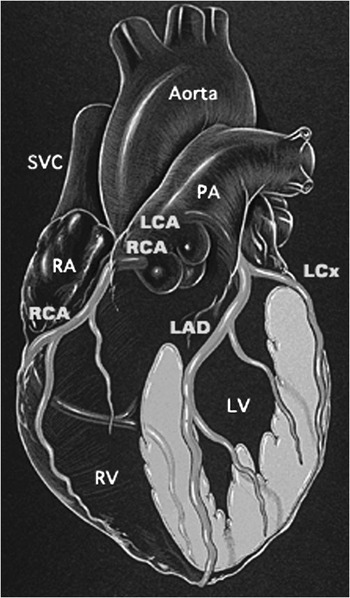
The coronary arteries are the vessels through which both substrate and oxygen are provided to the cardiac muscle. In the normal situation, there are two separate coronary arteries, right and left, each arising from a separate and distinct sinus of the aortic valve. The right coronary artery arises from the right sinus of Valsalva and enters the right atrioventricular groove, terminating in the majority of instances in the inferior interventricular groove. The main stem of the left coronary artery arises from the left sinus of Valsalva, and then bifurcates into the anterior descending, or interventricular, and the circumflex branches. The former terminates at the apex of the anterior interventricular sulcus, while the latter courses in the left atrioventricular groove, stopping short of the crux in most instances, but giving rise to the inferior interventricular artery in about one-tenth of individuals. Each of these coronary arteries provides branches, which dive perpendicularly into the myocardium and arborize in complex terminal patterns (Fig. 1).
This illustration shows the origin and course of the coronary arteries and their branches. Courtesy Patrick Lynch

Variations in the distribution of the coronary arteries are common, but anomalies are rare. Among the more commonly encountered variations is the situation in which the anterior descending artery and the circumflex artery arise by separate orifices from the left sinus of Valsalva. This is thought to occur in 1% of the population.Reference Roberts1 It is also not uncommon to find the infundibular artery arising as a separate branch from the right sinus of Valsalva.
As already stated, in 90% of the normal population, the inferior interventricular coronary artery, often said to be “posterior descending”, although it is certainly not posterior when viewed in attitudinally appropriate orientation, and may even ascend as it course towards the cardiac apex, arises from the right coronary artery. This arrangement is described as right coronary arterial dominance. About one-tenth of people have a left dominant system, in which the inferior interventricular artery is the terminus of the circumflex artery. An even smaller number of people have a balanced system, in which both the right and left coronary arteries supply arteries to the inferior interventricular region of the diaphragmatic surface of the heart.
Anomalies of the coronary arteries can be considered the result of a rudimentary persistence of an embryologic coronary arterial structure, a failure of normal coronary development, a failure of the normal atrophic process of development, or the misplacement of a connection of an otherwise normal coronary artery. Thus, one can consider anomalies of the coronary arteries in terms of:
• Abnormal origin
• Abnormal course
• Abnormal number of coronary arteries
• Lack of patency of the orifice of a coronary artery
• Abnormal connections of the arteries
We will discuss examples of each of these abnormalities.
Origin of the left coronary artery from the pulmonary trunk
While one of the more common coronary arterial abnormalities, this lesion occurs with an incidence of approximately 1 in 300,000 children,Reference Keith, Keith, Rowe and Vlad2 and males are affected more commonly than females, with a ratio of 2.3 to 1.Reference Neufeld and Schneeweis3 The fetus is typically unaffected by this lesion, as the diastolic pressure in the pulmonary trunk is high, thus providing an ample pressure of perfusion. Thus, the typical age of presentation for those affected by anomalous origin of the left coronary artery from the pulmonary trunk is during the first few months of life, when the pulmonary diastolic pressure falls concomitant with the diminishing pulmonary vascular resistance. As a result, the perfusion pressure through the left coronary arterial system decreases, leading to insufficient perfusion of the left ventricle, and resultant dysfunction. The degree of left ventricular dysfunction likely relates to the development of collateral vessels that arise from the right coronary artery, and provide flow into the left system. Patients with insufficient collateral vessels are prone to present during early infancy with symptoms of left ventricular failure, mitral regurgitation, and shock from ischemia or infarction. Those with prominent and well-developed collateral vessels may not present until later in childhood, when they can exhibit mitral regurgitation, cardiomegaly, and more progressive, indolent, left ventricular dysfunction. This latter group of patients is also at increased risk for unexpected sudden death, particularly during times of exercise, as they have increased myocardial demands with reduced coronary flow reserve.Reference Wesselhoeft, Fawcett and Johnson4
The presentation of irritability, and severe distress with feeding or defaecation, in the infant should alert the practitioner to the possibility of anginal pain, which can result from coronary arterial insufficiency when the left coronary artery arises anomalously from the pulmonary trunk. This combination of anatomy and symptoms is referred to as the Bland-White-Garland syndrome.Reference Hayashi, Kishida, Haneda and Mori5
Anomalous origin of a coronary artery from the opposite sinus of Valsalva
Either the right or the left coronary artery can arise from the opposite sinus of Valsalva. Such a course can be relatively innocuous, unless the anomalous artery takes an interarterial course between the pulmonary trunk and the aorta. In this latter setting sudden death, almost invariably during or immediately following strenuous exercise or competitive sporting events, may occur with no or few preceding symptoms. Although a mechanism for sudden death has not been definitively established, conjecture is that coronary arterial flow is compromised by compression of the coronary artery, so-called interarterial entrapment, restriction at the site of its orifice from the sinus of Valsalva, or kinking of the coronary artery itself. Although death from anomalous origin of the right coronary artery from the left coronary aortic sinus of Valsalva has been described, anomalous origin of the left coronary artery from the right aortic sinus of Valsalva appears to carry a higher risk of sudden death.Reference Roberts, Siegel and Zipes6, Reference Cheitlin, DeCastro and McAllister7 Other anatomical features associated with anomalous origin of a coronary artery from the opposite sinus of Valsalva, such as orificial stenosis, intramural coursing, and kinking may also play a role in the pathophysiology of this lesion.Reference Frommelt and Frommelt8
Cheitlin et al.Reference Cheitlin, DeCastro and McAllister7 reported death associated with anomalous origin of the left coronary artery from the right aortic sinus in one third of the military recruits who died suddenly. Eckart et al.Reference Eckart, Scoville and Campbell9 reported similar findings. It is the report from Cheitlin and his associatesReference Cheitlin, DeCastro and McAllister7 that is cited most frequently to substantiate the clinical relevance of anomalous origin of a coronary artery from the opposite sinus of Valsalva, and suggesting a high risk of death in the young patient,Reference Lorenz, Mookadam, Mookadam, Moustafa and Zehr10–Reference Maron, Roberts, Akhtar, Myerburg and Ruskin12 but such studies based on autopsy evidence do not define the risk of death because they provide no information about the population at risk, namely the denominator in the risk formula.
One clinical report has estimated the prevalence of anomalous origin of a coronary artery from the opposite sinus of Valsalva in children at 0.17%.Reference Davis, Cecchin, Jones and Portman13 Given the incidence of sudden death in young athletes, at about one in 220,000, and assuming that anomalous origin of a coronary artery from the opposite sinus of Valsalva represents the cause of death in one third of these cases, Mirchandani and PhoonReference Mirchandani and Phoon14 calculated the risk of sudden death for the patient with anomalous origin of a coronary artery from the opposite sinus of Valsalva to be approximately one in 650. Put in terms of an annual risk, Corrado et al.Reference Corrado, Basso, Pavei, Michieli, Schiavon and Thiene15 have determined the risk of death due to anomalous origin of a coronary artery from the opposite sinus of Valsalva to be 0.24 per 100,000 person years.
A single coronary arterial system arising from either the right or the left sinus of Valsalva is again not a pathologic lesion unless part of its course involves an intravascular or intramural pathway. When it does, sudden death may be associated with heavy exertion.Reference Cheitlin, DeCastro and McAllister7, Reference Maron, Roberts, Akhtar, Myerburg and Ruskin12, Reference Corrado, Thiene, Cocco and Frescura16
Coronary arterial fistulas
Distal anomalies of the coronary arteries most commonly involve abnormal connections, or fistulas, between the right or left coronary arterial systems and a chamber or vessel. Most commonly, the connection is to the right ventricle or the right atrium, though connections to the superior caval vein, coronary sinus, pulmonary arteries, left atrium, and left ventricle can occur. The coronary arterial fistula can be direct in its course and connection, or it can take a convoluted and worm-like path to its site of drainage. Occasionally, the fistulous connections can become aneurysmal. The involved coronary artery is typically dilated when the distal fistula is large, as there is preferential flow, or “steal”, from the coronary arterial system into the lower resistance chamber or vessel. The receiving structure is sometimes referred to as a low pressure “sink”. If the volume of blood delivered away from the coronary artery and into the sink is sufficiently large, it may result in myocardial ischaemia or infarction. About half of those affected by coronary arterial fistulas are asymptomatic, and are only recognized by the presence of a continuous murmur at examination. This is the ausculatory result of the constant pressure gradient between the coronary artery and the pressure sink, and the turbulence that results from the flow through the fistula.
Imaging of the coronary arteries in children
A disciplined interpretation of any modality used for imaging, whether invasive or non-invasive, is the positive recognition of the origin of each coronary artery, its course, size, and termination or connection. All too often one considers “ruling out” a particular diagnosis, such as the anomalous origin of the main stem of the left coronary artery from the pulmonary trunk in the young infant with severe ventricular dysfunction. The most important diagnosis, however, is not to “rule out” the anomaly, but to “rule in” the exact location of the origin of the coronary arteries, and the direction of flow within them. If this cannot be done non-invasively, then the clinician is obligated to investigate the precise details of myocardial perfusion with invasive imaging.
Echocardiography
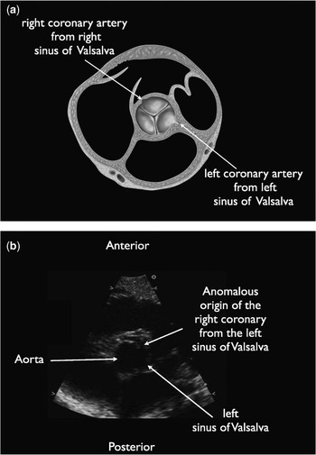
Imaging of the coronary arteries is a salient component of the routine echocardiographic assessment of cardiovascular evaluation of the infant and child. Routine bedside echocardiography remains the most commonly available and practical tool to perform this assessment. With the addition of colour flow mapping to the tomographic cross-sectional imaging, echocardiography can be a sensitive tool to identify the child with abnormal coronary arterial anatomy. The application of colour flow mapping provides important information about the patency and contour of the coronary arteries, as well as the direction of the flow of blood within them. The portability of echocardiography, particularly in the critically ill patient with congestive heart failure and shock, make this bedside tool the primary modality for imaging patients whose clinical presentation suggests the possibility of a coronary arterial abnormality. Continued advances in transducer technology, and in the sensitivity and resolution of echocardiography, have improved the ability to image coronary arteries in even the smallest and youngest of patients (Fig. 2).
This short axis two-dimensional echocardiogram is obtained at the level of the aortic valve. In Figure 2a*, the normal origins of the coronary arteries is illustrated. Figure 2b, demonstrates the origin of the right coronary artery from the opposite (left cusp) of the sinus of Valsalva in an infant. *Courtesy of Patrick Lynch.

Magnetic resonance imaging
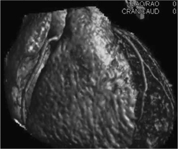
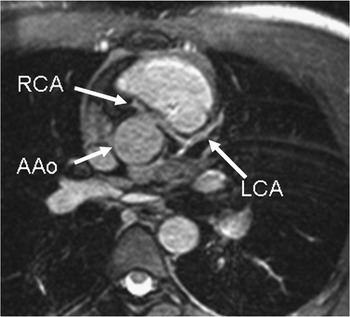
There have, of course, also been tremendous advances in other forms of non-invasive cardiac imaging. For example, cardiac magnetic resonance has been used to image coronary arteries for a number of years. Initial attempts in the 1980s met with only moderate success. Indeed, one of the first major studies of modern cardiac magnetic resonance used to image coronary arteries did not appear until 1995, when McConnell et al.Reference McConnell, Ganz, Selwyn, Li, Edelman and Manning17 identified 14 of 15 anomalous coronary arterial lesions correctly as validated against angiography. This was then followed rapidly by numerous other reports, such as that from Bunce et al.,Reference Bunce, Lorenz and Keegan18 where cardiac magnetic resonance identified the anomalous course of the coronary arteries in 26 patients, with 15 confirmed by angiography, and 11 where angiography could not define the anatomy with certainty, 8 of these running an intramural course. Su et al.Reference Su, Chung and Muthupillai19 had examined a larger series of 65 children, providing unambiguous diagnosis of coronary arterial anomalies in 62, with the other 3 having significant arrhythmias which precluded imaging. The march towards improved imaging with cardiac magnetic resonance has led to the development of whole heart 3-dimensional cardiac magnetic resonance techniques.Reference Sakuma, Ichikawa and Suzawa20 These can visualize the entire heart, and be performed in less than 30 minutes (Fig. 3). There are also multiple reports of cardiac magnetic resonance performed for lesions other than anomalous left coronary arteries in children, such as Kawasaki’s disease,Reference Babu-Narayan, Cannell and Mohiaddin21, Reference Mavrogeni, Papadopoulos and Douskou22 and in the setting of other congenital cardiac malformations.Reference Taylor, Dymarkowski and Hamaekers23
This magnetic resonance image demonstrates the origins and course of the normal coronary arteries.

“State of the art” techniques involving cardiac magnetic resonance to visualize the coronary arteries generally use a “free breathing navigator” approach, where the motion of the diaphragm is monitored, and imaging data is obtained only at certain points in the respiratory cycle. In addition, acquisition of imaging data is timed to the quiescent period of the cardiac cycle during diastasis. Both these methods are used to image the coronary arteries when there is little motion and, in the case of imaging in diastasis, where the artery contains the most blood. Since nearly all state-ofthe-art techniques use bright blood imaging, such as gradient echo, or more commonly, steady state free precession, tactics such as T2 preparation and fat suppression are used to enhance the contrast of the blood-filled artery with the surrounding tissue.
The appeal of cardiac magnetic resonance goes beyond visualizing the coronary arterial system in children with congenitally malformed hearts, as it also makes it possible to assess the effect of coronary arterial flow of blood. Cardiac magnetic resonance can evaluate the myocardium for scar tissue that may have resulted from coronary ischaemia utilizing delayed enhancement after injection with gadolinium, a magnetic contrast agent. Indeed, the technique has been used for that purpose in Kawasaki’s disease,Reference Mavrogeni, Papadopoulos and Douskou24 and in patients subsequent to repair of transposition using the arterial switch.Reference Taylor, Dymarkowski and Hamaekers23 Adenosine stress perfusion cardiac magnetic resonance is complementary to delayed enhancement, and is used to detect regional defects in myocardial perfusion.Reference Prakash, Powell, Krishnamurthy and Geva25 Another complementary technique is measuring the coronary flow reserve by estimating flow in the coronary sinus with phase-encoded velocity mapping. Further, the use of cine cardiac magnetic resonance and myocardial tagging makes it possible to identify regional abnormalities of wall motion, which may be related to the affected coronary artery.
The place of cardiac magnetic resonance in the assessment of the coronary arteries in children, and in those with congenitally malformed hearts, is clear. It is a “one stop shop” imaging modality that does not employ ionizing radiation or iodinated contrast. With even further advances in hardware and software, which will increase spatial and temporal resolution, it is obvious that cardiac magnetic resonance is an established technique for the imaging techniques not only of today, but also tomorrow (Figs 4 and 5).
This magnetic resonance image demonstrates anomalous origin of the left coronary artery from the right coronary cusp.

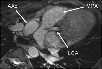
This magnetic resonance image demonstrates anomalous left coronary artery (LCA) from the pulmonary artery (MPA). Note the mild loss of signal in the MPA where the LCA inserts, signifying turbulent flow from the LCA into the MPA.

Computed tomography
Cardiac computed tomography with multidetector-row computed tomography technology is the most recent modality that can be used as a “one stop shop” to non-invasively evaluate the coronary arteries, cardiac morphology, and cardiac function. Advantages include its high spatial resolution, its fast acquisition, and patient comfort. Common paediatric applications include congenital coronary anomalies and Kawasaki’s disease. Cardiac computed tomography is also useful to assess coronary morphology and patency prior to and following surgical intervention for congenital heart disease (such as the arterial switch for transposition).
By the nature of the exam, patients are exposed to radiation and iodinated contrast medium. An understanding of fundamental cardiac computed tomography principles is therefore paramount to minimizing patient risk. A multidetector-row computed tomography scanner with at least 64-channels is recommended given its superior spatial and temporal resolution as compared to earlier generation multidetector-row computed tomography scanners. Depending on whether the exam is to evaluate morphology, function, or both, cardiac computed tomography may be acquired as a routine computed tomography angiogram or as a computed tomography angiogram with electrocardiogram-gating. Retrospective electrocardiogram-gating is currently offered by most vendors and is required to reduce cardiac motion and optimize coronary artery depiction. Prospective gating is recommended by other vendors to reduce radiation exposure, particularly when functional analysis is not required. If the scan acquisition for a 64-channel cardiac computed tomography is less than 2 seconds, electrocardiogram-gating may not be required to depict coronary origins, given the high temporal resolution. In this instance, radiation exposure is further reduced. In all paediatric cardiac computed tomography applications, voltage and amperage are prescribed according to the patient’s weight. Prospective gating is recommended when functional analysis is not required, as radiation exposure is reduced. To minimize the contrast volume, contrast medium is dosed according to the patient’s body weight. High concentration contrast medium can be used to prescribe a lower volume. The rate of contrast medium injection will depend on the caliber of the peripheral vein and catheter as well as the scan acquisition duration.
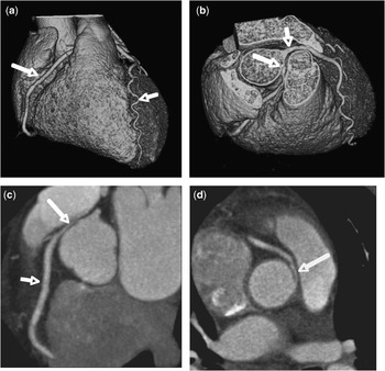
Accurate cardiac computed tomographic coronary artery assessment requires real time application of advanced two and three-dimensional visualization techniques on a workstation. Integration of volume rendered and thin-slab maximum intensity projection techniques can robustly display the origins and course of benign and malignant coronary anomalies, while multiplanar and curved planar reformations can reliably demonstrate ostial kinking, intramural course, and intra-myocardial extension (Fig. 6). Multiphase reconstruction datasets facilitate dynamic cine qualitative and quantitative evaluations of anomalous coronary arteries. Stepping through systolic and diastolic phases of the cardiac cycle is used to assess the relative degree of fixed versus compliant coronary artery narrowing.
Images (a--d) are from a retrospective ECG-gated 64-channel Coronary CT Angiogram, performed in a teenager who had exertional chest pain. Figure a is a volume rendered (VR) 15° LAO, 15° cranialcaudad view and demonstrates normal atrioventricular and interventricular courses for the RCA (long arrow) and LAD (short arrow), respectively. However, the RCA origin is not seen to arise from the expected right coronary cusp. Figure b is a steep RAO, cranialcaudad VR projection and identifies the RCA (long arrow) to arise off of the left coronary cusp with acute angulation, adjacent to the left main coronary artery (short arrow). The RCA post-ostial segment courses retropulmonic with moderate stenosis. Figure c is a 60° LAO maximum intensity projection (MIP) image, confirming the anomalous RCA with a retropulmonic course. Note again the acute angulation at its origin with post-ostial short segment stenosis (long arrow), relative to the normal caliber of the second segment of the RCA (short arrow). Figure d is an oblique multiplanar reformation through the anomalous RCA. It demonstrates an intramural post-ostial course for the RCA, accounting for the stenosis seen on the angiographic VR and MIP views. Note again the acute angulation and kinking at the RCA origin.

Angiography
Coronary angiography may be required when the precise diagnosis of the coronary arterial abnormality, and its dynamic implication, cannot be ascertained entirely from non-invasive modalities. With this algorithm, angiography should be considered to be a collaborative imaging tool, not a competitive one. Selective injections into the coronary arteries provide the most detailed anatomic information.
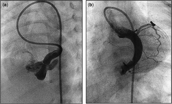
Angiography can be particularly useful in those with anomalies of coronary arterial connection. Connections to bronchial and mediastinal vessels are common, and usually are not of clinical consequence.Reference Angelini26 Clinically relevant coronary arterial fistulas can be exquisitely delineated with angiography. Their functional significance is often related to size of the connection and the resulting shunt, which can also be measured in the catheterization laboratory. Significant connections can be addressed with concurrent transcatheter embolization (Fig. 7), such that surgical ligation is now rarely necessary.Reference Okubo, Nykanen and Benson27 In the current era of non-invasive imaging, these embolizations should be undertaken with materials that do not interfere with subsequent imaging with magnetic resonance.
(a) Selective injection in the left main coronary artery demonstrates a fistulous connection to right atrium in close proximity to the coronary sinus. Note the dilation of the coronary artery and lack of angiographic contrast filling the myocardial vessels. (b) Immediately following transcatheter occlusion of the fistulous connection the myocardial perfusion is improved as evidenced by filling of the normal coronary circulation.

Functional assessment of the coronary arteries in children
The tools and techniques currently available to assess coronary arterial function are varied and many. Better to understand these various modalities used in the functional assessment of coronary arteries in children, we need an understanding of the factors that determine and regulate myocardial blood flow. The capillary beds of the coronary arteries are located within a muscular myocardium that undergoes intermittent contraction, with impedance of flow during systole. Impedance to flow is greater for the higher pressure systemic ventricle, usually but not always of left morphology, than for the lower pressure ventricle. Subsequently, 70–85% of flow of blood to the systemic ventricle occurs during diastole. In contrast, in the normal state, flow to the pulmonary ventricle is primarily a systolic phenomenon.Reference Guyton and Hall28, Reference Garson, Bricker, Fisher and Neish29 Resistance to flow is less in the sub-epicardial region than in the sub-endocardium, with subendocardial flow nearly zero during early systole. The subendocardium also has higher demands for oxygen. It is easy to see, therefore, how in the presence of significant hypertrophy, such as seen with severe aortic stenosis or asymmetric septal hypertrophy, the sub-endocardial region is at significant risk for ischaemia. Autoregulation, that is the ability of the coronary arterial bed to maintain a constant flow despite changes in the pressure of perfusion, is a unique feature of the coronary vascular bed. Autoregulation may not be as uniform throughout the heart as previously believed, as some regions may not be as protected from ischaemia as others.
Electrocardiographic assessment
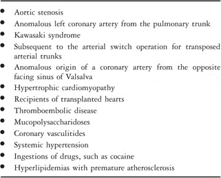
The electrocardiogram is the initial diagnostic tool to be used in the evaluation of possible coronary arterial insufficiency. Dysfunctional or ischaemic myocardium does not depolarize in a normal fashion, and therefore will also have abnormal repolarization. It is this characteristic of abnormal repolarization that was first recognized and documented in 1918, when BousfieldReference Bousfield30 noted depression of the ST segment during a spontaneous anginal attack in an adult who had a history of syphilis, and clinically had aortic insufficiency. Electrocardiographic criterions for ischaemia in children have been extrapolated from changes noted in the ST segments in adults. Horizontal or downward depression of 1 mm or more for greater than 60–80 milliseconds have been universally accepted as that criterion which suggests myocardial ischaemia. Historically, such changes have been observed in many disorders that are characterized as having either abnormal coronary vasculature, or increased myocardial demands for oxygen (Table 1). Numerous conditions, nonetheless, may produce a false positive test, including hyperventilation, therapy with digoxin, antidepressants or oestrogen, hypokalemia, mitral valvar prolapse, or pectus excavatum. It is also not uncommon to have a false negative electrocardiogram in the face of myocardial ischaemia given that changes in the ST segments appear to be a relatively late finding in the evolution of the ischaemic process. Furthermore, the magnitude of the changes does not always correlate with the severity of the disease process. Therefore, although historically tried and true in adults, the electrocardiogram does not appear to be the most accurate test for detecting coronary arterial insufficiency in children.
Disorders in which changes may be found with electrocardiographic signs of myocardial ischaemia.

Myocardial perfusion imaging
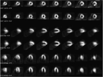
Imaging of myocardial perfusion has been used extensively in the assessment of myocardial ischaemia, infarction, and viability in both congenital and acquired cardiac disease. These studies are performed by injecting a radioisotope, which is a metastable element that emits photons, and is taken up by functioning cardiac myocytes. These myocytes then emit a signal that can be captured by a gamma scintillation camera and processed with a digital computer system. By arranging a series of rotating head cameras that acquire images in multiplane slices, one is able to generate a tomogram of the photon emitting myocardial tissue, hence the name single photon emission computed tomography. The magnitude of the uptake of the isotope correlates with the thickness of the tissue. Hence, the right ventricle is not typically imaged with this modality unless it is hypertrophied from pressure overload. Injured, diseased or dead myocardial cells will not take up the radioisotope, resulting in a perfusion defect on resting and/or stress images (Fig. 8). Two isotopes are commonly used. The tracer Thallium-201 was once commonly used in most major paediatric centers, but has given way to technetium, a radioactive element and product of uranium decay. Thallium has the advantage of rapid clearance, allowing for less time between resting images and images obtained at peak exercise. Its rates of washout, however, have been quite variable in children.Reference Flynn, Wernovsky, Summerville, Castaneda and Treves31 Furthermore, there is a relatively high incidence of false positives with this element during pharmacologic testing in adults with coronary arterial disease.Reference Yamazumi, Kobayashi and Horie32 Technetium 99 m has a few advantages over thallium, principally its improved radiation dosimetry, with less absorption of radiation by the gonads.Reference Flynn, Wernovsky, Summerville, Castaneda and Treves31 The disadvantage with technetium is its low hepatic clearance. Commonly one must wait at least one hour to avoid marked hepatic activity that can obscure results. Technetium linked to methoxy-isobutyl-isonitrile, nonetheless, is employed in many centers.
SPECT pre-operative images of an 8 year old who had anomalous right coronary from the left facing sinus. Note the reversible, inferior basal perfusion defect (top right frames at 8 o'clock).

Stress echocardiography
Kraunz and KennedyReference Kraunz and Kennedy33 were the first to report on the motion of the ventricular walls subsequent to exercise using echocardiography. In 1986, Berthe and colleaguesReference Berthe, Pierard and Hienaux34 were the first to report the use of pharmacological stress of the myocardium using dobutamine and then assessed by echocardiography. There is ample experience with this modality in adults with coronary arterial disease, but it remains a relatively infrequently used modality in children.
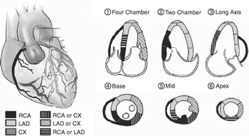
Stress echocardiography utilizes standardized echocardiographic views before and after stress. The stress can be the result of exercise, using a treadmill or cycle ergometer, or from the use of pharmacologic agents. The pharmacologic agents most commonly employed include dobutamine, adenosine and dipyridamole. Dobutamine mimics exercise by increasing heart rate and blood pressure, whereas adenosine causes vasodilation of normal coronary vasculature that leads to steal away from diseased segments, and the subsequent development of abnormalities of mural motion. Dipyridamole inhibits cyclic nucleotide phosphodiesterase, and inhibits the uptake of adenosine. Wall motion is analyzed either globally or by grading 16 myocardial segmentsReference Lang, Bierig and Devereux35, Reference Kimball36 (Fig. 9). Ischaemia is manifest as either new or worsened abnormalities of wall motion.
Segmental analysis of left ventricular walls. Adapted from J Am Soc Echocardiogr 2005;18:1440--1463.

Dobutamine stress echocardiography has been validated as an accurate diagnostic tool in patients with Kawasaki Disease, and those who have undergone the arterial switch operation for transposition. Numerous studies of this technique in these acquired coronary arterial processes suggest that the technique offers excellent sensitivity and specificity, albeit that its predictive value in children with congenital abnormalities of the coronary arteries remains unknown.
Positron emission tomography
Positrons are positively charged electrons, generated with the use of a medical cyclotron. Commonly employed elements such as oxygen, nitrogen, and rubidium are made unstable in medical cyclotrons, producing very unstable isotopes that are safe for medical use. When injected into the targeted tissue, the positrons from these elements combine with the electrons in surrounding tissue and ‘annihilate’ each other. This annihilation reaction emits gamma ray photons, which are visible and therefore recordable with tomographic equipment. These tracers are excellent choices for myocardial blood flow studies. Advantages of positron emission tomography compared to single photon emission computerized tomography include higher count rates, better quality of images, superior attenuation correction algorithms, and greater ability to quantify myocardial blood flow and flow reserve, the latter with F-18 fluorodeoxyglucose.Reference Noto37 The disadvantages of positron emission tomography are that the tracers are very unstable and must be used immediately, requiring an on-site cyclometer and medical physicist. When compared to stress echocardiography, one must also consider the additional risk of exposure to ionizing radiation. As mentioned above, cardiac magnetic resonance imaging techniques can be used to in the functional assessment of coronary perfusion while avoiding the added risk of ionizing radiation exposure.
Management of coronary arterial abnormalities in children
In the current era, surgical correction of significant coronary arterial abnormalities can be undertaken with excellent results. Re-implantation of the anomalous left coronary artery arising from the pulmonary trunk, and either transcatheter or surgical obliteration of the haemodynamically significant coronary arterial fistula, are part of the routine care for children treated in paediatric cardiac centers. There are, however, controversies concerning the management of anomalous origin of either the left or right coronary artery from the opposite sinus of Valsalva with a subsequent course between the arterial trunks. These controversies include the technique of surgical management, and in whom surgery is indicated. The debate is particularly heated when considering management of the asymptomatic patient with anomalous origin of a coronary artery from the opposite sinus of Valsalva with no evidence of ischaemia.
At the centre of the discussion is the proper identification and stratification of risk, which likely will depend on the degree to which, if any, the coronary artery runs an intramural course. If present, the shared wall between the anomalous coronary artery and the much larger aorta places the coronary artery at risk for compression as predicted by the law of LaPlace. The law of LaPlace states that the wall tension within the vessel is proportional to the radius. As a result, the smaller coronary artery may be compressed by the larger aorta. This compression, or stretching, of the coronary artery within the wall of the larger aorta results in a change of its cross-sectional shape from circular to oval. For any given circumference, the largest cross-sectional area is described by a circle, and as the coronary artery assumes an oval shape, the caliber is reduced. Such compression can also result in an abnormal or slit-like orifice that is also a common feature of this anomaly. With an increase in blood pressure, such as occurs with exercise, the mural tension in the aorta can increase acutely, leading to acute coronary arterial compression, which may occur on top of chronic deformity. This compromise in coronary arterial flow occurs during times of increased myocardial demand for oxygen. The combination of coronary arterial deformity and increased myocardial demand for oxygen results in a critical reduction in the flow of blood through the coronary artery, leading to ischaemia, arrhythmias, and perhaps, sudden death.
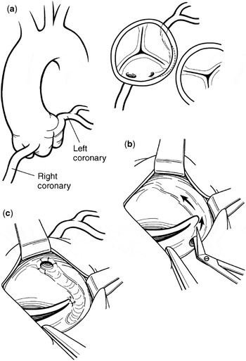
As a result, some have advocated surgical treatment that may include re-implantation, coronary arterial bypass grafting and coronary aterial unfoofing for these patients when the coronary artery takes an interarterial course.Reference Lorenz, Mookadam, Mookadam, Moustafa and Zehr10, Reference Erez, Tam, Doubin and Stakes38, Reference Frommelt, Frommelt, Tweddell and Jaquiss39 Coronary arterial bypass grafting is unlikely to be successful as the coronary vascular bed at risk is well supplied most of the time. Therefore, arterial grafts will fail to develop, and will not provide adequate protection during episodes of ischaemia. Furthermore, the procedure of grafting itself can lead to injury. Re-implantation is difficult because the intramural course makes it difficult to mobilize the proximal part of the coronary artery. The unroofing procedure is straightforward, and eliminates the ischaemic potential by removing the intramural course, at the same time eliminating any contribution from an abnormal coronary arterial orifice. The procedure involves unroofing the anomalous coronary artery into the aortic lumen (Fig. 10).Reference Frommelt, Frommelt, Tweddell and Jaquiss39, Reference Mustafa, Gula, Radley-Smith, Durrer and Yacoub40 The course of the anomalous coronary artery is generally at the level of the top of the zone of apposition between the right and left coronary aortic leaflets. In general, the unroofing can be accomplished without injuring the aortic valve, although the so-called commissure may have to be resuspended from the aortic wall. For patients with a long intramural course, it may be possible to unroof the coronary artery directly within the correct sinus of Valsalva, thereby eliminating the potential for injury to the aortic valve.Reference Van Son and Mohr41
Surgical procedure for unroofing of an anomalous coronary artery arising from the wrong sinus of Valsalva. The at risk anatomy is shown (a) with both a long intramural course of the left coronary artery combined with a slit like coronary ostium. The common wall is incised between the anomalous coronary and the aortic lumen (b). This eliminates both the abnormal origin as well as the intramural course. Fine tacking sutures are then placed to approximate the intima between the lumen of the coronary artery and aorta (c). Finally, the commissure between the right and left sinuses of Valsalva is resuspended. Reprinted with kind permission from ElsevierReference Frommelt, Frommelt, Tweddell and Jaquiss39.

Data from surgical treatment of anomalous origin of a coronary artery from the opposite sinus of Valsalva is very limited.Reference Erez, Tam, Doubin and Stakes38, Reference Frommelt, Frommelt, Tweddell and Jaquiss39 At present, as far as we are aware, results are available for only 17 instances of anomalous origin of a coronary artery from the opposite sinus of Valsalva that were managed surgically. While short-term results have been encouraging, and remain so, according to a personal communication from PC Frommelt and E Erez, mid- and long-term prevention of sudden death is unknown. The potential benefit from the procedure must be balanced with the early surgical morbidity and mortality, and the long-term risk of coronary arterial manipulation.
The success of pre-participation screening programmes in lowering the risk of sudden death in young athletes raises the question of whether avoidance of athletics and military participation in at-risk individuals might be sufficient to lower the risk of sudden death.Reference Mirchandani and Phoon14 At the very least, such conservative management should be part of the discussion with affected patients and their families. The role of non-surgical intervention, such as placement of coronary arterial stents, has not yet been established.
Only prospective monitoring of these patients through a national database carries the hope of providing answers to the outstanding questions concerning management. Stratification of risk based upon anatomical, symptomatic, and more accurate assessment of vulnerable myocardium may provide a basis for multi-prong strategies for treatment. Intravascular ultrasonography, or noninvasive nuclear myocardial viability screening, will offer promise to future patients found to have anomalous origin of a coronary artery from the opposite sinus of Valsalva.
Summary
Congenital coronary artery anomalies pose an array of challenges for the practitioner. These disorders are rare, their presentations range from the infant with heart failure to the adult with sudden death, and diagnosis may be difficult. An understanding of the anatomy of each defect and the impact of the specific anatomy on cardiovascular physiology is the most important weapon in the clinician’s armamentarium. Great strides have been made in the non-invasive imaging of coronary anomalies. However, no single diagnostic algorithm can be advocated. The sensitivity and specificity of imaging studies are operator-dependent and thus best practice depends on the available expertise in each clinical setting. When clinical suspicion exists, what is important is a definitive diagnosis with positive identification of the origin, course, and connections of each coronary, not the means by which this diagnosis is obtained.