Introduction
Objective tinnitus, also known as somatosound, generally originates in structures in and around the ear, and stimulates the patient's hearing in the same way as external sound. The most common sources of objective tinnitus are vascular abnormalities, palatal myoclonus, patulous Eustachian tube and temporo mandibular joint problems.Reference Herraiz and Aparicio1 The most frequent type of objective tinnitus is vascular tinnitus, which is characterised by a vascular bruit, a pulsatile murmur temporally related to or in synchrony with the heartbeat.
Both arterial and venous abnormalities around the ear can cause pulsatile, vascular tinnitus. Arterial causes include dural arteriovenous fistula or malformation, carotid artery atherosclerotic stenosis or aneurysm, and carotid cavernous fistula. Frequent venous causes include jugular bulb anomalies, transverse-sigmoid sinus stenosis or tortuosity, and sigmoid sinus diverticula (also termed aneurysm; a recently reported aetiology).Reference Chan2–Reference Otto, Hudgins, Abdelkafy and Mattox4
The diagnosis is based on clinical history, confirmed by physical examination and imaging studies. Recent investigations indicate that clinicians should be aware of pulsatile tinnitus related to the transverse-sigmoid sinus.
In this paper, we report a new, previously undescribed cause of pulsatile tinnitus. We examined the records of patients with pulsatile tinnitus, excluding those with paragangliomas or benign intracranial hypertension. We found that three patients presenting with pulsatile tinnitus had no signs of transverse-sigmoid sinus abnormality, but did have a focal defect of the mastoid bone shell overlying the transverse-sigmoid sinus, revealed by high-resolution computed tomography (CT) or computed tomographic angiography and venography. This study reports the clinical course of these three patients, and their responses to reconstructive surgery of the mastoid bone shell.
Patients and methods
A retrospective study was undertaken of patients presenting to our otolaryngology department between June 2008 and December 2010 with the chief complaint of pulsatile tinnitus. On detailed questioning, all patients met the criteria addressed by the Tinnitus Handicap Questionnaire, including tinnitus characteristics and temporal correspondence of the pulsatile aspect of the tinnitus with the resting pulse.
Patients with a known diagnosis of benign intracranial hypertension or paraganglioma were excluded.
All patients underwent full otomicroscopic, audiometric and tympanometric evaluation. They also underwent imaging studies, including high-resolution CT, computed tomographic angiography and venography, digital subtraction angiography and magnetic resonance imaging (MRI).
Three patients were identified whose high-resolution CT scans revealed a focal defect of the mastoid bone shell in the region of the transverse-sigmoid junction. The defect resulted in the mastoid cell directly enclosing the transverse-sigmoid sinus. In all cases, the side of the imaging finding corresponded to the side of the pulsatile tinnitus.
These three patients were offered surgical reconstruction of the mastoid bone shell adjacent to the transverse-sigmoid sinus, as a means of eliminating their pulsatile tinnitus. The surgical goal was to separate the mastoid air cell from the transverse-sigmoid sinus, in order to eliminate audible turbulence.
The surgical method followed that reported previously.Reference Otto, Hudgins, Abdelkafy and Mattox4 In each case, the mucosa of the mastoid cell was removed, and the mastoid bone shell adjacent to the transverse-sigmoid sinus was reconstructed with extraluminal placement of temporalis muscle, fascia and bone paté. The repair was then covered with a superiorly based periosteal flap and Gelfoam (Beijing Yierkang Bioengineering Development Center, Beijing, China).
Patients returned to the clinic one week post-operatively for evaluation. Audiometry was performed after resolution of haemotympanum (four weeks afterwards in all cases). The patients were questioned about post-operative symptoms and tinnitus resolution. Post-operative examination findings and audiometric results were also recorded.
Ethical considerations
All patients were informed of their condition in detail, and of the results of their CT scans. They all opted for surgical treatment, with the expectation that their tinnitus would be resolved. All patients supplied written consent for surgery. Confidentiality was preserved for all patients.
Results
In all three cases, a focal defect of the mastoid bone shell was identified in the region of the transverse-sigmoid junction.
The mean age of the patients at presentation was 40 years (range, 30–54 years). All patients were female.
None of the three patients had an audible bruit on the symptomatic side, either in the area of the external auditory canal or at the mastoid process. Symptomatic improvement could be achieved by external compression of the internal jugular vein in the neck on the symptomatic side.
Computed tomographic angiography and venography showed the focal defect.
All three patients opted for surgical intervention. In each case, the focal defect of the mastoid bone shell in the region of the transverse-sigmoid junction was easily identified. The area of the defect ranged from 1 × 2 to 4 × 5 mm. In no case was there a diverticulum in the vicinity of or confluent with the mastoid emissary vein.
There were no post-operative complications. All three patients reported complete resolution of their pulsatile tinnitus. The mean follow-up time was 11 months (range, 3–18 months). Long-lasting symptom resolution was achieved in all cases.
Case summaries
Case one
A 32-year-old woman, with no other medical history, presented with a 17-year history of pulsatile tinnitus in her right ear. The sound was synchronous with her heartbeat, and could be eliminated by manual compression of the upper right neck. The patient complained that the tinnitus interfered with her sleeping pattern, but did not report any hearing loss.
Audiometry revealed normal hearing.
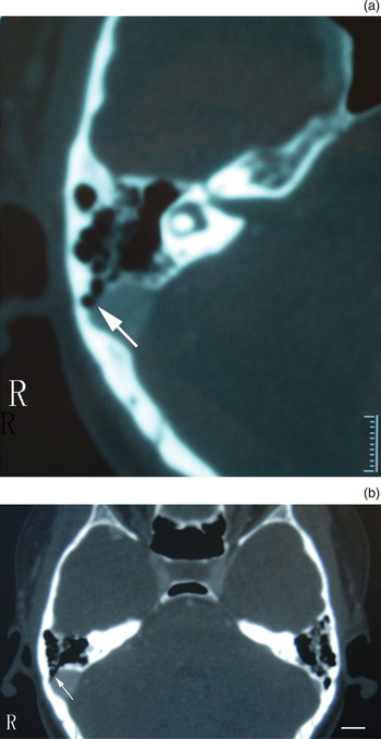
Computed tomographic angiography and venography identified a partial defect in the anterolateral mastoid bone shell of the right transverse-sigmoid sinus, at the posterior wall of the mastoid (Figure 1). No diverticulum or dural arteriovenous fistula was found.
Computed tomographic angiography and venography temporal bone scans for case one. (a) Bone window of a magnified axial image through the right mastoid, showing the focal defect (arrow) of the mastoid bone shell on the lateral surface of the right transverse-sigmoid sinus; the transverse-sigmoid sinus itself is complete and smooth. (b) Slightly more cephalic image showing contiguity of the focal defect (arrow) in the bony plate. R = right; scale bar = 1 cm

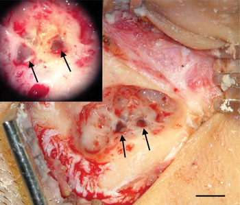
At surgical exploration, two defects were found, sized approximately 4 × 5 and 3 × 4 mm (Figure 2). The mastoid bone shell around the transverse-sigmoid sinus was reconstructed.
Intra-operative photograph of the mastoid bone shell defect around the transverse-sigmoid sinus, in case one. The defect creates two visible, bluish windows (arrows) just above the transverse-sigmoid junction. Inset shows a magnified view. Scale bar = 1 cm

The patient's pulsatile tinnitus was eliminated immediately after surgery, and remained so throughout her follow up. In the first night after her operation, the patient could not sleep without the tinnitus, which had been present for 17 years. Her post-operative audiogram was unchanged.
Case two
A 54-year-old woman had a six-year history of left-sided, pulsatile tinnitus which had recently worsened. There was no audible bruit over the left acoustic meatus or mastoid process.
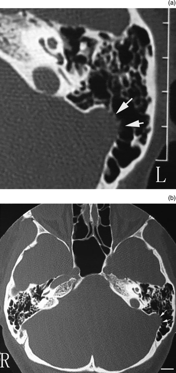
Computed tomographic angiography and venography demonstrated a defect of the left mastoid bone shell in the region of the transverse-sigmoid junction (Figure 3).
Computed tomographic (CT) arteriography and venography temporal bone scans for case two. (a) Bone window of a magnified, axial, high-resolution CT temporal bone scan, showing the focal defect (arrows) of the mastoid bone shell in the region of the left transverse-sigmoid junction. (b) Unmagnified image showing contiguity of the focal defect (arrows) in the bony plate. L = left; R = right; scale bar = 1 cm

Tinnitus evaluation indicated that the objective bruit volume was greater than the patient's subjectively perceived tinnitus loudness.
Audiography showed a mild, sloping, symmetrical sensorineural hearing loss between 4 and 8 kHz.
At surgical exploration, a 3 × 5 mm defect in the left mastoid shell was visible overlying the transverse-sigmoid sinus. The surgical method was the same as in case one.
Post-operatively, the patient reported resolution of her tinnitus, and her audiogram remained stable.
Case three
A 30-year-old woman complained of a 12-year history of left-sided, pulsatile tinnitus. She was able to eliminate the sound by pressing on her upper neck. No audible bruit was found over the left mastoid process or external auditory meatus.
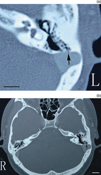
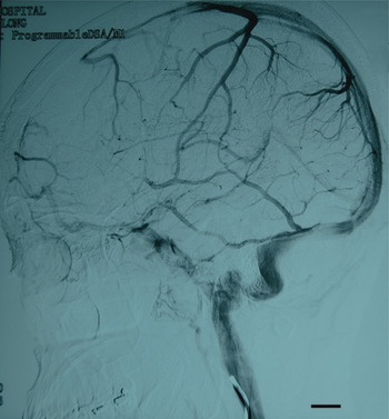
Computed tomographic angiography and venography demonstrated a small defect in the mastoid bone shell overlying the superior end of the left transverse-sigmoid sinus (Figure 4). Digital subtraction angiography showed no diverticulum or dural arteriovenous fistula (Figure 5).
Computed tomographic angiography and venography temporal bones scans for case three. (a) Bone window of an axial view showing a small defect of the adjacent mastoid bone shell on the lateral surface of the left sigmoid sinus (arrow). (b) Slightly more cephalic image showing contiguity of the focal defect (arrow) in the bony plate. L = left; R = right; scale bar = 1 cm

Digital subtraction angiogram of case three showing a normal transverse-sigmoid sinus, without venous aneurysm or diverticulum. Scale bar = 1 cm

Audiological assessment showed normal thresholds.
At surgery, the defect was easily found, and had an area of 1 × 2 mm. Because of its small size, the defect was covered by temporalis fascia alone.
Post-operatively, the patient reported complete alleviation of her pulsatile tinnitus.
Discussion
New findings and strengths of the study
Pulsatile tinnitus is frequently associated with identifiable and treatable causes with serious consequences, such as aneurysms and tumours. Thus, a high index of clinical suspicion is needed when assessing tinnitus patients. Sometimes, a detailed medical history alone will identify pulsatile tinnitus. An audible bruit may be detected by the diligent examiner, depending on the assessment techniques available. In the case of middle-ear disorders, otoscopy and audiometry are definitive in detecting changes. The distinction between arterial and venous bruits should be recognised by clinicians. Turning the head far to the contralateral side or compressing the upper ipsilateral neck will eliminate or attenuate most venous bruits.Reference Sismanis5
Radiological investigation is essential in cases of pulsatile tinnitus. Computed tomographic angiography and venography can also be valuable assessment tools, not only to distinguish arterial, venous, middle-ear and inner-ear causes of pulsatile tinnitus, but also to identify focal defects in the mastoid bone shell in the region of the transverse-sigmoid junction.Reference Otto, Hudgins, Abdelkafy and Mattox4, Reference Krishnan, Mattox, Fountain and Hudgins6 Non-contrast, high-resolution CT can be used to identify the defect, and MRI and magnetic resonance angiography may also be useful. Computed tomographic angiography and venography should not replace conventional angiography if dural arteriovenous fistula is suspected. Digital subtraction angiography can demonstrate a venous aneurysm or diverticulum of the transverse-sigmoid sinus.
In the current study, defects of the mastoid bone shell adjacent to the transverse-sigmoid junction were diagnosed with high-resolution CT or computed tomographic angiography and venography. Surgical inspection of the defects confirmed the radiological findings. Thus, we believe that, in such cases, and in the absence of vascular abnormalities, clinicians should pay more attention to the mastoid bone around the transverse-sigmoid sinus.
Comparison with other studies
As early as 1995, Mehall et al. reported a case with a ‘laterally placed sigmoid sinus’ and pulsatile tinnitus.Reference Mehall, Wilner and LaRouere7 Until recently, this structural abnormality of the sigmoid sinus has been referred to as a diverticulum or aneurysm.Reference Houdart, Chapot and Merland8, Reference Sanchez, Murao, de Medeiros, Kii, Bento and Caldas9
Cowley et al. reviewed the causes of tinnitus, and considered them to comprise flow velocity changes, aberrant flow and turbulent flow.Reference Cowley, Jones, Tuch and McAuliffe10 It is presumed that turbulence may be a contributing feature in all of these factors.Reference Cowley, Jones, Tuch and McAuliffe10 Most investigators agree that the turbulent blood flow associated with vascular diverticula is related to increased flow volume or lumen irregularity.Reference Herraiz and Aparicio1, Reference Otto, Hudgins, Abdelkafy and Mattox4, Reference Mehall, Wilner and LaRouere7, Reference Gologorsky, Meyer, Post, Winn, Patel and Bederson11
• Pulsatile tinnitus usually originates from vascular structures
• This paper reports a new cause of pulsatile tinnitus: a focal mastoid bone shell defect near the transverse-sigmoid junction
• Transmastoid reconstructive surgery of the mastoid bone shell was curative
In our patients, pulsatile tinnitus occurred in association with a normal transverse-sigmoid sinus, due to a defect in the mastoid bone shell. We infer from this that turbulent blood flow can be present in the normal transverse-sigmoid sinus, especially at or near the transverse-sigmoid junction. It is possible that the shape and angle of the transverse-sigmoid sinus and the resonating effect of the mastoid air cells are important in producing a venous bruit. A defect of the mastoid bone shell allows the sound to reach the middle ear, producing objective tinnitus. In all our patients, pulsatile tinnitus resolved post-operatively. From this, we conclude that a focal defect of the mastoid bone shell in the region of the transverse-sigmoid junction could be a previously unreported cause of pulsatile tinnitus.
Furthermore, we should consider whether pulsatile tinnitus is related to the size of the mastoid bone shell defect. What size of defect is too small or too large to cause tinnitus? In our three patients, the smallest mastoid bone shell defect was 1 × 2 mm (range, 1 × 2 to 4 × 5 mm). The sigmoid sinus is on occasion exposed to the mastoid cavity after middle-ear surgery, but we are unaware of any reports of pulsatile tinnitus in this clinical setting. Conversely, Duvillard et al. reported a case of pulsatile tinnitus of venous origin which resolved after mastoidectomy.Reference Duvillard, Ballester, Redon and Romanet12 In this latter case, possible explanations are that the defect was so large that the sound diffused post-mastoidectomy, or that mastoidectomy removed the resonance chamber amplifying the sound. Besides, it should be considered that the high-resolution computed tomography images behavior fuzziness due to the artifact of partial volume effects, in order to differentiate from the smaller size of the mastoid bone shell defect. All of these points deserve further study.
Clinical applicability of study findings
We have presented three patients with objective, pulsatile tinnitus who, on computed tomographic angiography and venography, were found to have a focal defect of the mastoid bone shell in the region of the transverse-sigmoid junction. We believe this report to be the first description of patients with a focal defect of the mastoid bone shell. Successful resolution of tinnitus was achieved in all three patients following transmastoid reconstructive surgery of the mastoid bone shell overlying the transverse-sigmoid sinus. We believe that a focal defect of the mastoid bone shell in the region of the transverse-sigmoid junction constitutes a previously unreported cause of pulsatile tinnitus.
Acknowledgements
This work was supported by the National Natural Science Foundation of China (Project 30872864). We wish to thank all the patients who kindly agreed to participate in this study, and their families, for their cooperation.