Introduction
Progression to lipid storage in adipocytes is characterised by two distinct phases. In the first step of hyperplastic expansion, progenitor cells are thought to become committed to the adipocyte lineage, after which they cannot revert back to a less differentiated ‘stem-like’ cell (Thompson et al., Reference Thompson, Lum, Nygaard, Kuestner, Kelly, Gimble and Moore1998; Boone et al., Reference Boone, Mourot, Gregiore and Remacle2000). Once committed, adipoblasts undergo an exponential replication phase that terminates and the cell cycle arrests at gap 1 (G1). Early markers of differentiation, such as lipoprotein lipase (LPL) are then expressed, and these cells, known as preadipocytes, may then undergo proliferation (Boone et al., Reference Boone, Mourot, Gregiore and Remacle2000). After preadipocytes stop proliferating, late markers of differentiation, such as glycerol-3-phosphate dehydrogenase (GPDH) and fatty acid synthetase (FAS) are detected. Cells then begin lipid accumulation in the cytosol at which time cells are termed adipocytes (Boone et al., Reference Boone, Mourot, Gregiore and Remacle2000).
Much of our understanding of these processes has developed through the use of in vitro models. A common approach when using in vitro models is to culture cells thought (or previously shown) to express adipogenesis lineage genes until they become confluent and then to apply a hormonal or xenobiotic agent ‘cocktail’ to ‘induce’ adipogenesis. Typically, these induction protocols include moderate to high concentrations of factors such as dexamethasone, methylisobutylxanthine and insulin in serum-containing media (Wu et al., Reference Wu, Bucher and Farmer1996; Sakaue et al., Reference Sakaue, Ogawa, Matsumoto, Kuroda, Takata, Sugimoto, Spiegelman and Kasuga1998; Habinowski and Witters, Reference Habinowski and Witters2001; Croissandeau et al., Reference Croissandeau, Chretien and Mbikay2002; Floyd and Stephens, Reference Floyd and Stephens2003; Shin et al., Reference Shin, Kim, Kim, Yoon, Choi and Yang2003). Both oleic (Guo et al., Reference Guo, Choi, Kirkland, Corkey and Hamilton2000) and linoleic (Kang et al., Reference Kang, Liu, Albright, Park and Pariza2003) acids were reported to induce differentiation in 3T3-L1 preadipocytes. However, octanoate appeared to attenuate differentiation in 3T3-L1 cells (Han et al., Reference Han, Farmer, Kirkland, Corkey, Yoon, Pirtskhalava, Ido and Guo2002). Conjugated linoleic acids have also been reported to inhibit differentiation in 3T3-L1 cells (Brodie et al., Reference Brodie, Manning, Ferguson, Jewell and Hu1999; Kang et al., Reference Kang, Liu, Albright, Park and Pariza2003).
One potential mechanism of enhanced adipogenesis from fatty acids (FA) may potentially be via peroxisome proliferator-activated receptors (PPARs) (Tontonoz et al., Reference Tontonoz, Hu and Spiegelman1994a and Reference Tontonoz, Hu, Graves, Budavari and Spiegelman1994b; Grimaldi, Reference Grimaldi2001; Bishop-Bailey and Wray, Reference Bishop-Bailey and Wray2003). During differentiation of preadipocytes to adipocytes, PPAR-γ, PPAR-α and CCAAT/enhancer-binding protein factors (C/EBP) α, β, δ and ζ are important transcription factors involved in the regulation of this mechanism (Lee et al., Reference Lee, Hausman and Dean1999; Kersten et al., Reference Kersten, Mandard, Tan, Escher, Metzger, Chambon, Gonzalez, Desvergne and Wahli2000; Lacasa et al., Reference Lacasa, Garcia Dos Santos and Giudicelli2001); PPAR-γ being activated prior to the activation of C/EBP-α. PPAR-γ is also required for survival of mature adipocytes (Imai et al., Reference Imai, Takakuwa, Marchand, Dentz, Bornert, Messaddeq, Wendling, Mark, Desvergne, Wahli, Chambon and Metzger2004), and is activated by a variety of factors, including glucocorticoids (Wu et al., Reference Wu, Bucher and Farmer1996), unsaturated FA such as oleic acid, linoleic acid, eicosapentaenoic acid and arachidonic acid (Amri et al., Reference Amri, Bonino, Ailhaud, Abumrad and Grimaldi1995) and insulin-sensitising reagents such as thiazolidinidiones (Spiegelman, Reference Spiegelman1998; Torii et al., Reference Torii, Kawada, Matsuda, Matsui, Ishihara and Yano1998; Yamauchi et al., Reference Yamauchi, Kamon, Waki, Murakami, Motojima, Komeda, Ide, Kubota, Terauchi, Tobe, Miki, Tsuchida, Akanuma, Nagai, Kimura and Kadowaki2001; Li and Lazar, Reference Li and Lazar2002). Hence, insulin is thought to be a factor in the increased expression of PPAR-γ. PPAR-γ activation also results in the initiation of expression of GLUT-4. This process also requires additional unknown factors, and appears to complete the regulatory pathway controlling glucose uptake in response to insulin (Wu et al., Reference Wu, Xie, Morrison, Bucher and Farmer1998).
Normal fasting concentrations of total plasma free FA fall approximately in the range 0.3 to 0.6 mmol/l and during exercise may be as high as 1.5 mmol/l. The contributions of each FA to total fasting FA is variable, but the major contributors are palmitic, 16 : 0 (20 to 25 mass %), oleic, 18 : 1 (15 to 25 mass %) and linoleic acids, 18 : 2 (22 to 30 mass %). In vivo, peripheral tissues, including adipose will utilise free FA from plasma and liberated FA from triglycerides facilitated by vascular endothelium LPL activity; the uptake of FA by adipocytes being favoured by the local concentration gradient. Thus, during periods of high nutrition (high energy intake) or exercise, peripheral tissues will be exposed to FA concentrations equal to or higher than the range shown above. It is difficult to emulate this model in vitro, but to better understand the mechanisms of utilisation of FA in cellular lipid accumulation, it may be necessary to provide FA in this physiological concentration range when performing in vitro studies.
In the present study, we report induction of lipid accumulation in 3T3-L1 cells. We asked the question: can 3T3-L1 cells be induced to accumulate lipid and ultimately progress through the process of adipogenesis in the absence of the effects of a hormone ‘cocktail’? The underlying hypothesis to the study was that supply of FA at physiological or slightly hyper-physiological concentrations would induce lipid accumulation and adipogenesis in confluent 3T3-L1 cells. This approach also facilitated investigation of the interaction of insulin effects. Thus, we also evaluated the response of this model to the presence or absence of insulin. To provide a model we considered physiologically appropriate in terms of energy substrate and lipid precursors, a standard defined medium (Dulbecco’s modified Eagle’s medium, DMEM), supplemented with individual FA at a concentration approximating total FA concentration in serum (0.3 mmol/l), protected by the addition of FA-free bovine serum albumin (BSA) (0.3 mmol/l) was used. We report the effects of treatment on cell number, cellular lipid accumulation and expression of adipogenic markers PPAR-γ and GLUT-4 proteins in this model.
Material and methods
Material
Cell-culture plasticware was supplied by Nunc (Naperville, IL, USA). DMEM, foetal bovine serum (FBS), penicillin-streptomycin, gentamicin, insulin and trypsin were purchased from Gibco BRL (Rockville, MD, USA). BSA (FA free), insulin, oil-red O stain, oleic and linoleic acid adsorbed to BSA were obtained from Sigma (St Louis, MO, USA). Rabbit anti-PPAR-γ (RDi-PPARGabrx) was obtained from Research Diagnostics, Inc. (Flanders, NJ, USA). Rabbit anti-GLUT-4 antibody (GLUT4ab654) was obtained from Abcam (Cambridge, MA, USA). Secondary antibody was Alexa Fluor 680 goat anti-rabbit IgG obtained from Molecular Probes (Eugene, OR, USA).
Cell culture
3T3-L1 preadipocytes were plated into 24-well plates at a density of 80 000 cells per well, and cultured at 37°C, 5% CO2 in DMEM + 10% FBS + antibiotics. The confluent cultures (2 to 3 days) were then rinsed three times with DMEM and transferred to a defined medium treatment, or control cultures were maintained in DMEM + 10% FBS + antibiotics. Media were changed every 48 h. Parallel cultures were grown in T-75 flasks for protein studies. Cell lysates were obtained at days 4 and 8 of each treatment.
Cells were cultured in DMEM with either 0.3 mmol/l oleic or linoleic acid plus 0.3 mmol/l FA-free BSA, and either in the presence or absence of 200 nmol/l insulin. FAs were purchased (non-covalently) adsorbed to BSA, protecting cells from the effects of unbound FA and providing similar FA availability to that in vivo.
Cell number and cell morphology
Cell number was determined by counting cells in each of 10 fields. Cultures from 24-well plates were fixed with 10% formalin and stained with oil-red O, to detect the presence of lipid, and counterstained with Harris haematoxylin.
Preparation of cell lysates and Western blot
Cells were freeze-fractured in liquid nitrogen, and cell lysates prepared in a buffer consisting of 50 mmol/l Tris, 1% NP-40, 0.25% sodium deoxycholate, 150 mmol/l NaCl, 1 mmol/l ethylenediamine tetraacetic acid (EDTA), 1 mmol/l sodium vanadate, plus Complete Mini-Cocktail (Roche Diagnostics; GmBH, Germany) pH 7.3 (Hill et al., Reference Hill, Strat, Hughes, Kokta, Dodson and Gertler2004). Total protein was determined using the BCA assay (Pierce Chemical Co., Rockford, IL, USA) and 20 μg protein was loaded per lane on SDS PAGE. Resolved proteins were transferred to nitrocellulose and detected in Western blot using (1/1000) polyclonal rabbit anti-PPAR-γ antibody or rabbit anti-GLUT-4 antibody, and secondary antibody (1/5000) Alexa Fluor 680 goat anti-rabbit IgG and scanned on an infra-red fluorescence imager (Odyssey; LI-COR, Inc., Lincoln, NE, USA).
Design and statistical analyses
Studies were designed as reported by others using cell line models (Chiaramonte et al., Reference Chiaramonte, Bartolini, Testolin and Comi1998; Schneider et al., Reference Schneider, Lanz, Ramer, Schaefer and GoppeltStruebe2001). Thus, each experiment was replicated using triplicate (or a greater number of replicate) assays from duplicate cultures for each test condition. Statistical analyses were conducted using the Statistical Analysis Systems Institute (2002). Lipid accumulation data were treated as binomial responses. Accordingly, cell counts were logit transformed, and analysed using a binomial probability distribution in PROC GENMOD. The model included effects of day (4 and 8), FA, insulin and the resulting interactions. Total cell numbers, PPAR-γ and GLUT-4 protein expression were analysed using the GLM procedure with a model including effects of day, treatment, and a day-by-treatment interaction. A single degree of freedom contrast was tested to compare controls with cultures treated with FA and insulin. Additional single degree of freedom contrasts were used to test the main effects of FA and insulin, and the resulting interaction. When a FA × insulin × day interaction was observed, main effects of FA and insulin and the resulting interaction were tested by day.
Results
Cell number
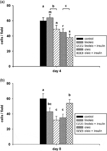
At day 4 of incubation, cell number per field of view was lower (P < 0.05) for insulin – compared with non-insulin-treated cultures and for oleic acid – compared with linoleic acid-treated cultures (Figure 1). A FA × insulin interaction was noted (P < 0.05) on day 8 of incubation in which there was a greater number of cells in cultures treated with oleic acid plus insulin compared with cultures treated with oleic acid alone or with linoleic acid plus insulin. It should be noted that one factor contributing to change in cell number was mean cell size. Although cell size was not quantified, it may be assumed that cells filling with lipid become larger and thus cell number expressed as cells/field will decrease.
3T3-L1 cells were cultured in 0.3 mmol/l oleic or linoleic acid, with or without 200 nmol/l insulin, for up to 8 days. Control cells were cultured in Dulbecco’s modified Eagle’s medium + 10% foetal bovine serum. For each treatment, cells were cultured in duplicate and 10 fields per culture were enumerated. Data show cell number per field (s.e.) for each treatment group cultured for (a) 4 days and (b) 8 days. There was a significant fatty acid (FA) effect on day 4 (a, b, c, P < 0.05) and a FA × insulin interaction on day 8 (P < 0.05). Also on day 4, there was a significant effect of the presence or absence of insulin (m, n and r, s, each P < 0.05).

Lipid accumulation
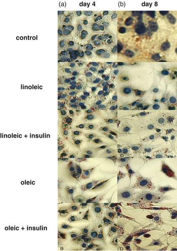
Qualitatively, lipid accumulation was difficult to detect in control cells cultured in DMEM + 10% FBS (Figure 2). In both oleic acid- and linoleic acid-treated cells, in the presence and absence of insulin on days 4 and 8, oil-red O stain-positive cells showed abundant lipid.
3T3-L1 cells were cultured, either in control medium (Dulbecco’s modified Eagle’s medium (DMEM) + 10% foetal calf serum) or in DMEM plus bovine serum albumin adsorbed fatty acids (0.3 mmol/l) as follows: either, linoleic acid, linoleic acid plus 200 nmol/l insulin, oleic acid or oleic acid plus 200 nmol/l insulin, and fixed and stained with oil-red O, after either 4 or 8 days.

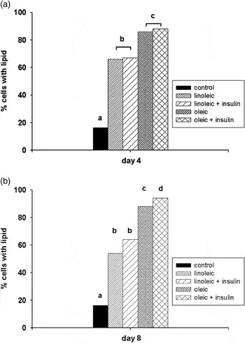
Quantitatively, the percentage of cells showing lipid accumulation was greater in FA-treated cultures compared with control cells grown in DMEM plus serum (P < 0.001). At day 4 of incubation, the percentage of cells showing lipid when cultured in FA in the absence of insulin, was lower compared with those cultured in FA in the presence of insulin (P < 0.01, Figure 3a). Also on day 4, the percentage of cells showing lipid, when cultured in linoleic acid was lower compared with those cultured in oleic acid (P < 0.01). On day 8, a FA × insulin interaction was noted (P < 0.05) in which lipid accumulation was greatest (P < 0.05) for cultures containing oleic acid plus insulin (Figure 3b). Additionally, cultures containing oleic acid alone showed greater (P < 0.05) lipid accumulation compared with cultures containing linoleic acid with or without insulin (Figure 3b).
3T3-L1 cells were cultured in 0.3 mmol/l oleic or linoleic acid, with or without 200 nmol/l insulin, for up to 8 days. Control cells were cultured in Dulbecco’s modified Eagle’s medium (DMEM) + 10% foetal bovine serum. For each treatment, cells were cultured in duplicate and 10 fields per culture were enumerated. Data are the percentage of cells showing lipid accumulation for each treatment group. (a) Day 4, the percentage of cells showing lipid accumulation was greater in fatty acid (FA)-treated cultures compared with control cells grown in DMEM plus serum (P < 0.001). Main effects of FA and insulin: the percentage of cells showing lipid, when cultured in FA in the absence of insulin, was lower compared with those cultured in FA in the presence of insulin (P < 0.01), and when cultured in linoleic acid was lower compared with those cultured in oleic acid (P < 0.01). Bars with different superscripts differ, oleic v. linoleic acid (insulin effect not denoted). (b) Day 8, the percentage of cells showing lipid accumulation was greater in FA-treated cultures compared with control cells grown in DMEM plus serum (P < 0.001). There was a FA × insulin interaction: lipid accumulation was greatest (P < 0.05) for cultures containing oleic acid plus insulin. Cultures containing oleic acid alone showed greater (P < 0.05) lipid accumulation compared with cultures containing linoleic acid with or without insulin.

PPAR-γ protein expression
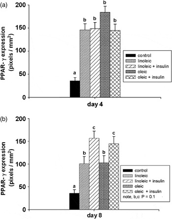
Treatment with both linoleic and oleic acid-containing media evoked higher levels of PPAR-γ than observed in DMEM plus serum on both days 4 and 8 (P < 0.05, Figure 4a and b). There were no differential effects of FA or insulin on PPAR-γ on day 4 (Figure 4a). After 8 days of culture, each FA showed a similar pattern in which the presence of insulin tended to increase PPAR-γ (P = 0.1, Figure 4b).
3T3-L1 cells were cultured in 0.3 mmol/l oleic or linoleic acid, with or without 200 nmol/l insulin, for up to 8 days. Control cells were cultured in Dulbecco’s modified Eagle’s medium+10% foetal bovine serum. For each treatment, cells were cultured in duplicate and peroxisome proliferator-activated receptor-gamma (PPAR-γ) protein was quantified (triplicate assays per culture) in Western blots of cell lysates using scanning laser excitation fluorescence (Odyssey, LI-COR). Data are expressed as pixels per mm2 (s.e.), from 20 μg protein per lane. (a) Day 4, overall treatment effect, PPAR-γ was increased v. control, (P < 0.001). There was no insulin or fatty acid (FA) effect. (b) Day 8, overall treatment effect, PPAR-γ was increased v. control (P < 0.001). Each FA showed a similar pattern in which the presence of insulin tended to increase PPAR-γ (P = 0.1).

GLUT-4 protein expression
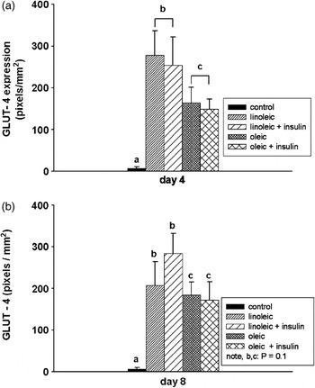
GLUT-4 protein appeared to be close to background levels in Western blots of cell lysates from control (DMEM plus serum) cultures. Treatment with both linoleic and oleic acid-containing media evoked higher levels of GLUT-4 than observed in DMEM plus serum on both days 4 and 8 (P < 0.001, Figure 5a and b). There was no effect of insulin on GLUT-4 on day 4, reflecting a similar observation of PPAR-γ. However, there was a FA effect in which cells cultured in linoleic acid showed higher GLUT-4 protein expression than cells cultured in oleic acid (P < 0.05, Figure 5a). Similar results were observed after 8 days of culture, in which there was no effect of insulin, and cells cultured in linoleic acid tended to show higher GLUT-4 expression than those cultured in oleic acid (P = 0.1, Figure 5b).
3T3-L1 cells were cultured in 0.3 mmol/l oleic or linoleic acid, with or without 200 nmol/l insulin, for up to 8 days. Control cells were cultured in Dulbecco’s modified Eagle’s medium+10% foetal bovine serum. For each treatment, cells were cultured in duplicate and GLUT-4 protein was quantified (triplicate assays per culture) in Western blots of cell lysates using scanning laser excitation fluorescence (Odyssey, LI-COR). Data are expressed as pixels per mm2 (s.e.) from 20 μg protein per lane. (a) Day 4, overall treatment effect, GLUT-4 was increased v. control, (P < 0.001). There was no insulin effect. Protein expression of GLUT-4 was higher in cells cultured in linoleic acid compared with oleic acid-containing media (P < 0.05). (b) Day 8, data were similar to day 4, for the overall treatment effect, GLUT-4 was increased v. control (P < 0.001). There was no insulin effect, and cells cultured in linoleic acid tended to show higher GLUT-4 expression than those cultured in oleic acid-containing media (P = 0.1).

Discussion
Cell number was lower in cultures treated with FA alone and FA plus insulin in comparison to the controls. Presumably, FA treatment results in withdrawal from cell cycle, whereas cells in control media continued to proliferate. These data are consistent with a study of human preadipocytes by McNeel et al. (Reference McNeel, Smith and Mersmann2003) who observed that the addition of either oleic acid or conjugated linoleic acid (in the presence of foetal calf serum (FCS), insulin, hydrocortisone and transferrin) resulted in cell number being 30% lower than in controls.
In the present study, effects of FA on cell number were not fully elucidated, and different FA may potentially have different effects. Others have found that oleic acid was better tolerated than palmitic acid, and may even have a role in rescuing cells from apoptosis in mouse pancreatic β-cells (Listenberger et al., Reference Listenberger, Han, Lewis, Cases, Farese, Ory and Schaffer2003) and rat ventricular myocytes (De Vries et al., Reference De Vries, Vork, Roemen, De Jong, Cleutjens, Van der Vusse and Van Bilsen1997) through the promotion of triglyceride accumulation instead of apoptotic pathways or alterations of the lipid membrane as seen by saturated FA. Evidence of apoptosis was not detected in the present study, caspase-3 activity being low and invariable across all treatments (data not shown). Another aspect of enumeration of these cells is the inevitable increase in cell size with lipid accumulation. Thus, part of the apparent decrease in cell number (expressed here as cells/field) may be attributed to larger cells occupying each field, and not loss of cells through mechanisms of cell death.
As most of the protocols used to study lipid accumulation of preadipocytes use high concentrations of insulin, there appears to be a lack of documented studies on the effects of the presence or absence of insulin in conjunction with physiological concentrations of FA. A novel finding of the present study is that treatment with oleic or linoleic acid, independent of insulin, was sufficient to induce lipid accumulation in 3T3-L1 cells. Many studies of adipocyte lineage cells report the addition of various hormone cocktails either with FCS (Brodie et al., Reference Brodie, Manning, Ferguson, Jewell and Hu1999; Guo et al., Reference Guo, Choi, Kirkland, Corkey and Hamilton2000; Kang et al., Reference Kang, Liu, Albright, Park and Pariza2003; McNeel and Mersmann, Reference McNeel and Mersmann2003; McNeel et al., Reference McNeel, Smith and Mersmann2003) or without FCS (Ding and Mersmann, Reference Ding and Mersmann2001; Ding et al., Reference Ding, McNeel and Mersmann2002; Hsu and Ding, Reference Hsu and Ding2003; Hutley et al., Reference Hutley, Newell, Joyner, Suchting, Herington, Cameron and Prins2003) in addition to FA to promote differentiation. These studies report that oleic and linoleic acids promote lipid accumulation in adipocytes. Oleic acid increased differentiation over controls in 3T3-L1 cells (Guo et al., Reference Guo, Choi, Kirkland, Corkey and Hamilton2000), and human preadipocytes (McNeel et al., Reference McNeel, Smith and Mersmann2003). Linoleic acid has been reported to increase differentiation over control in human preadipocytes (Brown and McIntosh, Reference Brown and McIntosh2003; Hutley et al., Reference Hutley, Newell, Joyner, Suchting, Herington, Cameron and Prins2003) and 3T3-L1 adipocytes (Kang and Pariza, Reference Kang and Pariza2001; Kang et al., Reference Kang, Liu, Albright, Park and Pariza2003). In each of these studies, cells were induced to differentiate with a hormone ‘cocktail’, and the resulting cultures were continuously maintained in media containing either no serum, but including a hormone ‘cocktail’ (Hutley et al., Reference Hutley, Newell, Joyner, Suchting, Herington, Cameron and Prins2003), or FCS plus or minus FA and apparently high concentrations of insulin for a further 2 to 12 days (Kang and Pariza, Reference Kang and Pariza2001; Hutley et al., Reference Hutley, Newell, Joyner, Suchting, Herington, Cameron and Prins2003; Kang et al., Reference Kang, Liu, Albright, Park and Pariza2003).
In the present study, oleic acid appeared to be more effective than linoleic acid in inducing lipid accumulation, with over 90% of cells accumulating lipid in response to oleic acid in comparison to about 60% of cells in response to linoleic acid. Oleic acid treatment retained higher efficacy on both days 4 and 8. This is apparently a more rapid response, although somewhat consistent with reports in the literature where oleic acid (plus FCS and hormone ‘cocktail’)-treated human adipocytes attained 90% differentiation by day 17 (McNeel et al., Reference McNeel, Smith and Mersmann2003), while another study reported about 65% differentiation in human preadipocytes treated with linoleic acid (plus hormone ‘cocktail’) on day 21 (Hutley et al., Reference Hutley, Newell, Joyner, Suchting, Herington, Cameron and Prins2003). It should be noted that each of these studies used primary cultures, and the remaining undifferentiated cells may have been non-adipocyte lineage cell types. In the present study, there were no differences in lipid accumulation between linoleic acid alone and linoleic acid plus insulin-treated cultures on either day 4 or day 8. Thus, perhaps surprisingly, it appears that insulin is not required to induce or maintain lipid accumulation in this model. The percentage of cells containing lipid in response to FA observed on day 4 of culture was only slightly modified by day 8, although the size and number of lipid droplets in each treatment may have increased (Figure 2, not quantified). Although insulin has been widely used in other studies of adipocyte development, in the present study, its addition to each of the FA media, had only a numerically small effect on lipid accumulation. Analysed as an overall main effect, the addition of insulin was significant (Figure 3). However, the numerically small effect observed suggests that in this in vitro model, insulin may be much less important for induction of lipid accumulation than indicated by other in vitro models.
PPAR-γ protein expression was increased by FA or FA plus insulin treatment by 2.5- to 4.5-fold. These data are consistent with a study by Kang et al. (Reference Kang, Liu, Albright, Park and Pariza2003) in 3T3-L1 cells where these authors reported that PPAR-γ expression was increased on days 4 and 7 compared with day 2 in response to linoleic acid. However, the additional effect of the use of a hormone ‘cocktail’ in the medium prevents a direct comparison with the data of the present study.
Further evidence that 3T3-L1 cells cultured in the presence of FA had entered the physiological state considered to be adipogenesis was demonstrated by greatly increased expression of GLUT-4. This observation is consistent with other studies in 3T3-L1 cells (El-Jack et al., Reference El-Jack, Kandror and Pilch1999; Gross et al., Reference Gross, Farmer and Pilch2004) in transition from preadipocyte to adipocyte morphology. Interestingly, in the present study no insulin effect on total cellular GLUT-4 expression was observed as might have been expected; although it may be assumed that GLUT-4 would be recruited to the plasma membrane in response to insulin. In all previously reported studies of GLUT-4 expression in adipocytes, insulin was used for the induction of lipid accumulation. Hence, the present study is the first to report the independent effects of FA (or FA plus insulin) on GLUT-4 in the absence of prior exposure to insulin. It appears that under the present experimental conditions, between culture days 4 and 8, these cells may be transitioning from preadipocytes to adipocytes. Thus, PPAR-γ and GLUT-4 are both expressed but additional, unknown intracellular regulatory mechanisms may not be fully activated.
Additionally, the effects of different FA on GLUT-4 expression have not yet been reported. We found linoleic acid to be initially more effective than oleic acid in inducing GLUT-4 expression. The differential response to linoleic and oleic acids was opposite in terms of lipid accumulation and GLUT-4 expression. This observation suggests that while oleic acid may be more lipogenic, linoleic acid up-regulated glucose transport, possibly reflecting a higher demand for ATP from glycolysis in its incorporation into lipid. The GLUT-4 response to oleic and linoleic acids is intriguing and suggests a direct PPAR-γ mediated mechanism. Further study is needed to clarify this issue. These data may be surprising, but indicate that insulin may not be essential for induction of lipid accumulation, or increased protein levels of PPAR-γ or GLUT-4.
This appears to be the first study to report lipid accumulation in adipocyte lineage cells in response to physiological concentrations of FA, in the absence of the influence of a complex hormone ‘cocktail’. Linoleic and oleic acids are considered to be ligands for PPAR-γ (Amri et al., Reference Amri, Bonino, Ailhaud, Abumrad and Grimaldi1995; Forman et al., Reference Forman, Chen and Evans1997; Margetic et al., Reference Margetic, Gazzola, Pegg and Hill2002; Ferré, Reference Ferré2004; Kokta et al., Reference Kokta, Dodson, Gertler and Hill2004), however there appear to be no other in vitro adipocyte lineage cell studies, which have investigated PPAR-γ expression or lipid accumulation in the absence of insulin. The strategy of the current study was to provide FA in the media at approximately physiological concentrations, as building blocks for lipid synthesis. Because FA form a regular component of the extracellular milieu in vivo, the interpretation of regulation of PPAR-γ expression in the absence of FA seems to be biologically irrelevant. In the present study, the effect of physiological concentrations of FA in the culture media on FA concentration in the intracellular or nuclear compartments were not investigated. This information would clarify the potential roles of these FA as PPAR-γ ligands.
Using the conventional hormone ‘cocktail’ adipogenesis induction approach, two pathways are rapidly activated in preadipocytes: in response to elevated cyclic AMP levels stimulated by methylisobutyl xanthine or glucocorticoids activating glucocorticoid response elements, expression of C/EBP-β and C/EBP-δ is rapidly increased while insulin signals to activate PPAR-γ transcription, via the 3-phosphoinositide-dependent protein kinase-1 (PDK-1) pathway following treatment (Ferré, Reference Ferré2004). An additional pathway regulating early events in differentiation appears to respond rapidly to FA and is regulated via PPAR-δ (Forman et al., Reference Forman, Chen and Evans1997). This pathway is independent of insulin signalling to activate PPAR-γ. It also appears that these pathways may be at least partially redundant (Grimaldi, Reference Grimaldi2001), and implies that insulin may not be essential for adipogenesis. The present study suggests that FA alone may induce the PPAR-δ pathway leading to progression of both PPAR-γ and GLUT-4 expression. Investigation of this hypothesis would require comprehensive study of role of FA in both the PPAR-δ and insulin-mediated pathways.
Conclusion
It appears that lipid accumulation occurs in response to either oleic or linoleic acids, and that both may act to increase PPAR-γ and GLUT-4 protein expression. These effects are independent of insulin. However, insulin appears to have a small enhancing effect on both lipid accumulation and PPAR-γ protein expression. Furthermore, 3T3-L1 cells can be induced to accumulate lipid in parallel to increased PPAR-γ and GLUT-4 protein expression in a serum-free medium supplemented with FA, without the use of typical induction protocols. The present report provides the basis of a novel model for the study of lipid accumulation and regulation of adipogenesis in adipocyte lineage cells. Because a complex hormone mix is not required, and energy and molecular building blocks are provided near to physiological levels for lipid synthesis, this model will provide additional insight into the regulation of adipogenesis.
Acknowledgements
This study was supported in part by National Research Initiative Competitive Grant numbers 2003-35206-12880 and 2003-35206-13617 from the USDA Cooperative State Research, Education and Extension Service.