INTRODUCTION
The flagellate protozoan Trypanosoma cruzi is the aetiological agent of Chagas disease or American trypanosomiasis. This parasite is mainly transmitted by the feces of triatomine insects and by other routes such as transfusion of blood products, organ transplantation, accidents in laboratories where parasites are handled live and by the congenital and oral routes, that will be addressed in the present review (Coura et al. Reference Coura, Junqueira, Fernandes, Valente and Miles2002; Hermann et al. Reference Hermann, Truyens, Alonso-Vega, Rodriguez, Berthe, Torrico and Carlier2004; Torrico et al. Reference Torrico, Alonso-Vega, Suarez, Rodriguez, Torrico, Dramaix, Truyens and Carlier2004; Yoshida, Reference Yoshida2008; Yoshida and Cortez, Reference Yoshida and Cortez2008; Raimundo et al. Reference Raimundo, Massad and Yang2009; Staquicini et al. Reference Staquicini, Martins, Macedo, Sasso, Atayde, Juliano and Yoshida2010). Trypanosoma cruzi transmission by vertical route and by transfusion of contaminated blood products have become the main mechanisms of Chagas disease transmission in non-endemic countries such as the USA and Spain, 2 of the most common destinations of Latin American people where there are estimations of more than 300 000 people infected with T. cruzi (Gascon et al. Reference Gascon, Bern and Pinazo2009; Schmunis and Yadon, Reference Schmunis and Yadon2009). The clinical features of T. cruzi infection range from the lack of symptomatology to the development of an inflammatory cardiomyopathy and enteric viscera dilation due to the damage of the enteric nervous system induced by the parasite in about 30–40% of people in the chronic stage of the infection (Coura, Reference Coura2007; Coura and Dias, Reference Coura and Dias2009; Moncayo and Silveira, Reference Moncayo and Silveira2009).
Carlos Chagas described the first case of congenital Chagas disease in 1901, in twins presenting convulsive episodes that consequently caused their death after 8 days of birth, revealing the parasites in their necropsies. Trypanosoma cruzi congenital infection can result in abortions, premature births or fetal organic lesions in liver, spleen, heart and central nervous system. According to reports by the Pan-American Health Organization (PAHO), it is estimated that in South and Central America, about 14 385 cases of congenital Chagas disease are presented annually. Trypanosoma cruzi infection by this route can be suspected in any newborn whose mother is infected, taking into account that congenital infection can occur in any moment of the pregnancy and in successive pregnancies. Although infection by this route cannot be prevented or treated during pregnancy (due to the lack of information about teratogenic risks of the available treatment and the high risk of adverse reactions), early diagnosis of infection in the newborn facilitates treatment as soon as possible, reaching a healing rate near to 100% and avoiding the progression of the disease to the chronic stage (World Health Organization, 2002; Carlier, Reference Carlier2005; Oliveira et al. Reference Oliveira, de Alencar, Tzelepis, Klezewsky, da Silva, Neves, Cavalcanti, Boscardin, Nunes, Santiago, Nobrega, Rodrigues and Bellio2010; Carlier et al. Reference Carlier, Torrico, Sosa-Estani, Russomando, Luquetti, Freilij and Albajar Vinas2011a, Reference Carlier, Truyens, Deloron and Peyronb; Yoshida et al. Reference Yoshida, Tyler and Llewellyn2011).
The first cases of T. cruzi oral transmission were reported for the first time in 1965 in Teutonia, Río Grande Del Sur (Brazil), where the affected population showed clinical features compatible with an acute infection with T. cruzi. Since then, multiple outbreaks of acute Chagas disease due to consumption of contaminated food and beverages have been reported in different states of Brazil, where the oral route emerges as the most important mechanism of transmission, as well as in other Latin American countries such as Argentina, Bolivia, Colombia, Ecuador and Venezuela (Toso et al. Reference Toso, Vial and Galanti2011). The source of contamination has been attributed to T. cruzi-infected insects that are macerated together with the fruits used for juice preparation and from meat from infected animals (Shikanai-Yasuda et al. Reference Shikanai-Yasuda, Marcondes, Guedes, Siqueira, Barone, Dias, Amato Neto, Tolezano, Peres, Arruda, Lopes, Shiroma and Chapadeiro1991; Silveira and Vinhaes, Reference Silveira and Vinhaes1999; Dias, Reference Dias2006; Steindel et al. Reference Steindel, Kramer Pacheco, Scholl, Soares, de Moraes, Eger, Kosmann, Sincero, Stoco, Murta, de Carvalho-Pinto and Grisard2008; Moncayo and Silveira, Reference Moncayo and Silveira2009; Pereira et al. Reference Pereira, Schmidt, Guaraldo, Franco, Dias and Passos2009, Reference Pereira, Schmidt, Barbosa, Guaraldo, Franco, Dias and Passos2010; Toso et al. Reference Toso, Vial and Galanti2011). All of these acute Chagas disease outbreaks associated with food and beverages consumption display as the common attribute more severe clinical characteristics compared with the clinical features of patients that have been infected with T. cruzi by other transmission routes (Coura et al. Reference Coura, Junqueira, Fernandes, Valente and Miles2002; World Health Organization, 2002; Pereira et al. Reference Pereira, Schmidt, Guaraldo, Franco, Dias and Passos2009, Reference Pereira, Schmidt, Barbosa, Guaraldo, Franco, Dias and Passos2010; Bisio et al. Reference Bisio, Seidenstein, Burgos, Ballering, Risso, Pontoriero, Moreau, Altcheh, Leguizamon, Freilij, Marceillac and Schijman2011; Vargas et al. Reference Vargas, Oporto and Espinosa2011).
Trypanosoma cruzi displays significant biochemical and genetic diversity. In 2009, during the most recent expert meeting held in Brazil, it was established that all T. cruzi strains should be classified into 6 DTUs (Discrete Typing Units) TcI–TcVI (Zingales et al. Reference Zingales, Andrade, Briones, Campbell, Chiari, Fernandes, Guhl, Lages-Silva, Macedo, Machado, Miles, Romanha, Sturm, Tibayrenc and Schijman2009, Reference Zingales, Miles, Campbell, Tibayrenc, Macedo, Teixeira, Schijman, Llewellyn, Lages-Silva, Machado, Andrade and Sturm2012). The poorly understood molecular epidemiology of congenital and oral transmission cases of Chagas disease have not been elucidated due to the absence of T. cruzi genotype discrimination involved in those cases. However, a few studies describe the genotypes involved in each transmission route and the possible association with disease outcome (Carlier, Reference Carlier2005; Virreira et al. Reference Virreira, Truyens, Alonso-Vega, Brutus, Jijena, Torrico, Carlier and Svoboda2007; Burgos et al. Reference Burgos, Altcheh, Petrucelli, Bisio, Levin, Freilij and Schijman2009; Corrales et al. Reference Corrales, Mora, Negrette, Diosque, Lacunza, Virreira, Breniere and Basombrio2009; Falla et al. Reference Falla, Herrera, Fajardo, Montilla, Vallejo and Guhl2009; Marcili et al. Reference Marcili, Valente, Valente, Junqueira, da Silva, Pinto, Naiff, Campaner, Coura, Camargo, Miles and Teixeira2009; Ramirez et al. Reference Ramírez, Guhl, Rendon, Rosas, Marin-Neto and Morillo2010; Bisio et al. Reference Bisio, Seidenstein, Burgos, Ballering, Risso, Pontoriero, Moreau, Altcheh, Leguizamon, Freilij, Marceillac and Schijman2011).
The aim of this review is to illustrate the recent research and advances in epidemiology, aetio-pathogeny, clinical features and diagnosis of oral and congenital Chagas disease in order to highlight the relevance of these two routes: Chagas congenital transmission spreading worldwide even in non-endemic countries, and oral Chagas disease as the responsible of multiple outbreaks of acute Chagas disease in regions where the vectorial route has been interrupted.
CONGENITAL CHAGAS DISEASE
Epidemiological aspects of congenital Chagas disease
Approximately 13 835 cases of congenital Chagas disease occur per year in Latin American countries (PAHO, 2006). As mentioned above T. cruzi infection congenitally acquired is responsible for many of the cases of Chagas disease in non-endemic countries. Considering the possible long-term consequences of Chagas disease attributed to the absence of an early reliable diagnosis and establishment of treatment, how convenient the screening of Chagas disease in pregnant immigrant women from endemic countries would be for a country´s economy. In a cost-effectiveness analysis performed by Sicuri et al. (Reference Sicuri, Munoz, Pinazo, Posada, Sanchez, Alonso and Gascon2011) in Spain, a comparison was performed between performing or not the necessary tests to detect Chagas disease in both, the pregnant woman and the newborn. A cost-effectiveness ratio of 22 €/QALYS gained was calculated in the newborn model in the case of performing screening and 125 €/QALYS gained in the opposite case. For the pregnant women model, a cost-effectiveness ratio of 96 €/QALYS gained was calculated in the case of performing the screening and a ratio of 1675 €/QALYS gained if the screening was not developed. This demonstrates the relevant impact of this transmission route and the reason why public health policies that allow early diagnosis and management of congenital Chagas disease should be established (Oliveira et al. Reference Oliveira, de Alencar, Tzelepis, Klezewsky, da Silva, Neves, Cavalcanti, Boscardin, Nunes, Santiago, Nobrega, Rodrigues and Bellio2010; Sicuri et al. Reference Sicuri, Munoz, Pinazo, Posada, Sanchez, Alonso and Gascon2011).
Clinical features and molecular epidemiology of congenital Chagas disease
At birth, most of the cases of congenital Chagas disease are asymptomatic. However, the presence of non-specific clinical features such as low birth weight, hepatosplenomegaly, anaemia, jaundice, symptoms of ARDS, pneumonia, gastrointestinal symptoms, meningoencephalitis and myocarditis as well as ocular lesions, intracranial calcification and microcephaly have been reported (Punukollu et al. Reference Punukollu, Gowda, Khan, Navarro and Vasavada2007; Schijman, Reference Schijman2006). Around the seventh month of life the congenitally infected patients develop their own antibody response being able to produce IgG antibodies, thus entering into the indeterminate phase of Chagas disease with subpatent parasitaemia. About 30% of T. cruzi congenitally infected patients will develop the symptomatic chronic phase of Chagas disease involving cardiac and digestive compromise (Carlier et al. 2011; Punukollu et al. Reference Punukollu, Gowda, Khan, Navarro and Vasavada2007; Rassi et al. Reference Rassi, Rassi and Marin-Neto2010; Schijman, Reference Schijman2006). In studies performed by Storino et al. (Reference Storino, Auger, Caravello, Urrutia, Sanmartino and Jörg2002), congenitally infected patients presented lower prevalence of cardiac alterations in comparison with patients infected by the vectorial route as well as a lower prevalence of cardiac eccentric hypertrophy (Bittencourt, Reference Bittencourt1963; Atias et al. Reference Atias, Morales, Munoz and Barria1985; Azogue et al. Reference Azogue, La Fuente and Darras1985; Storino et al. Reference Storino, Auger, Caravello, Urrutia, Sanmartino and Jörg2002;Torrico et al. Reference Torrico, Alonso-Vega, Suarez, Rodriguez, Torrico, Dramaix, Truyens and Carlier2004; Carlier, Reference Carlier2005; Schijman, Reference Schijman2006; Punukollu et al. Reference Punukollu, Gowda, Khan, Navarro and Vasavada2007).
In the reported cases of congenital Chagas disease, different T. cruzi genotypes have been reported as shown in Table 1. The DTU´s described in mothers are the same as those found in the neonates; in this sense the observed distribution of genotypes involved in congenital cases is similar to that observed in the infected local population (Carlier and Torrico, Reference Carlier and Torrico2003;Virreira et al. Reference Virreira, Truyens, Alonso-Vega, Brutus, Jijena, Torrico, Carlier and Svoboda2007; Burgos et al. Reference Burgos, Altcheh, Petrucelli, Bisio, Levin, Freilij and Schijman2009; Corrales et al. Reference Corrales, Mora, Negrette, Diosque, Lacunza, Virreira, Breniere and Basombrio2009; Falla et al. Reference Falla, Herrera, Fajardo, Montilla, Vallejo and Guhl2009; Bisio et al. Reference Bisio, Seidenstein, Burgos, Ballering, Risso, Pontoriero, Moreau, Altcheh, Leguizamon, Freilij, Marceillac and Schijman2011; Ramírez et al. Reference Ramírez, Guhl, Messenger, Lewis, Montilla, Cucunubá, Miles and Llewellyn2012). In order to determine whether T. cruzi congenital infection or whether the severity of congenital Chagas disease is determined by T. cruzi DTU's, several studies have been developed. Nevertheless, conclusive data associating T. cruzi genetic variability with the development or course of congenital cases are still lacking (Virreira et al. Reference Virreira, Truyens, Alonso-Vega, Brutus, Jijena, Torrico, Carlier and Svoboda2007; Burgos et al. Reference Burgos, Altcheh, Petrucelli, Bisio, Levin, Freilij and Schijman2009; Falla et al. Reference Falla, Herrera, Fajardo, Montilla, Vallejo and Guhl2009).
Trypanosoma cruzi genotypes involved in congenital Chagas disease

Laboratory diagnosis of congenital Chagas disease
The cases of congenital Chagas disease are set by the criteria previously established by different authors (Carlier, Reference Carlier2005; Carlier et al. 2011a). The serological diagnosis of a pregnant woman in the chronic phase of Chagas disease is based on 2 of 3 positive serological tests of different foundation (ELISA, IFAT, and IHA) that should be performed ideally on antenatal consultation during the pregnancy. However, in some circumstances (women in labour entering maternity wards without previous serological tests due to absence of antenatal controls and at primary health care facilities in rural endemic areas) the use of rapid diagnostic methods such as immunochromatographic, immunodot, and immunofiltration tests is an alternative; these tests present high sensitivity and specificity when the sample used is serum; however, the sensitivity of test decreases when the sample is whole blood, then the result must be confirmed by conventional serological methods (Ponce et al. Reference Ponce, Ponce, Vinelli, Montoya, de Aguilar, Gonzalez, Zingales, Rangel-Aldao, Levin, Esfandiari, Umezawa, Luquetti and da Silveira2005; Roddy et al. Reference Roddy, Goiri, Flevaud, Palma, Morote, Lima, Villa, Torrico and Albajar-Vinas2008; Ramirez et al. Reference Ramírez, Guhl, Umezawa, Morillo, Rosas, Marin-Neto and Restrepo2009; Carlier et al. 2011). In order to perform diagnosis of congenital Chagas disease in the newborn, a blood sample should be obtained either from the umbilical cord at birth or later by venopuncture. If a newborn has suggestive clinical features compatible with meningoencephalitis, a cerebrospinal fluid sample can be used to perform the diagnosis. Trypanosoma cruzi trypomastigotes can be detected by direct microscopic examination of blood samples, if these are negative, concentration techniques such as the Strout method or microscopic examination of buffy coat can be used; this latter technique is an inexpensive rapid test which can detect about 40 parasites/ml using about 4–6 tubes (Umezawa et al. Reference Umezawa, Nascimento, Kesper, Coura, Borges-Pereira, Junqueira and Camargo1996; Ponce et al. Reference Ponce, Ponce, Vinelli, Montoya, de Aguilar, Gonzalez, Zingales, Rangel-Aldao, Levin, Esfandiari, Umezawa, Luquetti and da Silveira2005; Virreira et al. Reference Virreira, Martinez, Alonso-Vega, Torrico, Solano, Torrico, Parrado, Truyens, Carlier and Svoboda2006; Roddy et al. Reference Roddy, Goiri, Flevaud, Palma, Morote, Lima, Villa, Torrico and Albajar-Vinas2008; Sosa-Estani et al. Reference Sosa-Estani, Gamboa-Leon, Del Cid-Lemus, Althabe, Alger, Almendares, Cafferata, Chippaux, Dumonteil, Gibbons, Padilla-Raygoza, Schneider, Belizan and Buekens2008; Ramirez et al. Reference Ramírez, Guhl, Umezawa, Morillo, Rosas, Marin-Neto and Restrepo2009).
Maternal antibodies disappear nearly 8–9 months after delivery. An alternative way to obtain a faster and reliable diagnosis is the detection of specific antibodies against SAPA (Shed Acute Phase Antigen) due to the disappearance of maternal IgG specific antibodies against this antigen at 3 months after delivery. Mallimaci et al. (Reference Mallimaci, Sosa-Estani, Russomando, Sanchez, Sijvarger, Alvarez, Barrionuevo, Lopez and Segura2010) evaluated a serological test using SAPA for the diagnosis of congenital infection and based on these results, the study concluded that it is possible to identify within 30 days of birth whether offspring born from infected mothers present congenital T. cruzi infection. When the mother is ELISA-SAPA non-reactive, the child is ELISA-SAPA reactive and the differential of ELISA-SAPA using matched samples of mother and child is ⩾1. In turn, the offspring does not present congenital T. cruzi infection within 30 days after birth when the child is SAPA non-reactive, both samples of the mother and child are reactive but the differential is <1 or the child is ELISA-SAPA non-reactive after 90 days (Umezawa et al. Reference Umezawa, Nascimento, Kesper, Coura, Borges-Pereira, Junqueira and Camargo1996; Torrico et al. Reference Torrico, Alonso-Vega, Suarez, Rodriguez, Torrico, Dramaix, Truyens and Carlier2004; Mallimaci et al. Reference Mallimaci, Sosa-Estani, Russomando, Sanchez, Sijvarger, Alvarez, Barrionuevo, Lopez and Segura2010). Polymerase chain reaction rarely detects T. cruzi DNA in the amniotic fluid, this being the reason why amniocentesis is not considered useful in the diagnosis of congenital infection with T. cruzi. Although this technique has not been validated for diagnosis yet, it can be used to follow-up patients under aetiological treatment or at risk of clinical re-activation (Carlier and Torrico, Reference Carlier and Torrico2003; Burgos et al. Reference Burgos, Begher, Freitas, Bisio, Duffy, Altcheh, Teijeiro, Lopez Alcoba, Deccarlini, Freilij, Levin, Levalle, Macedo and Schijman2005; Virreira et al. Reference Virreira, Truyens, Alonso-Vega, Brutus, Jijena, Torrico, Carlier and Svoboda2007; Duffy et al. Reference Duffy, Bisio, Altcheh, Burgos, Diez, Levin, Favaloro, Freilij and Schijman2009; Schijman et al. Reference Schijman, Bisio, Orellana, Sued, Duffy, Mejía-Jaramillo, Cura, Auter, Veron, Qvarnstrong, Deborggraeve, Hijar, Zulantay, Lucero, Velazquez, Tellez, Sanchez-Leon, Galvao, Nolder, Monje-Rumi, Levi, Ramírez, Zorrilla, Flores, Jercic, Grisante, Añez, De Castro, Gonzalez, Acosta, Yachelini, Torrico, Robello, Diosque, Triana, Aznar, Russomando, Buscher, Assal, Guhl, Sosa-Estani, Da Silva, Britto, Luquetti and Ladzins2011). Histopathology studies of the placenta can be performed after birth; however, is important to consider that the immune defences of the placenta are able to contain T. cruzi before it reaches the fetus, therefore the presence of parasites in the placenta does not confirm congenital transmission (Reyes et al. Reference Reyes, Lorca, Munoz and Frasch1990; Torrico et al. Reference Torrico, Alonso-Vega, Suarez, Rodriguez, Torrico, Dramaix, Truyens and Carlier2004; Luquetti et al. Reference Luquetti, Dias and Prata2005; Ponce et al. Reference Ponce, Ponce, Vinelli, Montoya, de Aguilar, Gonzalez, Zingales, Rangel-Aldao, Levin, Esfandiari, Umezawa, Luquetti and da Silveira2005;Virreira et al. Reference Virreira, Martinez, Alonso-Vega, Torrico, Solano, Torrico, Parrado, Truyens, Carlier and Svoboda2006; Roddy et al. Reference Roddy, Goiri, Flevaud, Palma, Morote, Lima, Villa, Torrico and Albajar-Vinas2008; Sosa-Estani et al. Reference Sosa-Estani, Gamboa-Leon, Del Cid-Lemus, Althabe, Alger, Almendares, Cafferata, Chippaux, Dumonteil, Gibbons, Padilla-Raygoza, Schneider, Belizan and Buekens2008; Carlier et al. 2011).
Physiopathogenic aspects of congenital T. cruzi infection
During pregnancy the immune system faces the dilemma between maintaining an immune regulatory state required for immune tolerance of the fetus and the ability to establish an adequate immune response to protect the fetus against pathogens, therefore the mechanisms underlying immune tolerance and their interaction with the virulence factors of T. cruzi should be understood (Munn et al. Reference Munn, Zhou, Attwood, Bondarev, Conway, Marshall, Brown and Mellor1998; Thellin and Heinen, Reference Thellin and Heinen2003; Mor and Cardenas, Reference Mor and Cardenas2010). Endocrine factors are an important part of the mechanisms underlying immune tolerance. Progesterone can interact with lymphocytes to stimulate the production of a 34 kDa protein named the progesterone-induced blocking factor (PIBF), which is able to induce a Th2 cytokine profile (involving the production of IL-4, IL-5, IL-6, IL-19 and TGF-B), and decrease the production of IL-2 and IFN-γ, leading to modulation of the immune response (Clark et al. Reference Clark, Ding, Yu, Levy and Gorczynski2001; Raghupathy et al. Reference Raghupathy, Al-Mutawa, Al-Azemi, Makhseed, Azizieh and Szekeres-Bartho2009; Thellin and Heinen, Reference Thellin and Heinen2003). Another mechanism involves the expression of Class I MHC as HLA-C, HLA-G and HLA-E instead of typical products of this group of genes such as HLA-A and HLA-B. Therefore the former can interact with the inhibitory receptor of Natural Killers (NKS) known as KIRs (Killer Inhibitory Receptors) avoiding its cytolytic activity. The production of indoleamine 2, 3-dioxygenase by syncytiotrophoblasts and the OX2 expression, also known as CD200, a glycoprotein that favours the Th1/Th2 balance towards Th2, inhibits macrophages and Fgl2 (fibrinogen-like protein 2), a pro-thrombinase capable of inducing abortion through placental thrombosis and complement activation (Munn et al. Reference Munn, Zhou, Attwood, Bondarev, Conway, Marshall, Brown and Mellor1998; Clark et al. Reference Clark, Ding, Yu, Levy and Gorczynski2001; Shakhawat et al. Reference Shakhawat, Shaikly, Elzatma, Mavrakos, Jabeen and Fernandez2010).
The placental cell population also plays an important role in the mechanisms of immune tolerance. Several studies developed by Mues et al. (1989), Heikkinen et al. (2003) and Svensson (2011), among others, have found that decidual macrophages are alternatively activated exerting an anti-inflammatory action with a high production of IL-10 and IL-1R. Regarding the defence mechanisms against infections during pregnancy, it is essential to mention some particularities. Human placental villi are covered by 2 layers of trophoblasts; an outer layer called the syncytiotrophoblast, which prevents intercellular penetration and remains interposed between the maternal blood and fetal tissues throughout the pregnancy, and an inner layer called the cytrotrophoblast (Langerhans cells) that also covers extravillous tissues such as the chorionic plate. The latter presents a high expression of TLRs 2 and 4 during the first quarter of pregnancy, which is related to a lower risk of congenital infection in comparison with other periods of pregnancy, together with the fact that the maternal blood supply is completed only after 12 weeks of pregnancy (the risk of congenital infection increases with the augmenting of pregnancy weeks. However, the severity of clinical features is inversely related to this latter) (Moya et al. Reference Moya, Villagra and Risco1979; Abrahams and Mor, Reference Abrahams and Mor2005; Carlier, Reference Carlier2005; Romero et al. Reference Romero, Gotsch, Pineles and Kusanovic2007; Carlier et al. 2011; Mor and Cardenas, 2010).
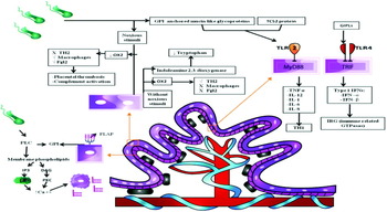
The risk of congenital infection is influenced by different factors; (i) the time: as mentioned before, with the increase of weeks of pregnancy the risk decreases; (ii) the parasitaemia in the pregnant woman: the risk of T. cruzi congenital infection is higher in pregnant women acutely infected with T. cruzi that show high parasitaemia in comparison to chronically infected women with a hardly detectable parasitaemia; (iii) the presence of an alteration in maternal-fetal barrier: a microorganism is considered a threat to the fetus if the cell layer of syncytiotrophoblasts is discontinued or if the pathogen gains access to the decidual compartment or to the placental villi (Bittencourt, Reference Bittencourt1963; Burgos et al. Reference Burgos, Begher, Freitas, Bisio, Duffy, Altcheh, Teijeiro, Lopez Alcoba, Deccarlini, Freilij, Levin, Levalle, Macedo and Schijman2005; Carlier, Reference Carlier2005; Redline, Reference Redline2006; Schijman, Reference Schijman2006; Carlier et al. 2011; Mor and Cardenas, 2011). After describing the main components participating in the pregnant woman immune state, this is the moment at which the question arises of how virulence factors of T. cruzi interact with these components in order to infect its host successfully. In an infected pregnant woman, the trypomastigotes located at the intervillous space are able to penetrate the trophoblasts where they turn into amastigotes and multiply mainly in the Hofbauer cells, creating a nest that is lysed later, releasing the parasites into the interstitial space causing an inflammatory reaction and necrosis of the chorionic villi (Schijman, Reference Schijman2006; Carlier et al. 2011). In the invasion of trophoblasts, Placental Alkaline Phosphatase (PLAP), a tetrameric protein anchored to the outer leaflet of the plasma membrane lipid bilayer of the syncytiotrophoblast by a molecule of glycosylphosphatidylinositol (GPI), plays an important role. Trypanosoma cruzi invasion seems to occur by hydrolysis of GPI by phospholipase C leading to generation of molecules such as diacylglycerol and inositol 1,4,5 triphosphate which are involved in the increase of intracellular calcium concentration, the re-arrangement of actin filaments of host cells and the release of human placental alkaline phosphatase (PLAP) in the dimeric form. This correlates with the findings of several studies that have shown a decrease of PLAP activity in T. cruzi-infected pregnant women (Sartori et al. Reference Sartori, Lin, Frank, Malchiodi and de Fabro2002; Schijman, Reference Schijman2006; Priotto et al. Reference Priotto, Sartori, Repossi and Valentich2009). As previously mentioned, cytrotrophoblasts are able to express TLRs 2 and 4. Activation of TLRs-2 triggers an intracellular signalling pathway dependent on MyD88, resulting in production of nitric oxide, pro-inflammatory cytokines such as IL-12, and induction of a Th1 response, this latter, mediated by GPI (glycosylphosphatidylinositol) anchored mucin-like glycoproteins and the released T. cruzi protein Tc52. The former molecules are distributed throughout the surface of the trypomastigotes and the latter, the Tc52, a protein composed of 2 domains; the first site called G (binding glutathione) responsible for the TLRs-2 activation and the second site called H (hydrophobic domain) associated with survival and virulence of the parasite (Buschiazzo et al. Reference Buschiazzo, Amaya, Cremona, Frasch and Alzari2002; Frasch, Reference Frasch2000; Jacobs et al. Reference Jacobs, Erdmann and Fleischer2009). Stimulation of TLR-2 seems to induce first quarter pregnancy trophoblasts to undergo apoptosis, leading to the barrier disruption and facilitating fetal infection (Abrahams and Mor, Reference Abrahams and Mor2005; Savino et al. Reference Savino, Villa-Verde, Mendes-da-Cruz, Silva-Monteiro, Perez, Aoki Mdel, Bottasso, Guinazu, Silva-Barbosa and Gea2007; Kayama and Takeda, 2009; Palm and Medzhitov, Reference Palm and Medzhitov2009). On the other hand, TLR-4 activation is mediated by The GIPLs (glycoinositolphospholipids) leading to activation of TRIF (domain-containing adapter-inducing interferon-β) to trigger an intracellular signalling pathway independent of MyD88 leading to the production of type 1 IFNs (IFN-α and IFN-β) and activation of IRG (immune-related GTPases) proteins, also known as p47 GTPases (also stimulated by IFN-γ) involved in the immune response against intracellular pathogens (Oliveira et al. Reference Oliveira, Torrico, Muñoz and Gascon2010a, b; Palm and Medzhitov, Reference Palm and Medzhitov2009; Taylor et al. 2004). The switch to the Th1 cytokine profile (IFN-γ, TNF-α, IL-12) activates NKS into LAK (Lymphokine-Activated Killer) which can exert cytolytic actions by stimulating the production of immunoglobulins (capable of activating the complement cascade) and by decreasing OX2 (CD200) leading to Fgl2 production. This promotes production of IL-8 (which attracts and activates neutrophils that attack the feto-placental structures) inducing the formation of clots that occlude the maternal-fetal circulation causing ischaemia, necrosis and death of fetal tissues. This can be seen in histopathological analysis of placentas of women infected with T. cruzi who have presented abortions, premature babies (who died in the neonatal period) or stillborn (Bittencourt, Reference Bittencourt1963; Raghupathy, Reference Raghupathy1997; Thellin and Heinen, Reference Thellin and Heinen2003; Torrico et al. Reference Torrico, Alonso-Vega, Suarez, Rodriguez, Torrico, Dramaix, Truyens and Carlier2004; Kayama and Takeda, 2009). The described immune aspects are summarized in Fig. 1.
Immunological aspects of Trypanosoma cruzi congenital infection.

TRYPANOSOMA CRUZI INFECTION BY THE ORAL ROUTE
Epidemiological aspects of T. cruzi infection by the oral route
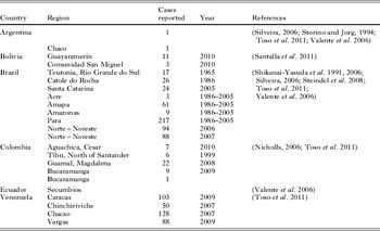
Nowadays the oral route is considered to be the most important transmission mechanism of T. cruzi in the Brazilian Amazon after the Pan-American Health Organization declared the interruption of vectorial transmission by Triatoma infestans (Yoshida, Reference Yoshida2006; Yoshida and Cortez, Reference Yoshida and Cortez2008; Pereira et al. Reference Pereira, Schmidt, Guaraldo, Franco, Dias and Passos2009, Reference Pereira, Schmidt, Barbosa, Guaraldo, Franco, Dias and Passos2010; Toso et al. Reference Toso, Vial and Galanti2011; Yoshida et al. Reference Yoshida, Tyler and Llewellyn2011). Food and beverages can be contaminated in areas where infected triatomine insects and T. cruzi reservoirs are found (Pereira et al. Reference Pereira, Schmidt, Guaraldo, Franco, Dias and Passos2009, Reference Pereira, Schmidt, Barbosa, Guaraldo, Franco, Dias and Passos2010; Bisio et al. Reference Bisio, Seidenstein, Burgos, Ballering, Risso, Pontoriero, Moreau, Altcheh, Leguizamon, Freilij, Marceillac and Schijman2011). Between 1968 and 2005 about 311 cases of food-borne Chagas disease have been reported. Therefore different studies have attempted to establish a means to prevent food contamination. The use of substances like 1% sodium hypochlorite, gentian violet and 70% ethanol as well as the use of methods such as food pasteurization have shown efficiency in eliminating T. cruzi-infective cells (Ferreira et al. Reference Ferreira, Martinho, Amato Neto and Cruz2001; Dias, Reference Dias2006). Multiple outbreaks of Chagas disease associated with food consumption, beverage drinking and consumtion of meat from hunting animals, have been reported as shown in Table 2 (Carlier and Torrico, Reference Carlier and Torrico2003; Pereira et al. Reference Pereira, Schmidt, Guaraldo, Franco, Dias and Passos2009, Reference Pereira, Schmidt, Barbosa, Guaraldo, Franco, Dias and Passos2010; Toso et al. Reference Toso, Vial and Galanti2011).
Cases reported of acute Chagas diseases associated with food/beverages consumption

Clinical features of T. cruzi infection by the oral route
Cases of acute Chagas disease due to food consumption present a wide range of symptoms that must be considered by the attending physician to develop an accurate diagnosis and initiate an appropriate management. The determinant factor in evaluation and diagnosis of patients with acute Chagas disease is the development of a complete medical history to establish links with the aetiological agent of this disease. In order to carry out the latter, the physician must take into account aspects such as the provenance and residency of the patient, and the presence in the personal background of events such as blood transfusions and organ transplants during the previous 4 months. It should be determined whether the patient has consumed uncooked food or not, food exposed to the feces of vectors or mammalian reservoir fluids, processed food using traditional methods, and also inquire about the occurrence of febrile illness in people with whom the patient has shared food in order to establish the mode of transmission involved (Yoshida, Reference Yoshida2006; Yoshida and Cortez, Reference Yoshida and Cortez2008; Yoshida et al. Reference Yoshida, Tyler and Llewellyn2011). An important aspect to consider is the incubation period, defined as the time between exposure to the pathogen and the signs and symptoms onset that will guide medical personnel to establish a possible diagnosis. In the case of acute Chagas disease, this period varies according to the transmission route. For the vectorial route, the incubation period is about 4 to 15 days, for the transfusional route it is about 8 to 120 days, for the vertical is about 3 to 22 days, varying according to T. cruzi strain and mother`s parasitaemia, and finally the incubation period for the oral route is about 3 to 22 days (Bisio et al. Reference Bisio, Seidenstein, Burgos, Ballering, Risso, Pontoriero, Moreau, Altcheh, Leguizamon, Freilij, Marceillac and Schijman2011). Trypanosoma cruzi infection by the oral route is associated with a high mortality rate in the first 2 weeks after infection. Mortality has been estimated to be about 8–35%, surpassing the calculated mortality by the vectorial route that has been estimated at less than 5–10%. Mortality is mainly associated with acute heart failure, myocarditis, and meningoencephalitis (Coura et al. Reference Coura, Junqueira, Fernandes, Valente and Miles2002; Punukollu et al. Reference Punukollu, Gowda, Khan, Navarro and Vasavada2007). In studies performed by Hoft et al. (Reference Hoft, Farrar, Kratz-Owens and Shaffer1996) a severe inflammation that even was extended to the gastric mucosa and to adjacent mesentery was observed at day 14 after infection. Trypomastigotes were observed in peripheral blood smears and inflammatory lesions were found in the pancreas, spleen, liver, bone narrow, heart, duodenum, brain and skeletal muscle, all of which demonstrated that the inflammatory response was unsuccessful in containing the local infection within the gastric mucosa. This led to the development of a systemic infection favoured by the disruption of the stomach serosal surface, allowing trypomastigotes released from infected cells to enter into the peritoneal cavity, that was correlated with the higher mortality in comparison with other routes of transmission. The clinical features of acute infection are variable. Within the reported symptoms are the presence of fever, dyspnea, myalgia, periorbital oedema, headache, heart murmurs, nausea, abdominal pain, hepatomegaly, chest pain, vomiting, palpitations, lower limb oedema, anasarca, abortion in pregnant women, syncope, rash, gastrointestinal tract bleeding, jaundice, elevated aminotransferases, enteritis, acute abdomen and multiple changes in diagnostic tests such as chest radiograph, electrocardiogram and echocardiogram (World Health Organization, 2002; Bisio et al. Reference Bisio, Seidenstein, Burgos, Ballering, Risso, Pontoriero, Moreau, Altcheh, Leguizamon, Freilij, Marceillac and Schijman2011). After the confirmation of a case of acute Chagas disease the next step is to determine the transmission route involved.
Molecular epidemiology of T. cruzi infection by the oral route
There are few reports regarding the incrimination of T. cruzi DTU´s in outbreaks of acute Chagas disease associated with food consumption because, in the vast majority of reported cases, the genotype characterization has not been performed. In studies developed by Marcili et al. (2009), T. cruzi DTU´s were characterized from wild primates, triatomines and people infected with T. cruzi associated with food consumption. The isolates found in primates and triatomines from Brazilian Amazon were genotyped as TcI and TcII and were very similar to those found in humans infected by the oral route with T. cruzi (Marcili et al. 2009). Andrade et al. (2011) examined T. cruzi strains from patients with oral Chagas disease reported in Santa Catarina in 2005 and 2 isolates that were obtained from Didelphis aurita and Triatoma tibiamaculata. This study found the presence of mixtures of different DTU´s (TcI, TcII and TcVI). According to several studies this could interfere with the course and severity of infections and with the host immune response, suggesting a strong association with the unusually high mortality index in this outbreak. However, there is stil not a convincing link between the severe clinical features of food-borne Chagas disease and T. cruzi genotypes found in this transmission route (Guhl and Vallejo, Reference Guhl and Vallejo1999; Campbell et al. Reference Campbell, Westenberger and Sturm2004; Macedo et al. Reference Macedo, Machado, Oliveira and Pena2004; Ramirez et al. Reference Ramírez, Guhl, Rendon, Rosas, Marin-Neto and Morillo2010, Reference Ramírez, Duque and Guhl2011). In unpublished observations performed by Ramirez et al. (Reference Ramírez, Duque and Guhl2011) 5 stocks (LJVP, EH, LER, NcH, XcH) were isolated from 2 cases of acute Chagas disease associated with food consumption reported in Colombia in 2009; these 5 stocks were characterized as TcI (Guhl and Ramirez, Reference Guhl and Ramirez2011). In the study, 2 of the 5 isolated stocks, LER and XcH showed a high degree of divergence including mixed TcI genotypes, demonstrating a pattern of contaminative transmission between transmission cycles of Chagas disease. On the other hand, mtMLST strategies demonstrated a recombination pattern in clones isolated from oral infection outbreaks, suggesting the importance of this infection route in association with the clinical manifestations of Chagas disease (Ramírez et al. Reference Ramírez, Guhl, Messenger, Lewis, Montilla, Cucunubá, Miles and Llewellyn2012).
Physiopathogenic aspects of T. cruzi infection by the oral route
Mucosal surfaces are highly susceptible to colonization and invasion by several pathogens. Gastric mucosa contains multiple components to maintain its integrity despite the stressor agents to which it is constantly exposed, such as gastric enzymes and hydrochloric acid, among others. A barrier formed by gastric mucin, bicarbonate, epithelial cells and surfactant phospholipids is present to permit the secreted bicarbonate to maintain a neutral microenvironment and an adequate state and structure of gastric mucosa (McGhee et al. Reference McGhee, Mestecky, Dertzbaugh, Eldridge, Hirasawa and Kiyono1992; Tulassay and Herszenyi, Reference Tulassay and Herszenyi2010). Nevertheless, how is T. cruzi able to survive in this hostile environment? Throughout its life cycle T. cruzi expresses large amounts of TcMUC (Trypanosoma cruzi surface mucins) that are highly resistant to degradation by proteases, glycosidases and human stomach acid, such that the viability of the parasite is not altered. Some T. cruzi strains resist digestion by the presence of a surface glycoprotein known as gp35/50. However, due to its weak signalling activity, T. cruzi strains that express it tend to be poorly invasive (Yoshida, Reference Yoshida2006; Yoshida and Cortez, Reference Yoshida and Cortez2008; Yoshida et al. Reference Yoshida, Tyler and Llewellyn2011).
When T. cruzi is ingested, it needs to move along the structures of the gastrointestinal tract that are found prior to the stomach. Subsequent replication in these parts might be predicted. However, in studies performed by Hoft et al. (Reference Hoft, Farrar, Kratz-Owens and Shaffer1996) parasite replication occurred only in the gastric mucosa and not in other parts of the gastrointestinal tract of mice (oral mucosa, oesophageal mucosa, etc.). In order to establish the infection, metacyclic trypomastigotes invade the gastric mucosa epithelium where they turn into amastigotes and replicate (Acosta-Serrano et al. Reference Acosta-Serrano, Almeida, Freitas-Junior, Yoshida and Schenkman2001; Pereira et al. Reference Pereira, Schmidt, Guaraldo, Franco, Dias and Passos2009, Reference Pereira, Schmidt, Barbosa, Guaraldo, Franco, Dias and Passos2010). The mechanisms involved in cell invasion during oral infection have been studied by Yoshida et al. (Reference Yoshida2008, Reference Yoshida, Tyler and Llewellyn2011) and the results suggest that the most outstanding molecules associated are glycoproteins gp82 and gp30 (McGhee et al. Reference McGhee, Mestecky, Dertzbaugh, Eldridge, Hirasawa and Kiyono1992; Acosta-Serrano et al. Reference Acosta-Serrano, Almeida, Freitas-Junior, Yoshida and Schenkman2001;Yoshida, Reference Yoshida2006; Yoshida and Cortez, Reference Yoshida and Cortez2008; Yoshida et al. Reference Yoshida, Tyler and Llewellyn2011).
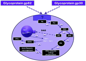
The glycoprotein gp82 is part of the trans-sialidase gp85 superfamily that is expressed by the trypomastigote stage and interacts with an unknown receptor on gastric mucosa epithelial cells coupled with a Gq protein, activating the phospholipase C pathway to generate inositol 1,4,5 triphosphate (IP3) which binds to its receptor in the endoplasmic reticulum promoting calcium output to the intracellular space.This allows diacilglicerol (DAG) to activate the protein kinase C which goes on to phosphorylate other molecules involved in actin filament rearrangement of the target cell (Docampo and Pignataro, Reference Docampo and Pignataro1991; Yoshida et al. Reference Yoshida, Favoreto, Ferreira and Manque2000). Intracellular pathways that involve an increase of intracellular calcium concentration induced by the parasite can participate in the generation of an immune response by a calmodulin/calcineurin-dependent activation of NFATc1 (Nuclear Factor of Activated T-cells). This has as function in the activation of genes encoding IFN-γ, a key cytokine in the development of a Th1 cytokine profile, essential for the host response against intracellular pathogens (Kayama et al. Reference Kayama, Koga, Atarashi, Okuyama, Kimura, Mak, Uematsu, Akira, Takayanagi, Honda, Yamamoto and Takeda2009) (Fig. 2).
Mechanisms involved in the invasion of stomach epithelial cells by Trypanosoma cruzi.

The studies performed by Yoshida et al. (Reference Yoshida and Cortez2008, Reference Yoshida, Tyler and Llewellyn2011), show that T. cruzi strains deficient in gp82 express gp30, a glycoprotein similar to gp82 that allows cell invasion by activating intracellular pathways that lead to the increase of intracellular calcium concentration but unlike the latter, present a low affinity for gastric mucosa. Therefore, T. cruzi strains expressing it generate low parasitaemia (Acosta-Serrano et al. Reference Acosta-Serrano, Almeida, Freitas-Junior, Yoshida and Schenkman2001; Yoshida, Reference Yoshida2006; Yoshida and Cortez, Reference Yoshida and Cortez2008; Villalta et al. Reference Villalta, Scharfstein, Ashton, Tyler, Guan, Mukherjee, Lima, Alvarez, Weiss, Huang, Machado and Tanowitz2009; Yoshida et al. Reference Yoshida, Tyler and Llewellyn2011). Another glycoprotein involved in the pathogenesis of T. cruzi infection by the oral route is gp90, which negatively regulates the infectivity of the parasite due to a phosphatase kinase that inactivates tyrosine kinase causing the inhibition of calcium mobilization into the intracellular space and preventing the internalization of the parasite. This glycoprotein displays different isophorms with different proteolysis resistance patterns (Acosta-Serrano et al. Reference Acosta-Serrano, Almeida, Freitas-Junior, Yoshida and Schenkman2001; Cortez et al. Reference Cortez, Silva, Neira, Ferreira, Sasso, Luquetti, Rassi and Yoshida2006; Yoshida, Reference Yoshida2006; Yoshida and Cortez, Reference Yoshida and Cortez2008; Staquicini et al. Reference Staquicini, Martins, Macedo, Sasso, Atayde, Juliano and Yoshida2010; Yoshida et al. Reference Yoshida, Tyler and Llewellyn2011).
The components of the gastric mucosa immune response have not been described yet. The expression of TLRs 2, 4, 5 and 9 by epithelial cells of the gastric mucosa has been demonstrated and based on this premise, the recognition would be performed by TLRs type 2, 4, and 9. TLRs 9 recognizing the T. cruzi regions containing abundant CpG motifs, TLRs 4 recognizing GIPLs (glycoinositolphospholipids) and TLRs 2 recognizing mucin-like proteins attached to glycosylphosphatidylinositol (GPI)(Cortez et al. 2006; Kayama and Takeda, 2009; Palm and Medzhitov, Reference Palm and Medzhitov2009). Nevertheless, the vast majority of immune responses produced throughout the gastrointestinal tract are thought to be mediated by direct or indirect stimulation of mucosa-associated lymphoid tissue found primarily in the gut, specifically Peyer's patches in the small bowel (Hoft et al. Reference Hoft, Farrar, Kratz-Owens and Shaffer1996; Pereira et al. Reference Pereira, Schmidt, Guaraldo, Franco, Dias and Passos2009, Reference Pereira, Schmidt, Barbosa, Guaraldo, Franco, Dias and Passos2010; Yoshida, Reference Yoshida2006; Yoshida and Cortez, Reference Yoshida and Cortez2008; Yoshida et al. Reference Yoshida, Tyler and Llewellyn2011). One possibility is that cytokine production participates in the recruitment of B lymphocytes found in lymphoid tissue of Peyer's patches. Another possibility is that antigens released from dead parasites can reach the small bowel where they are transported through M cells in the Peyer´s patches preparing B cells to circulate and proliferate when they reach the infected gastric tissue (Hoft et al. Reference Hoft, Farrar, Kratz-Owens and Shaffer1996; Carlier and Torrico, Reference Carlier and Torrico2003; Schmausser et al. Reference Schmausser, Andrulis, Endrich, Lee, Josenhans, Muller-Hermelink and Eck2004; Macian, Reference Macian2005; Cortez et al. Reference Cortez, Silva, Neira, Ferreira, Sasso, Luquetti, Rassi and Yoshida2006; Tarleton, Reference Tarleton2007; Yoshida and Cortez, Reference Yoshida and Cortez2008; Kayama et al. Reference Kayama, Koga, Atarashi, Okuyama, Kimura, Mak, Uematsu, Akira, Takayanagi, Honda, Yamamoto and Takeda2009; Palm and Medzhitov, Reference Palm and Medzhitov2009; Oliveira et al. Reference Oliveira, de Alencar, Tzelepis, Klezewsky, da Silva, Neves, Cavalcanti, Boscardin, Nunes, Santiago, Nobrega, Rodrigues and Bellio2010; Yoshida et al. Reference Yoshida, Tyler and Llewellyn2011).
CONCLUSIONS
Several microorganisms can be transmitted by the vertical route. TORCH (Toxoplasmosis, Others: syphilis, hepatitis B, Coxsackie virus, Epstein-Barr virus, human parvovirus and varicella zoster, Rubella, Cytomegalovirus and Herpes simplex virus) is an acronym used for a group of infections that can be congenitally transmitted and may cause a wide range of clinical presentations, from asymptomatic to abnormal fetus development and death of this latter. Therefore, screening must be conducted on every pregnant woman attending antenatal consultation at any moment of pregnancy as well as in their neonates if the infection is suspected (Greenough, Reference Greenough1994; Shet, Reference Shet2011). However, this acronym does not represent the entire variety of congenital infections, for example it only includes 1 parasite: Toxoplasma gondii, without including the other 3 parasites that can also be vertically transmitted: Plasmodium spp., Trypanosoma cruzi and Leishmania spp.(Ganga-Zandzou et al. Reference Ganga-Zandzou, Nguema, Mbo and Ondimba el2005; Kravetz Reference Kravetz2010; Lagerberg, Reference Lagerberg2008; Jones et al. Reference Jones, Krueger, Schulkin and Schantz2009; Kashyap, Reference Kashyap2010; Elmore et al. Reference Elmore, Jones, Conrad, Patton, Lindsay and Dubey2010; Kodjikian, Reference Kodjikian2010; Sankar et al. Reference Sankar, Menon and Kottarathara2010). Plasmodium spp. and Leishmania spp., probably due to the lack of information about these infections by the vertical route as well as their very low incidence, and T. cruzi probably because the vast majority of newborns infected with this parasite are asymptomatic, without considering that about 30% of these newborns eventually will develop the symptomatic chronic stage of disease which results in cardiac and enteric visceral compromise, all generate a great impact on the life quality of the patient and generate several costs that will affect the economy of the patient and the economy of health system (Gascon et al. Reference Gascon, Bern and Pinazo2009; Schmunis and Yadon, Reference Schmunis and Yadon2009; Bisio et al. Reference Bisio, Seidenstein, Burgos, Ballering, Risso, Pontoriero, Moreau, Altcheh, Leguizamon, Freilij, Marceillac and Schijman2011; Sicuri et al. Reference Sicuri, Munoz, Pinazo, Posada, Sanchez, Alonso and Gascon2011). As for gastric affectation, the pathogenic microorganisms involved are few; among these can be found bacteria such as Helicobacter pylori an aetiological agent that has been extensively studied (Wang and Peura, Reference Wang and Peura2011). Gastric infection by other agents such as Staphylococcus aureus and Streptococcus pyogenes causing phlegmonous gastritis. Mycobacterium tuberculosis, Treponema pallidum, Histoplasma capsulatum and Cytomegalovirus causing granulomatous gastritis and present in conditions such as malnutrition, alcoholism, HIV infection and other conditions all lead to immune suppression (Nevin et al. Reference Nevin, Eakins, Clarke and Carson1969; Tromba et al. Reference Tromba, Inglese, Rieders and Todaro1991; Ross and Vincenti, Reference Ross and Vincenti1994; Maeng et al. Reference Maeng, Lee, Choi, Kang and Kim2004; Corti et al. Reference Corti, Metta, Palmieri and Schtirbu2007; Hasegawa et al. Reference Hasegawa, Goto, Nishimura, Sukawa, Fujii, Suzuki, Yonezawa, Abe, Shinomura and Yoshida2009; Kakugawa et al. Reference Kakugawa, Kami, Matsuda, Saito, Kim, Fukuda, Mori, Shimoda, Tanosaki and Saito2010; Bhaijee et al. Reference Bhaijee, Subramony, Tang and Pepper2011). As well as 3 nematodes: Anisakis simplex, Pseudoterranova decipiens and Strongyloides stercoralis which also affect the gut (Ikeda et al. Reference Ikeda, Kumashiro and Kifune1989; Esteve et al. Reference Esteve, Resano, Diaz-Tejeiro and Fernandez-Benitez2000; Soto Tarazona et al. Reference Soto Tarazona, Meza Flores, Garrido Rivadeneyra and Cok Garcia2003; Meine et al. Reference Meine, Dietz, Rocha, Mattos, de Souza and Conteletti2004; Jofre et al. Reference Jofre, Neira, Noemi and Cerva2008; Fumarola et al. Reference Fumarola, Monno, Ierardi, Rizzo, Giannelli, Lalle and Pozio2009; Yaldiz et al. Reference Yaldiz, Hakverdi, Aslan, Temiz and Culha2009; Ozturk et al. Reference Ozturk, Aydinli, Celebi and Gursan2011). However, the only protozoan that has recently been involved in gastric infection is T. cruzi which can successfully establish the infection through gastric epithelial cells without requiring an immune-compromised host whereby replication apparently occurs only in the stomach and in no other location in the gastrointestinal tract (Hoft et al. Reference Hoft, Farrar, Kratz-Owens and Shaffer1996; Acosta-Serrano et al. Reference Acosta-Serrano, Almeida, Freitas-Junior, Yoshida and Schenkman2001; Pereira et al. Reference Pereira, Schmidt, Guaraldo, Franco, Dias and Passos2009, Reference Pereira, Schmidt, Barbosa, Guaraldo, Franco, Dias and Passos2010; Yoshida, Reference Yoshida2006, Reference Yoshida2008, Reference Pereira, Schmidt, Guaraldo, Franco, Dias and Passos2009; Yoshida and Cortez, Reference Yoshida and Cortez2008; Yoshida et al. Reference Yoshida, Tyler and Llewellyn2011) Most microorganisms causing gastrointestinal affectation interact directly with the bowel, explaining the lack of information about the gastric mucosa immune response.
Unlike vector and oral transmission of T. cruzi, that is only possible in endemic areas, infection through blood and organ transplantation or via vertical transmission can occur in any country (Basile et al. Reference Basile, Oliveira, Ciruela and Plasencia2011; Piron et al. Reference Piron, Verges, Munoz, Casamitjana, Sanz, Maymo, Hernandez, Puig, Portus, Gascon and Sauleda2008). Although systematic screening for T. cruzi among pregnant Latin American women in non-endemic countries is still uncommon, the World Health Organization's Programme on Control of Chagas disease highlights the need to promote the development of public health measures in disease-endemic and disease non-endemic countries. For the early diagnosis of congenital transmission and management of cases from endemic countries, a recommendation by the Catalonian Health Department, has implemented a systematic screening programme for Chagas disease among Latin American pregnant women and their children (Oliveira et al. Reference Oliveira, de Alencar, Tzelepis, Klezewsky, da Silva, Neves, Cavalcanti, Boscardin, Nunes, Santiago, Nobrega, Rodrigues and Bellio2010; Basile et al. Reference Basile, Oliveira, Ciruela and Plasencia2011). Finally, it is important to point out that, due to the great genetic and biochemical diversity of T. cruzi, the host immune response could be highly variable depending on the infecting strain, the patient immune status and the infection route used. Further studies that include comparison of clinical and paraclinical features, clinical outcome and T. cruzi genotypes involved in all transmission routes must be performed in order to establish possible links between T. cruzi DTUs, transmission mechanisms, clinical features, and treatment response.
ACKNOWLEDGMENTS
We thank D.C. Bautista from Universidad Militar Nueva Granada, Bogotá, Colombia for support with databases and the information search process, and M.C. Ortega from Hospital Militar Central, Bogotá, Colombia for reading the manuscript and providing valuable comments and suggestions about immunological aspects. We also thank Dila Angarita for the artwork of the figures.