Congenital heart disease (CHD), including atrial septal defect, ventricular septal defect, patent ductus arteriosus, tetralogy of Fallot, mitral regurgitation, aortic valvular regurgitation, and so on, occurs in approximately 10–14 of 1000 newborns in recent studies. The incidence varies according to geographic regions, the time of investigation, target population, and case definition.Reference Hoffman and Kaplan1–Reference Egbe, Uppu, Stroustrup, Lee, Ho and Srivastava4 It can occur as an isolated congenital anomaly independently, or with a wide variety of non-cardiac anomalies, such as microtia.Reference Calzolari, Barisic and Loane5 Microtia, the second most common congenital craniofacial abnormality after oral cleft, is a congenital anomaly of the ear that ranges in severity from mild structural abnormalities to complete absence of the ear and external auditory canal (aural atresia), and it can occur as an isolated birth defect or as part of multiple congenital defects or a syndrome. The incidence of microtia is reported varying from 0.83 to 17.4 per 10,000 births.Reference Castilla and Orioli6,Reference Harris, Källén and Robert7 Congenital heart malformations are one of the most common anomalies associated with microtia.Reference Harris, Källén and Robert7–Reference Mastroiacovo, Corchia and Botto9 Few studies have been done encompassing the characteristics and incidence of CHD in patients with isolated microtia (not associated with other craniofacial anomalies or syndromes). The purpose of our study was two-fold. First, we wanted to identify the characteristics and incidence of CHD in patients with isolated microtia and to determine whether the prevalence of CHD among patients with isolated microtia increases with the severity of microtia. Second, we sought to give some guidance to the paediatrician and plastic surgeon with respect to the timely detection and diagnosis of CHD in isolated microtia patients.
Methods
Patient selection
A total of 897 patients with microtia underwent individual treatment in the Department of Auricular Reconstruction at the Plastic Surgery Hospital of Peking Union Medical College from November, 2017 to January, 2019. Ninety-three patients with other craniofacial anomalies or syndromes, such as oculo-auriculo-vertebral syndrome, Treacher Collins syndrome, Klippel–Feil syndrome, Townes Brocks, hemifacial microsomia, other chromosomal abnormalities, and so on, were excluded from the study group. Thus, the study included the remaining 804 patients (with a range from 1 year to 31 years, mean age 9.1 years; 762 unilateral, 42 bilateral) with isolated microtia.
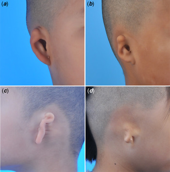
Among these patients, 461 cases (57.3%) were right-sided, 301 cases (37.4%) were left-sided, and 42 cases (5.8%) were bilateral. A total of 591 (73.5%) patients were male and 213 (26.5%) were female. According to Nagata’s definition,Reference Nagata10–Reference Nagata13 243 cases (30.2%) were grade I (concha-type microtia), 92 cases (11.4%) were grade II (small concha-type microtia), 449 cases (55.8%) were grade III (lobule-type microtia), and 20 cases (2.5%) were grade IV (anotia) (Table 1). The study was approved by the ethics committee of the Plastic Surgery Hospital, Peking Union Medical College, and Chinese Academy of Medical Sciences.
Characteristics of the patients with isolated microtia

Imaging materials
The colour Doppler echocardiographic image (LOGIQ E9, General Electric Company, Boston, MA, United States of America) was performed pre-operatively for every patient to evaluate cardiovascular anatomy and function. To determine the presence of CHD, three sonographers with more than 8 years of working experience reviewed all images in this double-blind study.
Statistical analysis
Commercially available software (Statistical Package for Social Sciences, version 19.0; SPSS Inc., Chicago, IL, United States of America) was used for the statistical analysis. The χ2 test was used to determine the relationship between isolated microtia and CHD. A p value of <0.05 was considered statistically significant.
Results
We observed 846 ears in 804 patients according to Nagata’s definition and classified into four types (concha-type microtia, small concha-type microtia, lobule-type microtia, and anotia) (Fig 1). A retrospective study was performed with the imaging data from 804 consecutive patients that received a pre-operative colour Doppler echocardiographic imaging.
Patients with isolated microtia varying from type I to type IV. ( a ) Type I, concha-type microtia, with the remnant ear, ear lobule, concha, acoustic meatus, tragus, and incisura intertragica. ( b ) Type II, small concha-type microtia, showing the remnant ear and lobule and a small indentation representing the concha. ( c ) Type III, lobule-type microtia, with the remnant ear and ear lobule and without the concha, acoustic meatus, and tragus. ( d ) Type IV, anotia with complete or almost complete absence of the external ear.

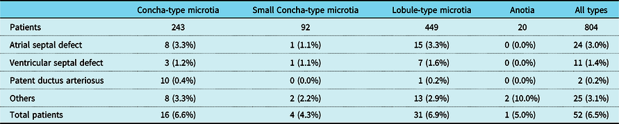
Overall, CHD, including atrial septal defect, ventricular septal defect, patent ductus arteriosus, and others, occurred in 52 of 804 patients (6.5%). Nine of 804 patients (1.1%) had two or more kinds of cardiac malformation. The incidences of the atrial septal defect, ventricular septal defect, patent ductus arteriosus, and other kinds of cardiac malformations were 3.0% (24/804), 1.4% (11/804), 0.2% (2/804), and 3.1% (25/804), respectively. We found atrial septal defect prevalence in patients with isolated microtia was significantly higher than ventricular septal defect (p < 0.05) and patent ductus arteriosus (p < 0.001). Ventricular septal defect prevalence in patients with isolated microtia was significantly higher than patent ductus arteriosus (p < 0.05).
The incidences of the CHD in concha-type microtia, small concha-type microtia, lobule-type microtia, and anotia patients were 6.6% (16/243), 4.3% (4/92), 6.9% (31/449), and 5.0% (1/20), respectively. All four types of isolated microtia had similar incidences of CHD and we found no differences in the incidences among these subtypes (p > 0.05). The incidences of the atrial septal defect, ventricular septal defect, patent ductus arteriosus, and other kinds of cardiac malformations with respect to the subtypes of microtia were, respectively, 3.3% (8/423), 1.2% (3/423), 0.4% (1/423), and 3.3% (8/423)in concha-type microtia; 1.1% (1/92), 1.1% (1/92), 0% (0/92), and 2.2% (2/92) in small concha-type microtia; 3.3% (15/449), 1.6% (7/449), 0.2% (1/449), and 2.9% (13/449) in lobule-type microtia; and 0% (0/20), 0% (0/20), 10.0% (2/20), and 5.0% (1/20) in anotia patients (Table 2). Furthermore, this retrospective study found no significant differences in the incidence of the atrial septal defect, ventricular septal defect, or patent ductus arteriosus among the four subtypes (p > 0.05 for all comparisons).
Incidents of CHD in patients with isolated microtia

Discussion
In China, the total prevalence of microtia is 3.06 per 10,000 births (1933 cases with anotia/microtia were identified among 6,308,594 individuals), and the prevalence of isolated microtia is 2.25 per 10,000 births.Reference Deng, Dai and Yi14 CHD is the most common type of congenital malformation and contributes significantly to infant mortality and morbidity.Reference Hoffman and Kaplan1,Reference Bernier, Stefanescu and Samoukovic15–Reference Lee, Khoshnood and Chen17 It occurs in approximately 10–14 of 1000 newborns in recent studies.Reference Hoffman and Kaplan1–Reference Egbe, Uppu, Stroustrup, Lee, Ho and Srivastava4 Birth prevalence of the three most common subtypes of CHD reported among worldwide (per 1000 live births) was: atrial septal defect, 1.64; ventricular septal defect, 2.62; and patent ductus arteriosus, 0.87.Reference van der Linde, Konings and Slager18 Microtia, the second most common congenital craniofacial abnormality after oral cleft, is a congenital anomaly of the ear that can be easily diagnosed by simple physical examination and inspection immediately after birth, so misdiagnosis is rare. It can occur as an isolated birth defect or as a part of multiple congenital defects or a syndrome. CHD is one of the most common anomalies associated with microtia.Reference Harris, Källén and Robert7–Reference Mastroiacovo, Corchia and Botto9 As microtia may be associated with syndromes, careful general physical examination, sometimes with a multi-slice spiral computerised tomography (CT) scan with three-dimensional reconstruction, and colour Doppler ultrasonography would be done, so CHD and other associated deformities can be found and treated at the appropriate time. Of note for isolated microtia, CHD might not be found immediately after birth. Pre-operative examination is therefore required to diagnose CHD which may have serious implications in microtia patients who require surgical repair under general anaesthesia. However, there are very few studies about CHD in patients with isolated microtia. Thus, this retrospective study reports for the first time on the characteristics and incidence of congenital heart defects in patients with isolated microtia.
In our study, all the data were acquired from the Department of Auricular Reconstruction at the Plastic Surgery Hospital of Peking Union Medical College. The patients in this study were relatively young and all were Chinese. Isolated microtia patients or microtia patients associated with other craniofacial anomalies or syndromes were all diagnosed mainly based on clinical features and medical history. In agreement with previous studies about microtia,Reference Marelli, Mackie, Ionescu-Ittu, Rahme and Pilote2,Reference Yeh, Chen and Lu3,Reference Harris, Källén and Robert7–Reference Mastroiacovo, Corchia and Botto9 we found the overall incidences of CHD in patient with isolated microtia (52/804, 6.5%) and the three most common CHD subtypes (atrial septal defect, 3.0% (24/804); ventricular septal defect, 1.4% (11/804); and patent ductus arteriosus, 0.2% (2/804)) were also significantly higher than the general population. While, the overall incidence is lower than that reported in the previous study (70/672, 10.6%) from our department,Reference Zhang, Jiang and Yang19 the main reason might be that patient selection criteria are different and both studies are hospital-based rather than population-based, as well as the being over different time periods. It is also lower than the study reported by Ramprasad et al,Reference Ramprasad, Shaffer and Jabbour20 which indicated that 40.4% (19/47) of their microtia patients had CHD. The reason for this difference might be that subjects in their study included isolated microtia and those associated with other craniofacial anomalies or syndromes. In addition, the sample size (47) was small, and may not be representative.
In addition, our e study demonstrates that all four subtypes of microtia show no difference in the incidences of CHD overall, or in the three most common CHD subtypes (atrial septal defect, ventricular septal defect, and patent ductus arteriosus). That is to say, the mildest type microtia (concha-type microtia) through to the severest type microtia (anotia), all have the same risk of having CHD or these specific types of CHD. Children with microtia are usually treated with total ear reconstruction surgery under general anaesthesia, thus, for the plastic surgeon and anaesthesiologist, it is important to undertake pre-operative colour Doppler echocardiography to evaluate heart anatomy and function to ensure the safety of peri-operative period and help avoid complications of the cardiovascular system. Furthermore, it is important that obstetricians, paediatricians and plastic surgeons are aware of this association when meeting patients with isolated microtia in outpatients. We suggest that patients with any type of isolated microtia should if possible have a complete echocardiographic study soon after birth with the consent of their parents.
Our study showed that CHD occurred in a significantly greater proportion of patients with isolated microtia than the general population. The exact cause of these differences is unclear, but the disturbance of neural crest cells and vascular disruption are presumed to be involved.Reference Bartel-Friedrich21,Reference Sadler and Rasmussen22 So, further exploration is needed. From a developmental point of view, it is difficult to explain the co-occurrence of various congenital abnormalities because of the formation of different structures at different points during development. Therefore, it is necessary to investigate pathogenic mutations, along with potential causative genetic and molecular mechanisms of the specific phenotype with next-generation genome sequencing techniques and gene function analysis. In our clinical centre, gene testing was successively done in eligible isolated microtia patients with CHD to investigate the pathogenetic mechanism involved. The complexity and high incidence of CHD associated with isolated microtia should also prompt clinicians treating such children to take into account other malformations so as to improve diagnosis and treatment.
Conclusions
Our study has demonstrated the characteristics and incidence of CHD and the three most common CHD subtypes in patients with isolated microtia. Overall incidences of CHD in patient with isolated microtia are significantly higher than the general population. The prevalence of CHD among patients with isolated microtia does not increase with the severity of microtia. According to our experience and this study, we suggest that colour Doppler echocardiography be performed for isolated microtia patients immediately after birth if possible. Furthermore, for plastic surgeons and anaesthesiologists, it is important to have an echocardiographic study to evaluate heart function to ensure the safety of the peri-operative period. Future studies investigating CHDs associated with isolated microtia could focus on genetic and molecular mechanisms.
Acknowledgements
H.J., Q.Y., and F.G. conceptualised and designed the paper; B.P., L.H., L.L., J.Z., and F.G. carried out the data collection, reviewed and revised the manuscript, and approved the final manuscript as submitted. All the authors approved the final manuscript as submitted and agree to be accountable for all aspects of the work. We thank all the patients for their participation and permission in this study.
Financial support
This study was supported by the CAMS Innovation Fund for Medical Sciences (CAMS-2017-I2M-1-007).
Conflicts of interest
None.