


%20--%3e%3c!DOCTYPE%20svg%20PUBLIC%20'-//W3C//DTD%20SVG%201.0//EN'%20'http://www.w3.org/TR/2001/REC-SVG-20010904/DTD/svg10.dtd'%3e%3csvg%20version='1.0'%20id='Layer_1'%20xmlns='http://www.w3.org/2000/svg'%20xmlns:xlink='http://www.w3.org/1999/xlink'%20x='0px'%20y='0px'%20width='64px'%20height='64px'%20viewBox='5.5%20-3.5%2064%2064'%20enable-background='new%205.5%20-3.5%2064%2064'%20xml:space='preserve'%3e%3cg%3e%3ccircle%20fill='%23FFFFFF'%20fill-opacity='0'%20cx='37.785'%20cy='28.501'%20r='28.836'/%3e%3cpath%20fill='%23595959'%20d='M37.441-3.5c8.951,0,16.572,3.125,22.857,9.372c3.008,3.009,5.295,6.448,6.857,10.314%20c1.561,3.867,2.344,7.971,2.344,12.314c0,4.381-0.773,8.486-2.314,12.313c-1.543,3.828-3.82,7.21-6.828,10.143%20c-3.123,3.085-6.666,5.448-10.629,7.086c-3.961,1.638-8.057,2.457-12.285,2.457s-8.276-0.808-12.143-2.429%20c-3.866-1.618-7.333-3.961-10.4-7.027c-3.067-3.066-5.4-6.524-7-10.372S5.5,32.767,5.5,28.5c0-4.229,0.809-8.295,2.428-12.2%20c1.619-3.905,3.972-7.4,7.057-10.486C21.08-0.394,28.565-3.5,37.441-3.5z%20M37.557,2.272c-7.314,0-13.467,2.553-18.458,7.657%20c-2.515,2.553-4.448,5.419-5.8,8.6c-1.354,3.181-2.029,6.505-2.029,9.972c0,3.429,0.675,6.734,2.029,9.913%20c1.353,3.183,3.285,6.021,5.8,8.516c2.514,2.496,5.351,4.399,8.515,5.715c3.161,1.314,6.476,1.971,9.943,1.971%20c3.428,0,6.75-0.665,9.973-1.999c3.219-1.335,6.121-3.257,8.713-5.771c4.99-4.876,7.484-10.99,7.484-18.344%20c0-3.543-0.648-6.895-1.943-10.057c-1.293-3.162-3.18-5.98-5.654-8.458C50.984,4.844,44.795,2.272,37.557,2.272z%20M37.156,23.187%20l-4.287,2.229c-0.458-0.951-1.019-1.619-1.685-2c-0.667-0.38-1.286-0.571-1.858-0.571c-2.856,0-4.286,1.885-4.286,5.657%20c0,1.714,0.362,3.084,1.085,4.113c0.724,1.029,1.791,1.544,3.201,1.544c1.867,0,3.181-0.915,3.944-2.743l3.942,2%20c-0.838,1.563-2,2.791-3.486,3.686c-1.484,0.896-3.123,1.343-4.914,1.343c-2.857,0-5.163-0.875-6.915-2.629%20c-1.752-1.752-2.628-4.19-2.628-7.313c0-3.048,0.886-5.466,2.657-7.257c1.771-1.79,4.009-2.686,6.715-2.686%20C32.604,18.558,35.441,20.101,37.156,23.187z%20M55.613,23.187l-4.229,2.229c-0.457-0.951-1.02-1.619-1.686-2%20c-0.668-0.38-1.307-0.571-1.914-0.571c-2.857,0-4.287,1.885-4.287,5.657c0,1.714,0.363,3.084,1.086,4.113%20c0.723,1.029,1.789,1.544,3.201,1.544c1.865,0,3.18-0.915,3.941-2.743l4,2c-0.875,1.563-2.057,2.791-3.541,3.686%20c-1.486,0.896-3.105,1.343-4.857,1.343c-2.896,0-5.209-0.875-6.941-2.629c-1.736-1.752-2.602-4.19-2.602-7.313%20c0-3.048,0.885-5.466,2.658-7.257c1.77-1.79,4.008-2.686,6.713-2.686C51.117,18.558,53.938,20.101,55.613,23.187z'/%3e%3c/g%3e%3c/svg%3e)
%20--%3e%3c!DOCTYPE%20svg%20PUBLIC%20'-//W3C//DTD%20SVG%201.0//EN'%20'http://www.w3.org/TR/2001/REC-SVG-20010904/DTD/svg10.dtd'%3e%3csvg%20version='1.0'%20id='Layer_1'%20xmlns='http://www.w3.org/2000/svg'%20xmlns:xlink='http://www.w3.org/1999/xlink'%20x='0px'%20y='0px'%20width='64px'%20height='64px'%20viewBox='5.5%20-3.5%2064%2064'%20enable-background='new%205.5%20-3.5%2064%2064'%20xml:space='preserve'%3e%3cg%3e%3ccircle%20fill='%23FFFFFF'%20fill-opacity='0'%20cx='37.637'%20cy='28.806'%20r='28.276'/%3e%3cg%3e%3cpath%20fill='%23595959'%20d='M37.443-3.5c8.988,0,16.57,3.085,22.742,9.257C66.393,11.967,69.5,19.548,69.5,28.5c0,8.991-3.049,16.476-9.145,22.456%20C53.879,57.319,46.242,60.5,37.443,60.5c-8.649,0-16.153-3.144-22.514-9.43C8.644,44.784,5.5,37.262,5.5,28.5%20c0-8.761,3.144-16.342,9.429-22.742C21.101-0.415,28.604-3.5,37.443-3.5z%20M37.557,2.272c-7.276,0-13.428,2.553-18.457,7.657%20c-5.22,5.334-7.829,11.525-7.829,18.572c0,7.086,2.59,13.22,7.77,18.398c5.181,5.182,11.352,7.771,18.514,7.771%20c7.123,0,13.334-2.607,18.629-7.828c5.029-4.838,7.543-10.952,7.543-18.343c0-7.276-2.553-13.465-7.656-18.571%20C50.967,4.824,44.795,2.272,37.557,2.272z%20M46.129,20.557v13.085h-3.656v15.542h-9.944V33.643h-3.656V20.557%20c0-0.572,0.2-1.057,0.599-1.457c0.401-0.399,0.887-0.6,1.457-0.6h13.144c0.533,0,1.01,0.2,1.428,0.6%20C45.918,19.5,46.129,19.986,46.129,20.557z%20M33.042,12.329c0-3.008,1.485-4.514,4.458-4.514s4.457,1.504,4.457,4.514%20c0,2.971-1.486,4.457-4.457,4.457S33.042,15.3,33.042,12.329z'/%3e%3c/g%3e%3c/g%3e%3c/svg%3e)
Introduction
Alzheimer’s disease (AD) is a progressive neurodegenerative disorder that is the most prevalent cause of dementia globally, accounting for 60%–80% of all dementia cases. With about 5 million new cases occurring annually, it is estimated that the number of people with AD and AD-related dementia (ADRD) will increase by more than 3-fold (∼131 million) by 2050 [Reference Tiwari, Atluri, Kaushik, Yndart and Nair1]. AD is indeed a significant burden on the burden and public health [Reference Skaria2,Reference Tobore3], as the number of patients is rapidly increasing. Thus, early diagnosis and treatment methods are crucial in addressing this challenge. AD is clinically characterized by a progressive cognition decline and dementia, in addition to the two required pathologies hallmarks, amyloid beta (Aβ) plaque aggregation and P-tau tangles [Reference Murphy and Levine4]. The traditional clinical criteria for AD diagnosis were originally founded upon cognitive assessments, based on the belief that there are no motor, sensory, or coordination deficits early in the disease, and cannot be determined by laboratory tests [Reference McKhann, Drachman, Folstein, Katzman, Price and Stadlan5]. However, accumulating clinical evidence indicates that AD may have early symptoms other than cognitive decline. In 2011, the National Institute on Aging and Alzheimer’s Association (NIA-AA) created separate diagnostic recommendations for the preclinical, mild cognitive impairment (MCI), and dementia stages of Alzheimer’s disease, labeled as “research framework” [Reference Jack, Bennett and Blennow6]. This proposed AD diagnostic criterion focuses on the diagnosis of AD with biomarkers grouped into amyloid-β deposition, pathologic tau, and neurodegeneration, also known as the AT(N) classification system [Reference Petersen, Wiste and Weigand7]. Based on the AT(N) system, several methods can be conducted to diagnose AD prior to cognitive symptomatology, including cerebrospinal fluid (CSF) analysis, positron emission tomography (PET), and magnetic resonance imaging (MRI) [Reference Mckhann, Knopman and Chertkow8]. Although capable of identifying early changes during the asymptomatic stage, these core clinical criteria are more generally reliable and provide high diagnostic accuracy in most patients for the advanced stages of AD. In early diagnosis, these methods are still premature and have drawbacks such as overdiagnosis, increased cost, and invasiveness of the assessment [Reference Ferreira, Perestelo-Perez, Westman, Wahlund, Sarria and Serrano-Aguilar9].
Recently, accumulated evidence revealed that sensory changes, including olfactory, auditory, visual, tactile, and gustatory dysfunction, are associated with AD pathological processes and usually emerge prior to the mild cognitive symptoms [Reference Martín, Barato and Oliva10–Reference Füllgrabe15]. Thus, the impairment in sensory systems could be potentially used as biomarkers for the early detection and intervention of AD. However, the examination of sensory function alone may not provide sufficient accuracy for the clinical AD diagnosis since it can also be caused by various non-AD-related factors, such as neutral aging, environmental factors, and other neurological disorders like Parkinson’s disease [Reference Permezel, Alty, Harding and Thyagarajan16]. Thus, integrating comprehensive sensory alteration across the pathological progression could lead to reliable and early detection of AD and pave the way to establish an early noninvasive AD diagnostic method. Furthermore, sensory deficits may also promote AD pathology, while early interventions and corrections may alleviate cognitive decline and slow down the AD pathological process. Recent studies in both human and animal models revealed that visual impairments or hearing loss (induced by cochlear ablation in mouse models) is associated with an increased likelihood of developing dementia [Reference Kuźma, Littlejohns, Khawaja, Llewellyn, Ukoumunne and Thiem17–Reference Zhao and Yang19]. Preventive actions for vision problems, such as eye exams, eyeglasses, and cataract surgery, may reduce the risk for AD and ADRDs [Reference Ehrlich, Goldstein, Swenor, Whitson, Langa and Veliz20]. Hearing intervention might also reduce cognitive change over 3 years in populations of older adults at increased risk for cognitive decline [Reference Lin, Pike and Albert21]. These findings suggest the promise of implementing several early interventions targeting sensory alterations to potentially reduce the risk of AD.
This review aims to collate current research on sensory changes caused by AD in the olfactory, visual, and auditory systems in both patients and transgenic mouse models of AD, exploring their relationship with established AD sensory biomarkers during the AD pathologic process and outlining a potential integration diagnostic technique for AD identification at early stages.
Pathogenesis and diagnosis of AD
The pathogenesis of AD
AD is an irreversible, highly complex, and progressive neurodegenerative disease. It was first described in 1907, with neurotic plaques, neurofibrillary tangles (NFTs), and amyloid angiopathy in the cerebral cortex [Reference Bondi, Edmonds and Salmon22]. These autopsy characterizations were then become the hallmarks of AD. Over the past several decades, numerous hypotheses have been made for AD pathogenesis. The amyloid cascade hypothesis postulates that neurodegeneration in AD is caused by abnormal accumulation of Aβ plaques in the cerebral cortex [Reference Barage and Sonawane23]. Aβ is cleavage from the amyloid precursor protein (APP), the defective intracellular cleavage of APP that is thought to be the predominant cause of plaque formation. The tau theory focuses on the aggregation of intracellular NFTs formed by accumulated tau protein in a hyperphosphorylated state [Reference Hampel, Mesulam and Cuello24]. The tau hypothesis suggests that the hyperphosphorylation of tau (P-tau) induces the formation of NFTs, which is another hallmark pathology of AD [Reference Mohandas, Rajmohan and Raghunath25,Reference Kametani and Hasegawa26]. Calcium dysregulation seems to generate large amounts of tau [Reference Arnsten, Datta, Del Tredici and Braak27]. The inflammatory hypothesis is based on the observation that the increased inflammatory markers in AD patients, such as pro-inflammatory cytokines interleukin-1β, interleukin-6, and tumor necrosis factor α [Reference Aberathne, Kulasiri and Samarasinghe28]. Increased oxidative stress in the AD brain is characterized by increased lipid peroxidation, increased protein, and DNA oxidation; these data support the oxidative stress hypothesis [Reference Markesbery29,Reference Pratico30]. Mitochondrial disturbances are also associated with both oxidative stress and AD plaque formation [Reference Aberathne, Kulasiri and Samarasinghe28].
The diagnosis of AD
In 2011, the NIA-AA conceptualized the pathophysiological process of AD in two stages, that is, preclinical stages and clinical stages based on whether the patients have overt symptoms of dementia and cognitive impairment [Reference Petersen, Wiste and Weigand7]. The diagnosis of AD in the clinical stage is clear in the guidelines, but the recommendations for preclinical AD which could potentially be decades before the symptoms become noticeable [Reference Monsell, Mock and Hassenstab31] were not established. Furthermore, accumulated evidence indicates that the neurodegeneration and cognitive decline in AD are a continuous pathophysiological process that should be regarded as a continuum rather than two distinct preclinical and clinical entities [Reference Jack, Bennett and Blennow6,Reference Dubois, Hampel and Feldman32]. This concept is recognized widely, indicating the importance of early AD diagnosis methods.
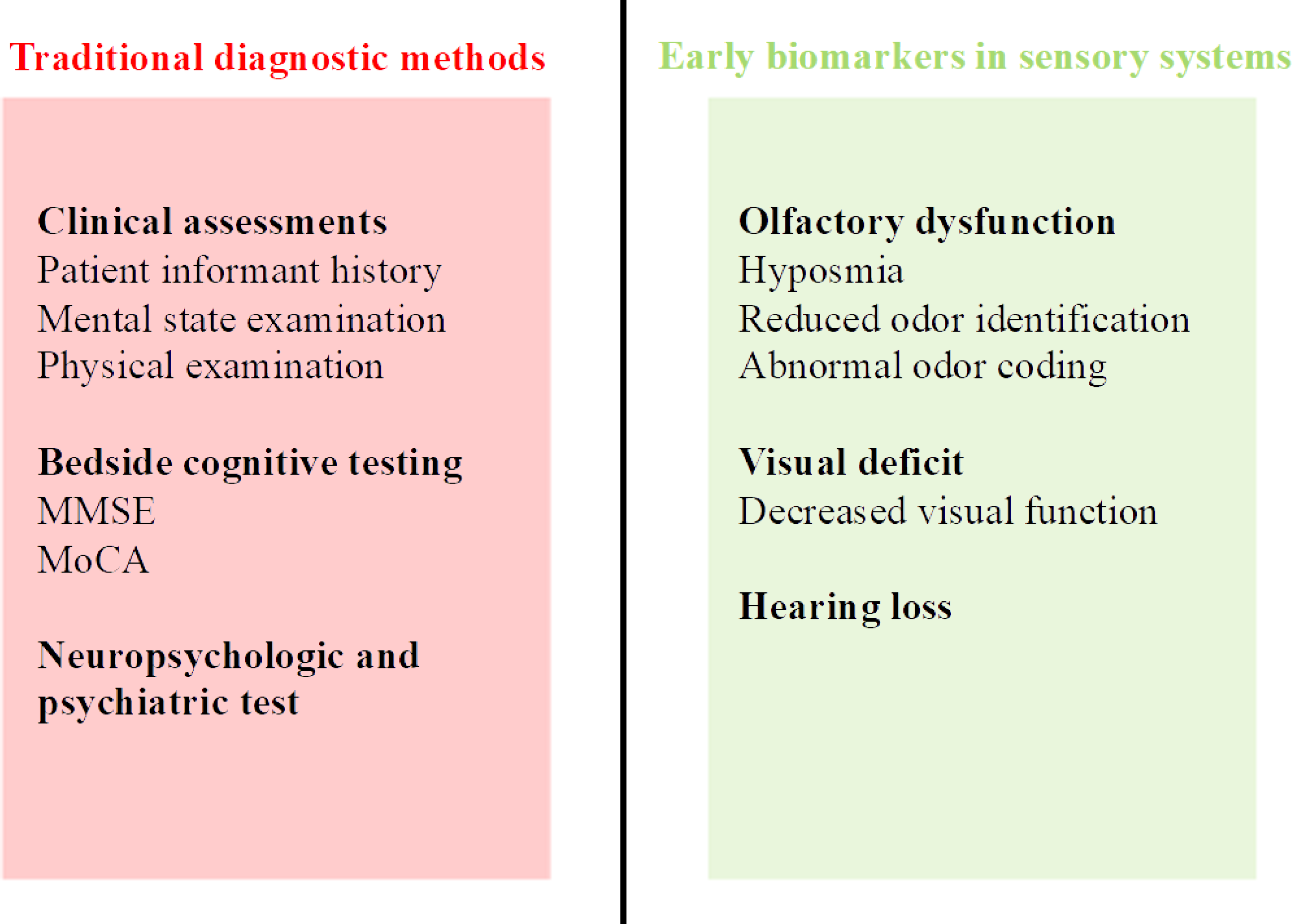
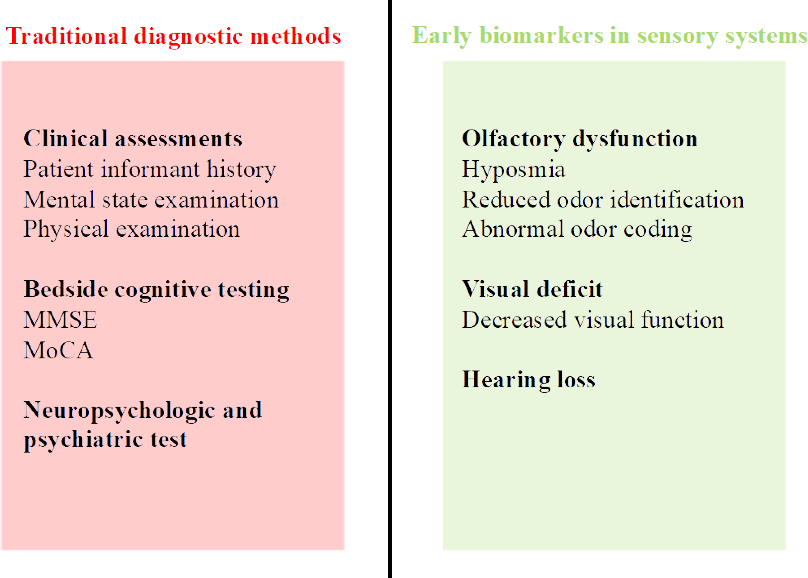
In 2011 NIA-AA guidelines, several methods have been established for identifying and assessing the progression of AD in clinical stages (Fig. 1). These methods can be clustered into two divisions: clinical assessments and neuroimaging techniques. The clinical assessments of AD rely on patient informant history, mental state examination, thorough physical examination, bedside cognitive testing including mini-mental state examination and Montreal cognitive assessment (MoCA), and some neuropsychologic and psychiatric tests such as memory, attention, language, problem-solving skills, and attention [Reference Eratne, Loi, Farrand, Kelso, Velakoulis and Looi33]. AD can be further diagnosed with firm clinical evidence provided by neuroimaging techniques, such as MRI and PET scans [Reference Islam and Zhang34,Reference Valotassiou, Malamitsi and Papatriantafyllou35], or direct biomarker tests including CSF analysis and blood tests [Reference Bouwman, Frisoni and Johnson36,Reference Schneider, Hampel and Buerger37]. All these traditional methods have a certain limitation, as these methods always work after the symptoms presented, such as memory loss and cognitive decline. Furthermore, these diagnostic techniques could be invasive procedures or require radiation exposure, posing a risk of side effects. Also, certain methods need specialized and costly equipment that is less available. Additionally, AD can only be noticed by families 4 years before a firm diagnosis is made, and this delay is often longer and includes misdiagnosis in the variants and early-onset groups [Reference Draper, Cations and White38].
Overview of traditional diagnostic methods and early biomarkers in sensory systems. MMSE = mini-mental state examination; MoCA = Montreal cognitive assessment.
Early biomarkers of AD in the sensory system
In recent years, more and more results have shown that the sensory system can be affected in AD patients. For example, several studies have revealed that olfactory function was profoundly affected by AD in the clinical stages, revealing that the impairment of olfactory function could be a potential biomarker for cognitive impairment and neurodegeneration [Reference Jung, Shin and Lee39–Reference El Haj and Laroi43]. Visual manifestations can also be observed in AD patients, with decreased visual acuity, reduced contrast sensitivity, poor color discrimination, and abnormal ocular motor function [Reference Javaid, Brenton, Guo and Cordeiro44]. AD also manifests with hearing loss [Reference Li, Cheng and Lu45]; even though the causation between hearing loss and dementia remains unknown, evidence shows that the intervein of age-related hearing loss could prevent AD, making hearing loss another high-risk factor for AD [Reference Shityakov, Hayashi, Stork, Scheper, Lenarz and Forster46]. In this section, we started with a concise summary of the structure composition of different sensory systems, and then we aimed to provide an overview of early sensory biomarkers related to AD in preclinical research. Following this, we encapsulated the sensory deficits reported in the symptomatic stage of AD and concluded by describing the discoveries related to sensory impairments in preclinical AD patients.
Structural overview of sensory pathways
Olfaction is a highly conservative primary sensory system [Reference Ache and Young47]. Olfactory can be clustered as the peripheral and central system [Reference Smith and Bhatnagar48]. Odorants are first received by olfactory sensory neurons (OSNs) located on the olfactory epithelium, where the chemical signals are converted to neuroelectrical signals [Reference Duchamp-Viret, Chaput and Duchamp49]. The electrical potentials are then transferred to the mitral cells and tuft cells on the surface of the olfactory bulb (OB) via OSNs projection [Reference Firestein50] and then handed over to olfactory cortical areas, including the piriform cortex (PCX), the medial olfactory cortex, and the amygdala [Reference Giessel and Datta51].
About half of the neuronal pathway in the brain subserves visual function, light signals are transduced to the neuronal signals via photoreceptors, the cones, and rods which are located on the outer nuclear layer of the retina. It is then passed to the inner nuclear layer, including [Reference Chaya, Matsumoto and Sugita52], followed by ganglion cells on the last layer. The axon of ganglion cells projects to the brain cortex via the lateral geniculate nucleus and superior colliculus and finally reaches the visual cortex.
The auditory system can be divided into two main components – peripheral and central systems – both are essential for normal hearing function. The peripheral auditory system includes external, middle, and inner ears; approximately 70% of hearing loss is caused by various pathologies in the inner ear and auditory nerve [Reference Strenzke, Pauli-Magnus, Meyer, Brandt, Maier and Moser53]. A specialized hearing organ in the inner ear, the cochlea, transduces the sound vibrations to neuronal electrical signals; this process is also known as mechanotransduction [Reference Stover and Diensthuber54]. The main sensory cells for mechanotransduction in the cochlea are hair cells. The ion homeostasis of the cochlea and hair cells (e.g., K+ and Ca2+) is required for normal hearing function and the development of the auditory system [Reference Schmiedt55–Reference Yang, Murtha and Climer58]. After mechanotransduction, the spiral ganglion neurons transmit the acoustic information from the cochlea to the central auditory system via the superior olivary complex, lateral lemniscus, inferior colliculus, medial geniculate nucleus, and the auditory cortex [Reference Jayakody, Friedland, Martins and Sohrabi59].
Preclinical research revealed early sensory biomarkers related to AD
AD animal models, primarily rodent models, have provided valuable insights into the disease and contributed to preclinical research. Since rodents do not develop AD, introducing human AD pathogenic genes is essential to model the AD pathology in mice. Thus, mouse models of AD can be generally classified as follows based on what gene is engineered, and each of these models is slightly different in AD pathophysiology. Here, we briefly summarized the pathogenesis of five main AD mouse models, including the APP transgenic model, APP/PS1 transgenic model, 5xFAD transgenic model, Tau transgenic model, and 3xTg mouse model.
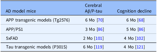
Interestingly, numerous studies have also demonstrated that early sensory impairments are present in AD mouse models, highlighting the utility of these AD mouse models for exploring sensory deficits as potential early indicators of AD. The impairment of sensory systems can be assessed through behavioral tests, such as odor discrimination, identification, threshold test [Reference Hummel, Sekinger, Wolf, Pauli and Kobal60,Reference Zhang, Zhao and Sun61], Morris water maze (MWM) [Reference Lissner, Wartchow, Toniazzo, Goncalves and Rodrigues62,Reference Leinonen and Tanila63], acoustic startle response (ASR), and pre-pulse inhibition (PPI) tests [Reference Wendt, Kuhn, Hamm and Lonsdorf64]. Behavioral tests, however, may suffer from certain limitations such as variability in sensitivity and environmental factors and may not always directly correlate to the function being assessed. Electrophysiologic tests of sensory functions provide a noninvasive diagnosis [Reference Danjou, Viardot and Maurice65]. Here, we highlighted the earliest point at which the cerebral Aβ deposition, coupled with cognition decline, can be detected in each AD mouse model, which is, as previously described, the hallmark for the transition from the preclinical to clinical stages of AD (Table 1). We also summarized the earliest time points at which sensory impairments manifest, based on the primary research papers we have reviewed; this includes Aβ/P-tau aggregation in different sensory systems, functional-related behavioral deficits, and electrophysiologic changes within sensory systems including olfactory, auditory, and visual impairment (Table 2).
Earliest detection points of symptoms in AD mouse models
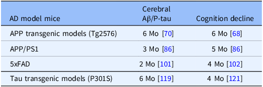
Table 1 Long description
A table comparing the earliest detection points of cerebral Aβ/P-tau and cognition decline in various Alzheimer’s disease (AD) mouse models. The table has four rows and three columns. The columns are labeled ’AD model mice’, ’Cerebral Aβ/P-tau’, and ’Cognition decline’. The rows are labeled with different AD mouse models: APP transgenic models (Tg2576), APP/PS1, 5xFAD, and Tau transgenic models (P301S). Each row lists the age in months at which cerebral Aβ/P-tau and cognition decline are first detected. For example, in APP transgenic models, cerebral Aβ/P-tau is detected at 6 months, and cognition decline is detected at 6 months. The table provides specific references for each detection point.
AD = Alzheimer’s disease; Aβ/P-tau = amyloid beta/hyperphosphorylated tau; APP/PS1 = amyloid precursor protein/presenilin 1; Mo = month of age.
Earliest detection points of sensory deficits in AD mouse models

Table 2 Long description
A table comparing the earliest detection points of sensory deficits in Alzheimer’s Disease (AD) mouse models across olfactory, visual, and auditory systems. The table has 4 rows and 12 columns. Column headers are: AD model mice, Olfactory APP/Aβ/P-tau aggregation, Olfactory Behaviors, Olfactory EP, Visual APP/Aβ/P-tau aggregation, Visual Behaviors, Visual EP, Auditory APP/Aβ/P-tau aggregation, Auditory Behaviors, Auditory EP. Row labels are: APP transgenic models (Tg2576), APP/PS1, 5xFAD, Tau transgenic models (P301S). Row 1: APP transgenic models (Tg2576), 2 Mo, 4 Mo, 3 Mo, 7.8 Mo, 9 Mo, NA, NA, NA, NA, NA. Row 2: APP/PS1, 3 Mo, 3 Mo, 3 Mo, 5 Mo, 8.5 Mo, 3 Mo, 2 Mo, 7 Mo, 2 Mo. Row 3: 5xFAD, 2 Mo, 3 Mo, 3 Mo, 6 Mo, 6 Mo, 3 Mo, 1 Mo, 2 Mo, 6 Mo. Row 4: Tau transgenic models (P301S), 1 Mo, 2 Mo, 3 Mo, 2 Mo, 2.5 Mo, NA, 4 Mo, 3 Mo, NA.
AD = Alzheimer’s disease; APP/Aβ/P-tau = amyloid precursor protein/amyloid beta/hyperphosphorylated tau; APP/PS1 = amyloid precursor protein/presenilin 1; EP = evoked potential; Mo = month of age; NA = not applicable.
APP transgenic model
APP transgenic models overexpress the mutation of human APP, which is associated with familial AD (FAD), such as the famous Tg2576 mouse model that overexpress Swedish mutation (K670M/N671L) [Reference Hsiao, Chapman and Nilsen66]. Tg2576 mice exhibit a large number of failures in the MWM test with visible platforms starting from 6 to 11 months [Reference Westerman, Cooper-Blacketer and Mariash67] and exhibit cognitive decline around 6 months of age [Reference Lesné, Koh and Kotilinek68]. At this age, the soluble Aβ levels in the brain cortex start to increase [Reference Nyul-Toth, Delfavero and Mukli69]. However, the actual cerebral Aβ plaques can be observed around 6–10 months old, typically by 9–12 months [Reference Kawarabayashi, Younkin, Saido, Shoji, Ashe and Younkin70]. Another APP transgenic mouse model is TgCRND8. Similar to Tg2576, TgCRND8 mice encode Swedish mutation (K670M/N671L) and Indiana mutations (V717F) APP protein [Reference Chishti, Yang and Janus71]. With the extra expression of mutant Aβ peptides, TgCRND8 shows an earlier onset of AD pathology compared with the Tg2576 model. The Aβ deposits can be detected as early as 3 months old, accompanied by the early onset of memory and learning impairment [Reference Chishti, Yang and Janus71].
Tg2576 transgenic mice exhibited sensory impairment at the early stage of AD. In the APP transgenic model Tg2576, the soluble human Aβ is detectable in the OB as early as 2 months old, which is before the deposition in the PCX, entorhinal cortex (EC), or hippocampus [Reference Duffy, Morales-Corraliza and Bermudez-Hernandez72,Reference Wilson and Yan73]. The nonfibrillar Aβ deposition can be observed within the OB around 3 months of age in Tg2576 mice [Reference Wesson, Levy, Nixon and Wilson74]. Mistargeted OSN connection in Tg2576 mice can be observed at ages from 12 months down to even postnatal day 1075. Furthermore, the decline of olfactory function is also revealed by the impairment of odor-related behaviors in Tg2576 mice. Four-month-old Tg2576 mice showed a significant impairment in the olfactory working memory task when more odors were applied [Reference Young, Sharkey and Finlayson76]. Four-month-old Tg2576 mice are also impaired at object placement, an EC-dependent cognitive task [Reference Duffy, Morales-Corraliza and Bermudez-Hernandez72]. These results indicate that the Aβ deposition in the olfactory system is earlier than it is in the brain cortex, and the impairment of olfactory function also emerges much earlier compared with the MCI and neurodegeneration. Tg2576 mice also show changed electrophysiology characters in the olfactory system before the cognitive impairment. Three-month-age Tg2576 mice exhibit hyperactive odor-evoked activity in the PCX, which is shown to be involved in higher-order olfactory functions, and increased OB-PCX functional connectivity [Reference Wesson, Borkowski, Landreth, Nixon, Levy and Wilson77]. The hyperactivity can be also seen in both OB and PCX, with significantly increased beta and gamma band power of spontaneous local field potential (LFP) activity [Reference Wesson, Borkowski, Landreth, Nixon, Levy and Wilson77]. The single-unit spontaneous activity in PCX shows a trend toward increased baseline firing starting from 3 months of age [Reference Xu, Lopez-Guzman and Schoen78]. This data reveals a hyperactive activity persisted during the early stage of AD, before converting to a hyporesponsive state [Reference Wesson, Borkowski, Landreth, Nixon, Levy and Wilson77]. However, in 18–21-month-old APP knock-in mice (harboring three FAD mutations from Swedish, Iberian, and Arctic), no significant differences were observed for the olfactory-related mRNA expression [Reference Narukawa, Mori and Nishida79], indicating that the correlation might only be observed at the early stage of AD and could be overlapped by the normal aging process. We discussed the aging factor during AD pathologies in the following section.
The APP transgenic model exhibited visual impairment at the early stage of AD. APP immunoreactivity can be observed as early as the 7.8-month-old Tg2576 mice ganglion cell layer and the inner nuclear layer of the retina [Reference Ning, Cui, To, Ashe and Matsubara80]. Aβ can be detected in the retina of 12-month-old Tg2576 mice [Reference Dutescu, Li, Crowston, Masters, Baird and Culvenor81,Reference Liu, Rasool and Yang82]. Tg2576 mice were impaired in visible platform recognition at 9 months of age and increased activity measures as early as 3 months old [Reference King and Arendash83]. Other research confirmed that at both 6 and 14 months of age, the Tg2576 mice were not affected using water maze spatial reference memory and T-maze working memory [Reference Zhuo, Prescott, Murray, Zhang, Baxter and Nicolle84].
APP/PS1 transgenic model
APP/PS1 is another AD mouse model that harbors a chimeric mouse/human APP (Mo/HuAPP695swe) and a mutant human presenilin 1 (PS1-dE9); both are associated with early-onset AD [Reference Jankowsky, Fadale and Anderson85]. Compared with APP transgenic mice, APP/PS1 mice show an early onset of Aβ plagues and AD pathology. The expression of cerebral Aβ deposition in APP/PS1 mice that express cerebral Aβ can be detected as early as 3 months of age – the significant memory deficits start from 5 months old [Reference Zhu, Wang and Zhang86].
APP/PS1 mice exhibit olfactory deficits but not cognitive impairment at 3–4 months of age, with soluble Aβ deposition aggregates and morphology changes in granule cells and mitral cells (MCs) in OB [Reference Li, Li and Shen87]. At this age, APP/PS1 mice show impaired odor-related behavioral performance in odor sensitivity tests and discrimination tests [Reference Li, Li and Shen87]. APP/PS1 mice also have an altered electrophysiology in the olfactory system at the early stage of AD. MCs exhibit increased spontaneous firing rates as early as 3 months induced by the inhibitory defects of pre-synapses in OB [Reference Hu, Geng, Guo, Liu, Zong and Hou88]. Three-month-old APP/PS1 mice show altered LFP activity, with a reduced theta band and increased low gamma oscillatory activity that can be detected in OB slices from 3-month-old APP/PS1 mice [Reference Chen, Chen and Huo89]. Others reported reduced low and high gamma oscillations with or without odor stimulation using the LFP analysis in the anterior piriform cortex from 3-month-old APP/PS1 mice [Reference Shen, Li, Long, Yang and Jiang90].
Aβ can be detected in 6 months of age APP/PS1 mice [Reference Dutescu, Li, Crowston, Masters, Baird and Culvenor81]. Another report indicates that Aβ levels in the retina correlate with cerebral levels that can be detected as early as 5 months [Reference Mei, Yang and Zhu91]. Substantial color and contrast-mode alternation deficits appear in APP/PS1 mice as early as 8.5 months of age [Reference Vit, Fuchs and Angel92]. APP/PS1 mice exhibit altered electroretinogram (ERG) and optical coherence tomography (OCT) starting from 3 months; the retinal pathology, with a reduced inner retinal thickness measured by OCT, can be observed as early as 9 months [Reference Georgevsky, Retsas, Raoufi, Shimoni and Golzan93].
It has been reported that APP/PS1 mice showed an early onset of hearing loss as early as 2 months old, starting from the high-frequency region, and appear to be whole-frequency range later on, similar to the age-related hearing loss [Reference Liu, Fang and Liu94,Reference Ruan, Chen and Panza95]. However, another study has reported that there is no significant change in both auditory brainstem response (ABR) and distortion product otoacoustic emissions (DPOAE) thresholds even in 13 months of age APP/PS1 mice. A recent study showed progressive age-related hearing loss in both WT and APP/PS1 mice after shame operation (SO) of cochlear ablation; the ABR thresholds were compatible at 4, 6, 9, and 12 months of ages between WT and APP/PS1 mice with SO [Reference Pan, Li and Meng18]. One possible reason for the different outcomes could be that different backgrounds of mice were utilized. C57 mice typically show a faster onset of age-related hearing loss, while CBA/CaJ mice exhibit a longer hearing lifespan due to the cdh23 mutation [Reference Kane, Longo-Guess, Gagnon, Ding, Salvi and Johnson96]. Thus, the difference may be obscured by normal aging, given the inherent age-related hearing loss in some mouse backgrounds. For the auditory-related behavioral test, there is no significant difference between APP and APP/PS1 mice and wild-type mice at 6 months old [Reference Pietropaolo, Delage, Lebreton, Crusio and Cho97]. This data is also confirmed using a 7-month-old APP/PS1 mouse model that indicates a nonsignificant trend of altered ASR, but no changes in PPI can be found [Reference Cheng, Logge, Low, Garner and Karl98,Reference Na, Zhang and Beaulac99]. Another report also showed that APP/PS1 mice exhibited normal ASR and PPI at 3 months old but significantly lower PPI starting from 7 months of age [Reference Wang, He and Zhang100]. APP protein expression in the auditory cortex can be detected as early as 2 months old, accompanied by decreased auditory-evoked cortical potential[Reference Mei, Liu, Chen and Zhao13]. These data indicate that hearing loss and reduced auditory cortical response could be early biomarkers for AD in this mouse model.
5xFAD transgenic models
5xFAD mouse model co-express five FAD mutations, three APP mutations (mutant human APP with Swedish [K670N, M671L], Florida [I716V], and London [V717I] mutations), and two PS1 mutation (M146L, L286V) (Oakley et al., 2006). 5xFAD mouse model is designed for the early onset of AD, as it displays strong Aβ pathologies, with plaques appearing in the brain from 2 to 4 months of age, resulting in robust synaptic and neuronal loss [Reference Jawhar, Trawicka, Jenneckens, Bayer and Wirths101]. Early cognitive deficits related to the frontal cortex appear at 4 months of age [Reference Girard, Baranger and Gauthier102].
The odor-related behavioral change can be detected in 3-month-old 5xFAD mice, with [Reference Son, Yoo and Kang103]. The Aβ deposition in this model can be seen at 2 months of age [Reference Ziegler-Waldkirch, Friesen and Loreth104] and is high in the OSNs located in the olfactory epithelial ectoturbinate and the ventral OB glomeruli at 3 months of age [Reference Son, Yoo and Kang103]. Another report shows that Aβ is co-localized with synaptic markers on OB glomeruli as early as 2–4 months of age, but the olfactory memory is not impaired from 3 to 15 months of age [Reference O’Leary, Stover, Mantolino, Darvesh and Brown105]. However, other research shows an impaired olfactory function starting from 3 months and can be detected at 6 and 8 months of age [Reference Zhang, Zhao and Sun61,Reference Girard, Jacquet and Baranger106]. OSNs from 5xFAD mice at 3 months of age show reduced responses to odorant stimulation measured by Ca2+ signals [Reference Son, Yoo and Kang103].
5xFAD mice show reduced ganglion cell responses to light as early as 6 months of age, with a thicker inner plexiform layer [Reference Lim, Li and He107]. This mouse model exhibits impaired spatial learning in water maze tests starting at 6 months of age [Reference Patil, Grossman, Kenney, Balcer and Galetta108]. The changed full-field ERGs can be observed as early as 3 months of age [Reference Patil, Grossman, Kenney, Balcer and Galetta108].
In 5xFAD mice, Aβ deposition and intracellular accumulation can first be found in the primary auditory cortex as early as postnatal day 21 (p21), and the extracellular plaques appear later by age p90 [Reference Weible and Wehr109,Reference Oakley, Cole and Logan110]. The Aβ accumulation occurs at cortical levels and the upper auditory brainstem as early as 3 months of age but at lower levels of the brainstem such as the cochlear nucleus [Reference Na, Zhang and Beaulac99]. Using the ASR and PPI test, 5xFAD mice exhibit an age-related decline in acoustic startle as early as 3 months of age before any hearing loss symptom [Reference Omata, Tharasegaran and Lim111], but no significant reduction of PPI at 3–4 months of age [Reference O’Leary, Shin and Fertan112]. Thirteen-month-old 5xFAD mice have increased ABR thresholds, indicating an early onset of hearing loss [Reference Omata, Tharasegaran and Lim111]. 5xFAD mice also exhibit gap detection deficits in startle response tests as early as 2 months of age [Reference Kaylegian, Stebritz, Weible and Wehr113], with degraded gap responses and baseline firing rates in the auditory cortex [Reference Weible, Stebritz and Wehr114]. All these results demonstrate early-onset impairments of the central auditory system in 5xFAD mice. However, it has also been reported that at 3 months of age, 5xFAD mice are indistinguishable from the control in central auditory activity and hearing threshold, but at 6 months of age, the central gain was significantly increased (p4:p1) in 5xFAD mice [Reference Na, Zhang and Beaulac99]. Overall, 5xFAD mice exhibit delayed ASR, which could be a potential biomarker for early AD diagnosis [Reference Story, Chan, Munro, Rossignol and Dunbar115].
Tau transgenic model
The tau pathology and synaptic failure correlate with cognitive decline [Reference Serrano-Pozo, Frosch, Masliah and Hyman116]. Thus, overexpression of incorrect tau protein could also induce AD in mouse models. Tau transgenic model exhibits mutant tau protein, the P301S (PS19) mice model, for example, expressing P301S mutation tau driven by mouse prion protein promoter (Prnp), gives rise to NFTs and neurodegeneration by 9–12 months of age [Reference Yoshiyama, Higuchi and Zhang117,Reference Allen, Ingram and Takao118]. Hyperphosphorylated tau (P-tau) can be detected in the hippocampus and cortex at 6 months of age [Reference Takeuchi, Iba and Inoue119]. Memory deficits can be identified around 5 months of age in P301S mice [Reference Xu, Rosler and Carlsson120]. Spatial learning deficits can be observed as early as 4 months of age [Reference Przybyla, Van Eersel and Van Hummel121]. P301S mice do not typically develop Aβ plaques since it is designed for tau pathology research. However, it has been reported that the intracerebral injection of Aβ1-42 increases tau phosphorylation, cleavage, and aggregation [Reference Hu, Li, Zhao, Gottesdiener, Luo and Paul122]. Co-expression of soluble Aβ (Aβ4-42) in the P301S tau transgenic model worsens spatial memory deficits and motor performance [Reference Zampar and Wirths123]. These data demonstrate that Aβ promotes tau pathogenesis.
Tau transgenic mice (P301S) show increased P-tau expression levels in the OB and PCX as early as 1 month of age; the MC layer is severely affected [Reference Li, Li and Wu124]. A progressive neuronal cell loss in OB and PCX can be observed during aging [Reference Yang, Kuan and Spillantini125]. The olfactory impairment starts as early as 2 months of age using the food-seeking test [Reference Zhang, Zhao and Sun61,Reference Li, Li and Wu124]. Three-month-old P301S mice show a reduction in gamma frequency oscillation and severe early impairments in the theta-gamma phase-amplitude coupling in the OB area, but the LFP oscillatory activities do not show any difference in EC [Reference Ahnaou, Rodriguez-Manrique, Biermans, Embrechts, Manyakov and Drinkenburg126].
P-tau starts to accumulate in the nerve fiber layer in the P301S mouse model at 2 months of age and aggregates into filamentous inclusions in retinal ganglion cells [Reference Gasparini, Crowther and Martin127]. P301S mice show a worse learning curve compared with wild-type mice at 2.5 months of age [Reference Xu, Rosler and Carlsson120]. Another study reports early and persistent spatial working memory deficits, with hyperactivity in the Y-maze test with less alternation, and this trend can be seen as early as 2 months of age [Reference Ahmad, Mein, Jing, Zhang and Liu128].
P301S mice express phosphorylated tau in the auditory cortex, amygdaloid nucleus, and ventral hippocampus as early as 4 months of age, with the enhancement of the PPI test [Reference Takeuchi, Iba and Inoue119]. Other studies show that P301L mice exhibit unaltered ASR but reduced PPI (or increased %PPI) at 3 months of age [Reference Takeuchi, Iba and Inoue119,Reference Koppel, Jimenez and Azose129]. Whether the tau pathway affects auditory electrophysiology needs further investigation.
Overall, in four commonly utilized AD mouse models, a consistent pattern can be observed that sensory system impairments were detectable earlier than the cerebral Aβ deposition and cognitive decline (Tables 1–2). Furthermore, most of these models exhibited multiple sensory deficits at the early stage of AD, suggesting the possibility of using an integrated assessment of sensory system impairments as a noninvasive diagnostic tool.
Sensory deficits and aging in the symptomatic stage of AD
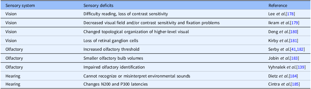
Transitioning from the exploration of sensory deficits in the research animal models to the symptomatic stage marks a critical juncture in understanding the disease’s progression. During the preclinical phase, subtle sensory changes may begin to emerge, often unnoticed or dismissed as benign age-related decline. However, as AD advances to its symptomatic stage, these sensory impairments become more pronounced, directly impacting patients’ quality of life and their ability to interact with the world around them (Table 3).
Sensory deficits in the symptomatic stage of Alzheimer’s disease
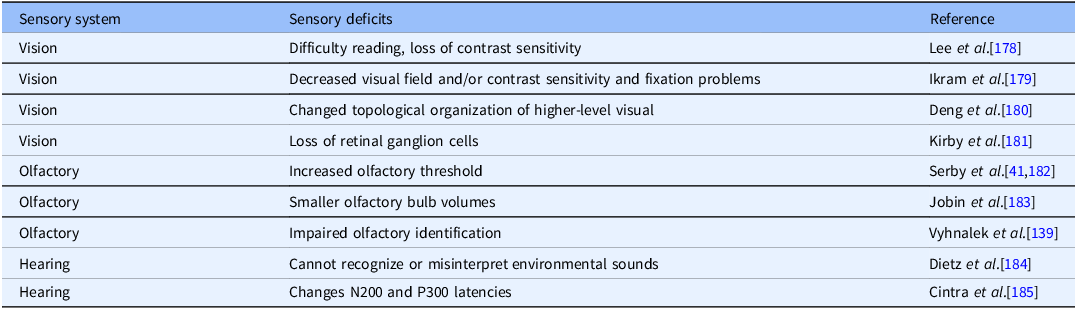
Table 3 Long description
A table with three columns and eight rows. The columns are labeled Sensory system, Sensory deficits, and Reference. The rows list different sensory systems and their associated deficits, along with references. Row 1: Vision, Difficulty reading, loss of contrast sensitivity, Lee et al. Row 2: Vision, Decreased visual field and/or contrast sensitivity and fixation problems, Ikram et al. Row 3: Vision, Changed topological organization of higher-level visual, Deng et al. Row 4: Vision, Loss of retinal ganglion cells, Kirby et al. Row 5: Olfactory, Increased olfactory threshold, Serby et al. Row 6: Olfactory, Smaller olfactory bulb volumes, Jobin et al. Row 7: Olfactory, Impaired olfactory identification, Vyhnalek et al. Row 8: Hearing, Cannot recognize or misinterpret environmental sounds, Dietz et al. Row 9: Hearing, Changes N200 and P300 latencies, Cintra et al.
A variety of ocular abnormalities have been observed in AD patients in the MCI stage, indicating a strong association between AD and visual impairment [Reference Papageorgiou, Tsirelis, Lazari, Siokas, Dardiotis and Tsironi130]. In the clinical stage, a large number of visual disorders and their association with Aβ burden have been reported in AD patients [Reference Golzan, Goozee and Georgevsky131], such as lower visual acuity in recognition of low luminance and low spatial frequency pictures [Reference Lee and Pai132], reduced visual contrast sensitivity [Reference Gilmore, Groth and Thomas133], altered color vision [Reference Kim, Ryou, Choi, Kim, Kim and Han134], retinal hemodynamic parameters [Reference Berisha, Feke, Trempe, Mcmeel and Schepens135], visual field loss [Reference Bateman136], and poor ocular motor function [Reference Crawford, Devereaux, Higham and Kelly137]. A deteriorated olfactory function can be observed in AD patients [Reference Dibattista, Pifferi, Menini and Reisert138]. Furthermore, olfactory dysfunction is associated with Aβ burden. Several studies have reported early olfactory impairment in AD patients also known as hyposmia [Reference Vyhnalek, Magerova and Andel139], where these patients exhibit a reduced ability to detect, discriminate, and identify odors, coupled with abnormal odor coding [Reference Yaffe, Freimer and Chen140,Reference Li, Howard and Gottfried141]. As many as 60% of older adults with AD and ADRD have hearing loss [Reference Powell, Reed and Wolff142]. All these sensory impairments could be used to evaluate the AD pathophysiology and should be taken into consideration for the AD clinical diagnosis. Multiple epidemiological studies have revealed that hearing loss is correlated with pathological cognitive decline and dementia [Reference Jayakody, Friedland, Martins and Sohrabi59,Reference Tarawneh, Menegola, Peou, Tarawneh and Jayakody143]. Hearing loss is a risk factor for cognition impairment. It has been reported that mild to severe hearing loss significantly increases the dementia risk [Reference Lin, Metter, O’Brien, Resnick, Zonderman and Ferrucci144,Reference Livingston, Huntley and Sommerlad145]. Elderly individuals with hearing loss symptoms have a higher risk of AD, which is also associated with loss of brainstem and cerebellar volume [Reference Llano, Kwok and Devanarayan146]. Furthermore, AD could cause a faster onset of hearing loss. Individuals’ AD risks can be evaluated using a polygenic risk score (PRS) [Reference Jansen, Savage and Watanabe147]. High PRS individuals are more likely to experience hearing difficulty than those with lower PRS [Reference Mitchell, Thorp, Evans, Nyholt, Martin and Lupton148]. However, the causation between AD and hearing loss is still controversial. Several hypotheses might explain the relationship between AD and hearing loss. First, hearing loss could cause cognitive decline and eventually dementia. Indeed, hearing loss induced by noise exposure exacerbates cognitive decline [Reference Paciello, Rinaudo and Longo149], and expressing Aβ derivatives in the cochlear hair cells leads to an early onset of hearing loss, especially in high-frequency regions, similar to age-related hearing loss [Reference Omata, Tharasegaran and Lim111]. Overexpression of tau synergistically worsens the Aβ-induced hearing defects [Reference Omata, Tharasegaran and Lim111]. These data indicate that Aβ and tau are both toxic to the peripheral auditory system, and overexpression of Aβ might accelerate the process of age-related hearing loss. Furthermore, hearing loss leads to less engagement in social and leisure activities which results in dementia. Lastly, both hearing loss and cognitive decline could be caused by a “common factor” [Reference Amieva and Ouvrard150] and might share the same pathway with age-related hearing loss, or presbycusis [Reference Chern and Golub151]. Despite the unclear causation between AD and auditory impairment, it has been reported that inducing gamma oscillations in the auditory cortex improved spatial and recognition memory and reduced Aβ in AC [Reference Martorell, Paulson and Suk152], indicating that the auditory pathway could be a potential target to ameliorate AD-associated pathology.
Noticeably, all the sensory impairments mentioned above could also be the comorbidities of the natural aging process [Reference Liu, Tan, Zhang, Yi, Zhu and Peng153]. This confluence of sensory impairments with aging complicates the differential diagnosis of AD from normal age-related changes, necessitating a nuanced understanding of pathological versus physiological decline. Moreover, the overlap of sensory impairments with AD is not uniquely induced by the aging process but could also be induced by other neurodegenerative diseases such as Parkinson’s disease (PD) and dementia with Lewy bodies (DLB) [Reference Lad, Sedley and Griffiths154,Reference O’Callaghan, Firbank, Tomassini, Schumacher, O’Brien and Taylor155]. Critical to advancing this differentiation is the longitudinal study of sensory deficits, utilizing both animal models and human subjects, to delineate the specific markers of AD-related sensory degradation. The challenge remains to disentangle the intricate web of aging and disease-specific sensory changes, requiring innovative methodologies and interdisciplinary approaches. Integrating multisensory assessments with cognitive evaluations and biomarker analysis could enhance the specificity of early AD diagnoses. Ultimately, this comprehensive approach will not only improve our understanding of AD progression but also pave the way for the development of targeted interventions to mitigate sensory deficits and improve the quality of life for individuals with AD.
Sensory impairments in preclinical AD patients
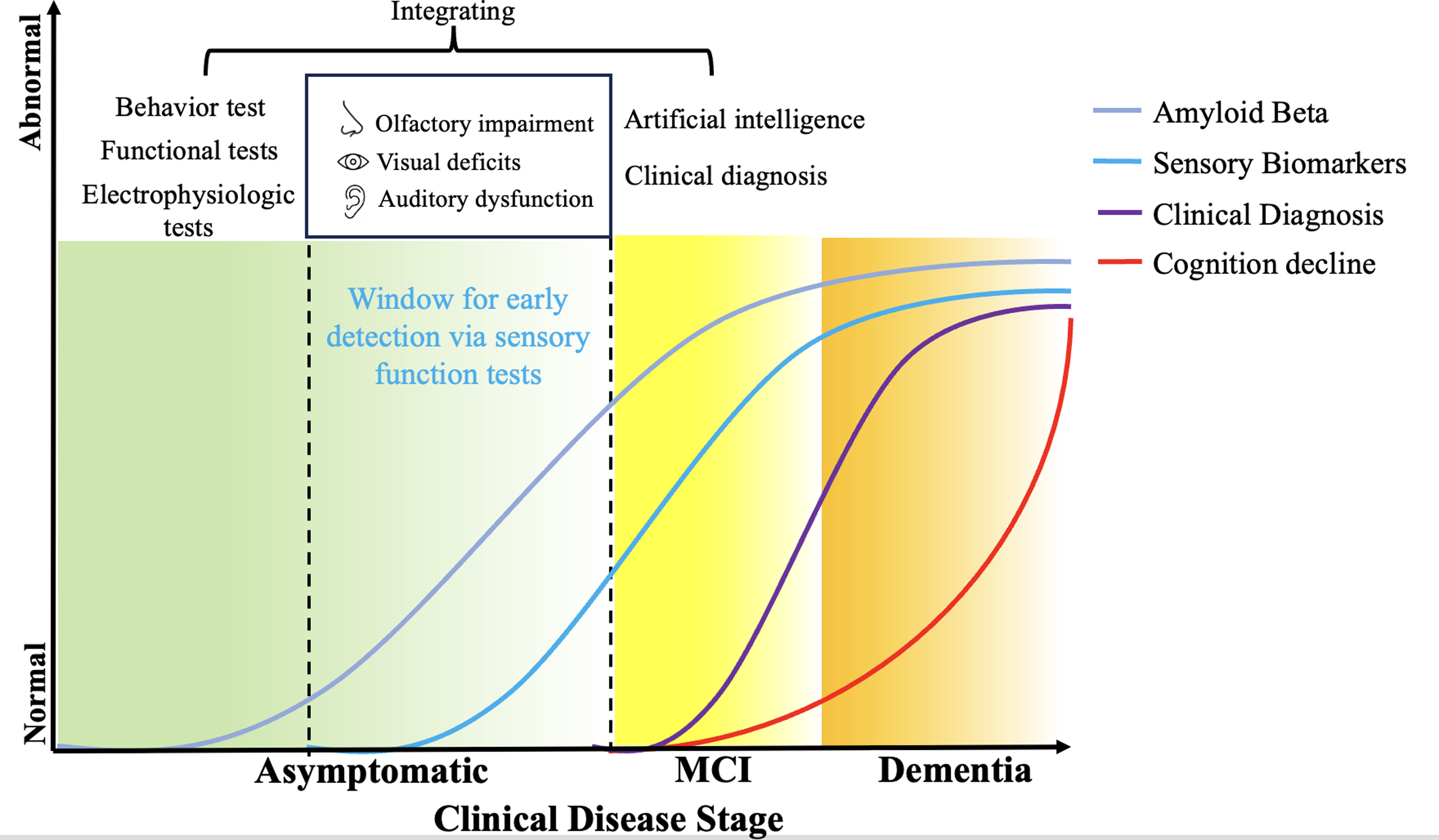
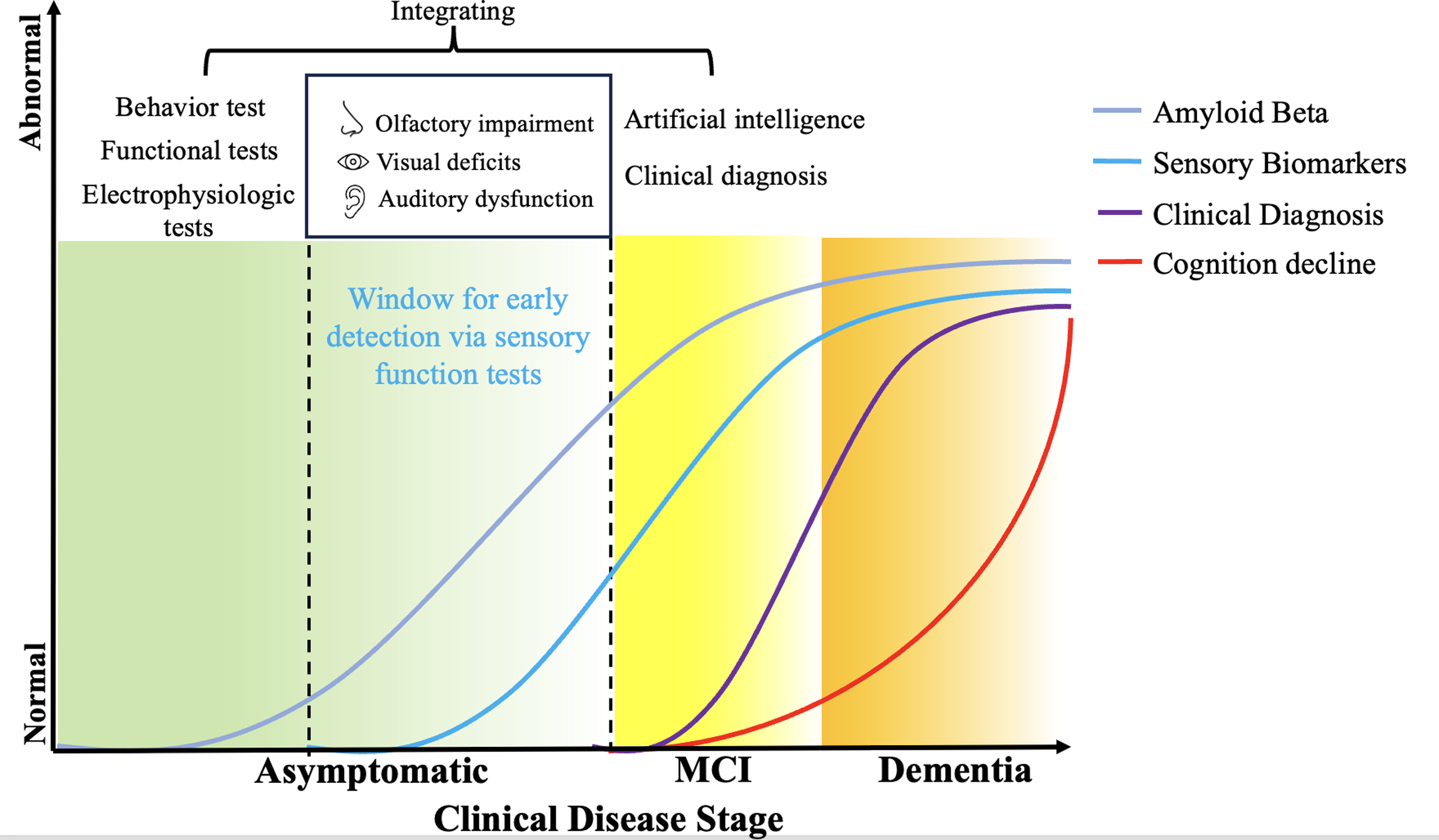
The impairment of the sensory system in AD patients may occur in the preclinical stage, well before the emergence of the onset of clinical symptoms (Fig. 1). Olfactory dysfunction is associated with a higher Aβ burden in older people. Early olfactory impairment in AD patients also known as hyposmia [Reference Vyhnalek, Magerova and Andel139], where patients exhibit a reduced ability to detect, discriminate, and identify odors, coupled with abnormal odor coding [Reference Yaffe, Freimer and Chen140,Reference Li, Howard and Gottfried141], elucidating the potential contribution of olfactory testing to detect preclinical AD [Reference Growdon, Schultz and Dagley156]. In clinically normal elderly individuals, worse olfactory function was associated with decreased hippocampal volume, thinner EC, and a trend associated with greater Aβ burden [Reference O’Callaghan, Firbank, Tomassini, Schumacher, O’Brien and Taylor155]. Another study showed that reduced odor identification (OI) was associated with lower cognitive score and older age and increased CSF tau and Aβ, revealing that OI could be an affordable biomarker of AD pathology [Reference Lafaille-Magnan, Poirier and Etienne157]. The visual deficit is considered a high-risk factor for AD and is increasingly supported by evidence linking visual system alterations to the early stage of AD [Reference Hawkins, Goyal and Sergio158]. For the preclinical stage, substantial visual deficits have been reported in AD patients [Reference Javaid, Brenton, Guo and Cordeiro44,Reference Katz and Rimmer159]. Potential measurable functional, structural, metabolic, and vascular changes have been identified in the retina during the early stages of AD [Reference Gupta, Chitranshi and den Haan160], indicating that vision could be another biomarker for early detection of AD. As another high-risk factor of AD, hearing loss was also associated with higher Aβ and tau burden. A study indicated that worsened hearing was related to the increased Aβ and tau burden [Reference Zheng, Yan and Hao161].
Above all, the evidence supporting sensory deficits as early indicators of AD suggests a paradigm shift in the approach to diagnosing and managing this complex condition in the preclinical stages.
Integrating sensory detection in early AD diagnosis
The imperative of incorporating sensory detection
In our comprehensive review of early changes in various AD mouse models, notable early alternations take place in sensory systems prior to the cognition decline and the cerebral Aβ/P-tau aggregation. Here we listed out the early time point of alternation in different sensory systems using different AD mouse models (Table 2). This trend is consistent in olfactory, visual, and auditory systems across different AD models. Further, we also found a trend of hyperactivity status across sensory systems before the onset of neurodegeneration and cognition decline. The soluble Aβ oligomers induced hyperactivity on memory performance, cell death, epileptiform activity, gamma oscillations, and slow wave activity can be observed in both AD mouse models and human patients [Reference Hector and Brouillette162], consistent with early detection of soluble Aβ in sensory organs. The hyperactivity status of sensory functions, also known as network hyperexcitability [Reference Tok, Ahnaou and Drinkenburg163], could be a biomarker for AD.
However, we also found results may vary in different research due to the diversity of methodologies and sensory systems. Indeed, some tests have certain limitations. For example, the behavioral test can be affected easily by handling and environmental factors and can be stressful for animals [Reference Gouveia and Hurst164,Reference Fonio, Golani and Benjamini165]. Additionally, the accurate measurement and diagnosis of AD using sensory deficits may be complicated by other variables, including natural aging processes, PD, DLB, and various other neurodegenerative conditions, which can introduce significant variability into the assessment of sensory impairments. The variability and uncertainty indicate the necessity of integrating sensory function tests and the combination of sensory tests and other test methods to increase the accuracy of early AD diagnosis. To achieve this, multidisciplinary approaches that incorporate neuropsychological, neurobiological, and genetic analyses are essential. Implementing these strategies will improve the accuracy of early AD diagnosis through sensory deficit detection and contribute to a more nuanced understanding of the disease’s pathophysiology, for instance, the combination of vision and hearing deficits in older people experience higher rates of cognitive impairment [Reference Davidson and Guthrie166]. Thus, the simultaneous application of visual and auditory assessments could potentially enhance the precision of preclinical AD diagnosis, offering a more robust framework for identifying early indicators of cognitive impairment. Above all, sensory tests have demonstrated potential as supplementary tools for the diagnosis of AD and MCI. For instance, odor identification screening (Sniffin’ Sticks Odor Identification Test, SS-OIT) can improve the diagnostic accuracy of AD and MCI when combined with MoCA, providing additional information for clinical categorization of AD and MCI [Reference Quarmley, Moberg and Mechanic-Hamilton167]. Visuospatial measures show a significant diagnostic and prognostic potential in dementia, which means visuospatial tests could also improve the diagnosis of AD [Reference Salimi, Irish, Foxe, Hodges, Piguet and Burrell168]. Central auditory testing might be another suitable biomarker for identifying people at risk for dementia combined with electrophysiologic testing [Reference Gates, Anderson, Feeney, McCurry and Larson169].
Nevertheless, before these approaches can be adopted clinically, extensive validation steps are essential. This encompasses replicating findings across diverse studies to ensure reproducibility, alongside rigorous comparisons with established diagnostic benchmarks to clinical implementation. Additionally, defining precise diagnostic thresholds for sensory tests is crucial to delineate AD-related sensory impairments from those attributable to other causes. Moreover, evaluating the predictive value of these tests in foreseeing disease progression is essential. The pathway toward clinical adoption involves meticulous steps, including the elaboration of a clear context of use, submission of a comprehensive qualification package to regulatory bodies, and achieving a consensus on standards for test performance [Reference Cummings and Kinney170–Reference Goldsack, Coravos and Bakker173]. These processes are instrumental in ensuring the reliability, efficacy, and clinical relevance of sensory detection methods, thereby facilitating the integration into the diagnostic repertoire for early AD.
Integrating sensory change detection with artificial intelligence
Utilizing artificial intelligence (AI) in predicting AD is a rapidly evolving field. AI aids early detection and diagnosis based on a variety of machine learning-trained models. Based on the cognitive tests, imaging techniques, and CSF test, AI can improve the accuracy of traditional diagnostic methods, such as analyzing PET and MRI scan images [Reference Aberathne, Kulasiri and Samarasinghe28], and new biomarkers analysis beside Aβ42 and P-tau using the CSF database [Reference Chang, Lin and Lane174]. AI can also do end-to-end detection without using additional diagnostic methods. A pre-trained data2vec model can perform a self-supervised algorithm that works for speech, vision, and text [Reference Agbavor and Liang175]. An integrating pre-trained model that can access multimodal sensory test data would be a potential method for rapid, noninvasive AD diagnosis during the early detection window (Fig. 2).
Schematic of alzheimer’s disease (AD) stages. Amyloid beta (Aβ) begins to accumulate many years before cognitive decline, marking the asymptomatic stage. This accumulation of Aβ starts to impair the sensory system’s function, including olfactory, visual, and auditory systems, before the onset of mild cognitive impairment (MCI). After the asymptomatic stage, some clinical diagnostic methods become applicable. However, by this point, patients start to experience cognitive decline, which, at this stage, is irreversible. Therefore, the window for early detection through sensory function tests is crucial. Integrating these tests with common clinical diagnostics and artificial intelligence could enhance the accuracy of early AD diagnosis before the onset of cognitive decline.
Conclusion
In conclusion, we have thoroughly reviewed the changes in multiple sensory systems in the early stage of AD, focusing on olfactory, visual, and auditory functions. Changes in these sensory systems manifest significantly in both AD patients and various AD mouse models, and the alternations of the sensory system appear much earlier than the typical neurodegeneration and cognitive decline. These results shed light on the importance of multiple sensory tests for early and noninvasive AD diagnosis. Moreover, the potential integration of multisensory examination and the combination with traditional diagnostic methods and/or AI prediction present a promising methodology for the improvement of accuracy and rapid AD detection in the future.
Author contributions
Y.Y takes responsibility for the manuscript.
Funding statement
None.
Competing interests
None.


 Open access

