Introduction
In intensive pig husbandry, pregnant sows are exposed to various stressors such as diet and space restriction, which may have negative consequences on their welfare, health (Salak-Johnson and McGlone, Reference Salak-Johnson and McGlone2007) and reproductive performances (Von Borell et al., Reference Von Borell, Dobson and Prunier2007). One potent stressor that pigs are exposed to is social stress, i.e. stress caused by composition or modification of their social group (de Groot et al., Reference De Groot, Ruis, Scholten, Koolhaas and Boersma2001; Otten et al., Reference Otten, Puppe, Kanitz, Schön and Stabenow2002, Coutellier et al., Reference Coutellier, Arnould, Boissy, Orgeur, Prunier, Veissier and Meunier-Salaün2007). In farms, the grouping of unfamiliar animals is usually accomplished to form cohorts of homogeneous age or body weight, but actual EU guidelines (2001/88/CE) also impose the housing of gestating sows in groups for welfare reasons. The formation of a new social group leads to fights during the first days to establish a social hierarchy (Puppe et al., Reference Puppe, Langbein, Bauer and Hoy2008). The physical and psychological stress associated with the fights activates stress systems such as the hypothalamo–pituitary–adrenal (HPA) axis and the sympathetic system, leading to increased plasma levels of cortisol and catecholamines (Otten et al., Reference Otten, Puppe, Kanitz, Schön and Stabenow2002), which in turn modulate animal physiology in order to cope with the new situation. In pigs, social stress or high cortisol concentrations can decrease weight gain (McGlone et al., Reference McGlone, Salak, Lumpkin, Nicholson, Gibson and Norman1993, Coutellier et al., Reference Coutellier, Arnould, Boissy, Orgeur, Prunier, Veissier and Meunier-Salaün2007), lymphocyte proliferation (Kanitz et al., Reference Kanitz, Tuchscherer, Puppe, Tuchscherer and Stabenow2004), natural killer cell activity (McGlone et al., Reference McGlone, Salak, Lumpkin, Nicholson, Gibson and Norman1993; Salak-Johnson et al., Reference Salak-Johnson, McGlone and Norman1996), neutrophilic chemotaxis (Salak-Johnson et al., Reference Salak-Johnson, McGlone and Norman1996) and antiviral immunity (de Groot et al., Reference De Groot, Ruis, Scholten, Koolhaas and Boersma2001). Most of the studies were performed on young animals and little is known about the influence of stress on the immune capacity of pregnant sows. Considering that pregnancy is a particular physiological state associated with immunological changes (Wegmann et al., Reference Wegmann, Lin, Guilbert and Mosmann1993) and a decreased sensitivity of the HPA axis to stress (Neumann et al., Reference Neumann, Johnstone, Hatzinger, Liebsch, Shipston, Russell, Landgraf and and Douglas1998), one can wonder whether chronic stress has similar immune effects in pregnant gilts as in younger non-pregnant females or males.
Evaluation of the endocrine and immune consequences of stress in gestating sows can be of particular importance because stress during pregnancy in mammals can affect not only maternal welfare and health but also embryonic and foetal development. Although Soede et al. (Reference Soede, Roelofs, Verheijen, Schouten, Hazeleger and Kemp2007) failed to observe any difference in fertilization rate and embryonic development in sows, data in rodents showed that stress during early gestation can lead to abortions (Wiebold et al., Reference Wiebold, Stanfield, Becker and Hillers1986). In species with multiple foetuses like pigs, maternal stress is suspected to reduce the number of youngs born alive, suggesting an increased foetal mortality (von Borell et al., Reference Von Borell, Dobson and Prunier2007). In addition, maternal stress during gestation can have detrimental effects on growth (Fowden et al., Reference Fowden, Dino and Forhead2006), neuroendocrine (Weinstock, Reference Weinstock1997) and immune (Merlot et al., Reference Merlot, Couret and Otten2008) functions in the offspring after birth. The deleterious effects of stress on pregnancy and foetal development seem to be related to maternal immune changes (Arck, Reference Arck2001; Joachim et al., Reference Joachim, Zenclussen, Polgar, Douglas, Fest, Knackstedt, Klapp and Arck2003; Coussons-Read et al., Reference Coussons-Read, Okun and Nettles2007) and to maternal neuroendocrine activity, especially to glucocorticoid secretions (Barbazanges et al., Reference Barbazanges, Piazza, Le Moal and Maccari1996).
The purpose of this study was to determine whether exposing pregnant gilts to repeated social stress was able to induce a prolonged state of stress as reported for non-pregnant growing pigs (Coutellier et al., Reference Coutellier, Arnould, Boissy, Orgeur, Prunier, Veissier and Meunier-Salaün2007) and therefore to determine the consequences of this stressor on their behaviour, endocrine and immune responses. The second aim was to investigate the effects of social stress on their reproductive performances when gestation is maintained despite stress. Because the risk of abortion is higher at the beginning of gestation, the social stress procedure was performed during the last third of pregnancy, and the litter characteristics were observed at birth.
Material and methods
Animals and treatments
A total of 36 nulliparous Large White × Landrace gilts from 11 litters were used for the experiment in three independent replicates of 12 females. Gilts were inseminated with fresh Pietrain semen after oestrus synchronisation with Regumate® (20 mg of 17 α-allyl-trenbolone; Roussel-Uclaf, Romainville, France). During gestation, gilts had free access to water. They were fed once a day at 0830 h with a standard pig diet for pregnant sows, which covered 145% of their metabolic requirements (1.1 kg per 100 kg live weight of a diet containing 13.5% crude protein, 2280 kcal/kg of metabolizable energy and 0.48% lysine). Animals were blocked in individual feeders for 30 min during the meal in order to prevent competition for food. Within each replicate, gilts were equally allocated to one of the two treatment groups (Stress group, S; Control group, C) at 77 ± 1 days of gestation (mean ± s.d.). They were 396 ± 9 days of age and weighed 215 ± 10 kg at the beginning of the experiment. Assignment of gilts to treatment groups was realized in order to balance litter of origin and live weight between S and C groups. At least 4 months before allocation to the treatment groups, all gilts were housed in stable groups of four in a single room on a concrete floor with straw bedding (11.5 m2). On the first day of the stress procedure, two gilts per pen were moved to new pens (11.5 m2) located in another room for allocation to the S group. The other two gilts, allocated to the C group, remained undisturbed in the initial pens. Gilts from the S group were exposed to a repeated social stress routine. Over a period of 4 weeks, between days 77 and 101 of gestation, pairs of S gilts were changed twice a week (on Mondays and Thursdays at 1400 h). In order to form pairs of unfamiliar animals at each grouping, six additional non-experimental gilts with similar age and weight as S gilts were used. Pairs of C gilts were left undisturbed during the experimental period. On day 105 of gestation, all gilts were moved to individual farrowing crates (5 m2) with straw bedding and stayed there with their litter until weaning of piglets at 4 weeks of age. Parturition was not induced.
On days 70 and 105 of gestation, gilts were weighed and back fat thickness was measured ultrasonically (back at 65 mm from the midline). All gilts were immunized intra-muscularly in the neck with a solution of 2.5 mg DNP-keyhole limpet haemocyanin (KLH) (2,4-dinitrophenyl conjugated to keyhole limpet haemocyanin; Merck, Nottingham, UK) diluted in 1 ml sterilized water and 1 ml Montanide ISA 206 adjuvant (SEPPIC; Paris, France) on days 81 and 95 of gestation.
No animal was severely injured, required veterinary treatment or died during the experiment. Piglets were weighed at birth and the average weight of piglets was calculated. Stillborn piglets and mummified piglets were numbered.
Blood and saliva collection
Blood samples were collected from the anterior vena cava of gilts restrained by snaring on days 70 and 105 of gestation. The sampling procedure took less than 2 min. EDTA-blood samples were collected for blood formula analyses, heparinized-blood samples for the proliferation assays and blood samples without anti-coagulant for immunoglobulins G (IgG) measurement. Blood was kept at 4°C until use.
Salivary samples were collected at 0900 h and 1500 h 1 week before the first grouping (day 72 of gestation). For the 1st, 4th and 8th groupings, samples were collected before (at 1330 h), after 1 h (at 1500 h) and after 19 h (at 0900 h) of grouping. Additional samples were collected 2 days after the 3rd and 7th groupings (i.e. the day before the 4th and 8th groupings) at 0700, 1100, 1500 and 1900 h in order to analyse diurnal variations of cortisol. Salivary samples were collected by allowing gilts to chew on cotton buds until they were moistened. Cotton buds were rapidly centrifuged at 3000 × g for 15 min at 4°C. Saliva samples were stored at −20°C until cortisol analysis.
Measurements
Agonistic behaviour and skin lesions
In the S group, behaviour was video recorded for the first 3 h of each grouping. Agonistic behaviour of the dyads was analysed from the videotapes by continuous event sampling to determine the number and duration of agonistic interactions, the initiating sow and the outcome (win, defeat or undecided). An agonistic interaction was defined as an overt fight or a displacement event with physical contact for more than 3 s. If an agonistic sequence lasted longer than 8 s, the next fight was classified as a new interaction. Agonistic interactions comprised ‘bitings’, ‘head-to-head knocks’, ‘head-to-body knocks’ or ‘parallel/inverse pressing’ (Puppe, Reference Puppe1998; Tuchscherer et al., Reference Tuchscherer, Puppe, Tuchscherer and Kanitz1998). A sow won the agonistic interaction when the other (defeated) sow first stopped fighting, turned away from an attack or tried to flee. The agonistic interaction was defined as undecided when there was no clear win or defeat. In addition, the initiating sow for each agonistic interaction was registered. For each S gilt a success value (SV) was evaluated for each grouping day according to the formula: (number of wins − number of defeats)/(number of wins + number of defeats) ranging from −1 to +1. An aggression value (AV) was calculated according to the formula: (number of agonistic interactions initiated by the gilt)/(total number of agonistic interactions) and ranged from 0 (not aggressive) to +1 (aggressive).
Skin lesions on the entire body (legs, sides, back and ears) were numbered 2 h after the beginning of each grouping in both S and C groups by an observing person in the pen. Only fresh lesions of more than 2 cm length were numbered.
Anti-KLH IgG
Blood samples without anti-coagulant were left to clot for 3 h at room temperature and were centrifuged at 3000 × g for 15 min at 4°C. Sera samples were stored at −20°C until analysis for anti-KLH IgG by ELISA as described previously (Schrama et al., Reference Schrama, Schouten, Swinkels, Gentry, De Vries Reilingh and Parmentier1997). Measurements were performed in duplicates. Briefly, serial diluted sera were applied to antigen-coated wells (KLH, 0.1 μg/150 μl per well) of a 96-well round-bottomed plate (Greiner, Courtaboeuf, France). After 2 h of incubation at room temperature and subsequent washing with phosphate-buffered saline (Tris 0.05% Tween20), a one-step conjugation was performed. Conjugation consisted of incubation for 1 h with a 1 : 8000 diluted peroxydase-conjugated rabbit anti-swine IgG Fc (RaSw-IgG (Fc)/PO; Nordic, Tilburg, The Netherlands). After washing, tetramethylbenzidine substrate (TMB; Sigma-Aldrich, Saint Quentin Fallavier, France) was added. After 10 min, the reaction was stopped with 2.5N sulphuric acid. Absorbance was read with a micro-plate reader at a test wavelength of 450 nm. The titer of immunoglobulins against KLH was calculated as the dilution that showed 50% extinction of Emax, where Emax represents the saturation optical density of a positive standard serum. In the absence of immunization, there was no detectable level of IgG anti-KLH in sera of gilts. Inter- and intra-assay CV were 9.9% and 2.2%, respectively. Gilts from the first replicate were not immunized.
Lymphocyte proliferation
Peripheral blood mononuclear cells (PBMC) were isolated from heparinized blood samples by density gradient centrifugation. Briefly, 3 ml of blood diluted in an equal volume of Hank’s balanced salts (HBSS) culture medium was layered on 3 ml histopaque 1077. After centrifugation (30 min, 400 × g, 4°C), PBMC were collected from the interface and washed twice in HBSS medium. Erythrocytes were lysed in 1 ml of sterilized water. Lysis reaction was stopped after 15 s by adding 8 ml of HBSS medium. PBMC were spun down (100 × g, 10 min) and re-suspended in plain growth medium (RPMI 1640 supplemented with 10% FBS, 1% penicillin/streptomycin and 1% l-glutamine). Viability of cells was determined by trypan blue dye exclusion and cell concentration was determined using a Malassez counting chamber. Cell viability was always higher than 90%. Cell concentration was adjusted to 5 × 10⁶ cells/ml in growth medium. T-cell-specific mitogen concanavalin A (ConA; 1.5 μg/ml), B-cell-specific mitogen lipopolysaccharid (LPS; 6.25 μg/ml), T-cell-dependent B-cell mitogen pokeweed (PWM; 1.5 μg/ml) mitogen and KLH (10 μg/ml) were used for the proliferation assays. All media and mitogens were obtained from Sigma-Aldrich, except KLH, which was purchased from Merck Chemicals (Nottingham, UK). Cells (0.5 × 10⁶ cells/well) were pipetted into 96-well flat-bottomed cell culture plates (Nunc, VWR International SAS, Fontenay sous bois, France) and 50 μl of the appropriate mitogen or growth medium was added in triplicates. After 68 h of incubation in a 5% CO2-humidified incubator at 37°C, cultures were pulsed with 10 μl of MTT labelling reagent (3-[4,5-dimethylthiazol-2-yl]-2,5-diphenyl tetrazolium bromide, final concentration 0.5 μg/ml) for 4 h at 37°C. A 10% sodium dodecyl sulphate buffer in 0.01 M HCl was used to solubilize the dark-blue crystals overnight at 37°C. The proliferation was proportional to the mitochondrial activity of viable cells evaluated by the reduction of tetrazolium salt into a blue formazan product. The optical density (OD) was measured by a micro-plate reader at a test wavelength of 550–600 nm and a reference wavelength of 630 nm. Results were expressed as proliferative index (PI) calculated according to the formula: PI = (OD (570/630 nm) of stimulated cells)/(OD (570/630 nm) of non-stimulated cells). Lymphocyte proliferative response to PWM was not assessed on day 70 of gestation in gilts from the first replicate and response to KLH was not assessed in gilts from the first replicate on days 70 and 105 of gestation.
Blood formula
Blood collected in EDTA tubes was used for blood formula determination. The total numbers of lymphocytes and granulocytes were measured with a haematology automatic cell counter calibrated for pigs (MS-9®, Melet Schloesing Laboratories, Osny, France).
Cortisol
Cortisol was measured in salivary samples with a luminescence immunoassay kit (LIA, IBL, Hamburg, Germany). The measurements were performed without replicates. The inter- and intra-assay coefficients of variation were 7.8% and 6.1% at 2.1 ng/ml, respectively. The assay sensitivity was 0.15 ng/ml.
Sub-classification of gilts
In order to explain the variability in the results obtained within the S group, we tried to determine whether particular behavioural or cortisol response patterns could be identified and used as main factors of variation in the statistical analyses. First, the S group was divided into three homogenous groups of similar size (High H, Medium M and Low L, n = 6/group) according to the average success value (AvSV) calculated on the eight groupings. Thereafter, it was determined whether behavioural, cortisol, immune and reproductive results differed between these groups by ANOVA (see section Statistical analysis). The same procedure was applied to classifications based on average aggression value (AvAV), acute cortisol response (mean of cortisol levels at 1 and 19 h after the 1st, 4th and 8th groupings), diurnal cortisol (mean of cortisol levels at 0700, 1100, 1500 and 1900 h 2 days after the 3rd and 7th groupings) or total cortisol (mean of all acute and diurnal levels). Differences between sub-groups were more marked for the classifications based on success and aggressive values. Both classifications led to nearly similar sub-groups. Therefore, the AvSV sub-classification was chosen.
Statistical analyses
Statistical analyses were performed with Statistical Analysis Systems Institute Software (SAS, 1999) on 36 gilts except for gestation length and litter data where one gilt of the S group was excluded because of a leg injury, which occurred during transfer to the farrowing crate. A square root transformation was used for skin lesions and behavioural data; a logarithmic transformation was used for cortisol concentrations and proliferative indexes to fit a normal distribution. Data measured only once per individual were analysed by ANOVA using the MIXED procedure of SAS, including replicate as a random effect in the model and treatment as a main effect. Litter size was included in the statistical model as a covariate for gestation length, number of stillborn piglets, mummified piglets, piglet weight and mean litter weight. For data measured on the same animals at different times, replicate was included as a main effect in the model and the repeated statement of SAS was used with individual females as experimental units. The best covariance structure was chosen for each variable, using the structure minimizing the absolute value of the Akaike and Bayesian information criteria (AIC and BIC). The complete model included the main effects of treatment, time, replicate, and the treatment × time interaction. The time effect refers either to the number of grouping (skin lesions, behavioural data, cortisol at 1 and 19 h after grouping), to the hour within a day (diurnal variations of cortisol) or to the stage of gestation (blood formula, proliferative indexes).
Statistical analyses for behavioural data and skin lesions were performed only within the S group because there were no behavioural observations in the C group and because the distribution of skin lesions in the C group was not normal since the lesions were observed in very small numbers and in a sporadic way. The different sub-classifications for S gilts were tested by including the gilt status (H, L, M or C) instead of the treatment as the main effect in the model of analysis. Correlation analyses were performed using the CORR procedure of SAS. When a significant effect was found, comparison between groups was made with the Bonferroni t-test. For all comparisons, P < 0.05 was considered significant and 0.05 < P < 0.1 as a tendency. Data are presented as mean ± s.e. before square root and logarithmic transformation.
Results
Agonistic behaviour and skin lesions
Within the C group, agonistic behaviour was not recorded and number of skin lesions was very low all over the experiment (Figure 1a). Within the S group, numerous agonistic interactions and skin lesions were observed during all groupings (Figure 1b–d). However, the number (F 7,109 = 4.90; P < 0.001) and duration (F 7,109 = 2.24; P < 0.05) of agonistic interactions and the number of skin lesions (F 7,105 = 3.72; P < 0.01) varied significantly over time. Generally, the number of skin lesions and agonistic behaviour was very high at the first grouping and decreased over the successive groupings. Compared to the first grouping, the number of agonistic interactions was lower from the 3rd to the 8th grouping (P < 0.05), the duration of agonistic interactions was shorter after the 7th grouping (P < 0.05) and the number of skin lesions was lower after the 6th (P = 0.07) and 8th (P < 0.01) groupings.
Number of skin lesions in C (a) and S (b) gilts 2 h after grouping. In the S group, number (c) and duration (d) of agonistic interactions (AI) were evaluated during the first 3 h of grouping. Values are mean ± s.e. Different letters (a, b, c) indicate significant differences at P < 0.05.

Success and aggressive values were tightly correlated at each grouping (r = 0.75 to 0.99 depending on the grouping, P < 0.05, n = 18/group), indicating that winning sows were also the most aggressive ones.
Cortisol
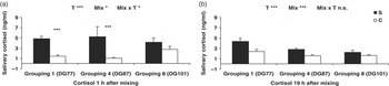
Before the beginning of the experimental procedure, salivary cortisol levels at 0900 and 1500 h were similar in both groups (data not shown). Cortisol responses were measured at the 1st, 4th and 8th groupings. The grouping day × treatment interaction was significant at 1 h (F 2,68 = 3.45; P < 0.05) but not at 30 min before and 19 h after grouping (Figure 2). One hour after grouping, cortisol was higher in S than in C gilts at the 1st and 4th groupings (P < 0.001). Nineteen hours after grouping, cortisol levels were higher in the S group regardless of the number of grouping (F 1,32 = 19.23; P < 0.001).
Salivary cortisol measured 1 h (a) and 19 h (b) after grouping. For cortisol measured 1 h after the beginning of grouping, a significant interaction between time (number of grouping) and treatment (T) was found, and comparisons between treatments within each time were realized using the Bonferroni t-test. Values are mean ± s.e. *P < 0.05, ***P < 0.0001.

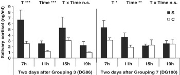
For the investigation of diurnal rhythm, four samples were taken between 0700 and 1900 h, 2 days after the 3rd and 7th grouping. The sampling hour × treatment interaction was not significant, which indicates that S and C groups followed the same pattern of diurnal variations. However, on both sampling days, salivary cortisol was higher in the S group than in the C group (F 1,30 = 13.73; P < 0.05, Figure 3). Compared to 0700 h, cortisol was lower at 1100, 1500 and 1900 h after the 3rd and 7th grouping (P < 0.05). After the 3rd grouping, cortisol was also lower at 1100 and 1900 h than at 1500 h (P < 0.05).
Salivary cortisol measured 2 days after the 3rd and the 7th groupings. Significant effects of treatment (T) and time but not time × treatment interaction were found. Values are mean ± s.e.

Immune parameters
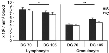
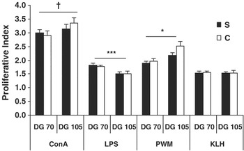
The treatment × stage of gestation interaction and the treatment effect were not significant for the numbers of lymphocytes and granulocytes (Figure 4). Towards the end of gestation, the number of lymphocytes decreased (F 1,23 = 7.07; P < 0.05) whereas the number of granulocytes increased (F 1,23 = 17.78; P < 0.05). Both groups of gilts had detectable levels of IgG against KLH on day 105 of gestation (3.7 ± 0.7 arbitrary units) but there was no difference between groups (F 1,20 = 1.56; P = 0.2). The treatment × stage of gestation interaction and the treatment effect were not significant for the proliferative index regardless of mitogen. Between days 70 and 105 of gestation, the proliferative index tended to increase for ConA (F 1,30 = 4.55; P = 0.08) and significantly increased for PWM (F 1,19 = 8.56; P < 0.01), but decreased for LPS (F 1,29 = 27.66; P < 0.001) (Figure 5).
Numbers of blood lymphocytes and granulocytes before and after the 4-week stress procedure (on days 70 and 105 of gestation, DG70 and DG105). Values are mean ± s.e. *P < 0.05, ***P < 0.0001.

Lymphocyte proliferative responses to lipopolysaccharide (LPS, 6.25 μg/ml), pokeweed mitogen (PWM, 1.5 μg/ml) or keyhole haemocyanin (KLH, 10 μg/ml) measured before and after the 4-week stress procedure (on days 70 and 105 of gestation). Values are mean ± s.e. †P < 0.1, *P < 0.05, ***P < 0.0001.

Reproductive performances
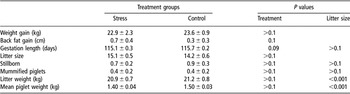
Weight and back fat gains during the experimental period were similar in both groups (Table 1). None of the sows aborted. Litter size, number of stillborn and mummified piglets as well as litter weight and the average piglet weight were also similar in both groups. However, there was a tendency for a shorter length of gestation (F 1,30 = 2.93; P = 0.09) in the S group.
Effect of repeated social stress on weight gain between days 70 and 105 of gestation and reproductive performances of pregnant gilts

Litter size was included in the statistical model as a covariate for gestation length, number of stillborn piglets, mummified piglets, litter weight and mean piglet weight. Values are mean ± s.e.
Sub-classification of S gilts according to their success values
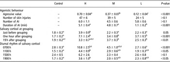
The mean AvSV in the different sub-groups of S gilts were as follows: H: 0.44 ± 0.12; M: −0.32 ± 0.13, L: −0.86 ± 0.06. The effects of AvSV classification on other behavioural variables and salivary cortisol are presented in Table 2. There was a significant effect of AvSV classification on AVs, showing that H gilts were the most aggressive ones (F 2,13 = 23.34; P < 0.001). The duration of agonistic interactions was significantly lower in L gilts compared to M and H gilts (P < 0.01) but AvSV did not influence the numbers of skin lesions and agonistic interactions.
Effect of average success value on behavioural parameters (aggressive value, number of skin injuries, number and duration of agonistic interactions by grouping) and salivary cortisol (before, 1 and 19 h after the 1st, 4th and 8th groupings and between 0700 and 1900 h 2 days after the 3rd and 7th groupings)

Values are mean ± s.e. Within a row, different superscript letters indicate significant differences at P < 0.05 and values with superscript t tend to differ each other (0.05 < P < 0.1).
H=high; M=medium; L=low; AI=agonistic interactions; AvSV=average success value.
n = 6/group.
For cortisol levels at 30 min before grouping, AvSV (F 3,30 = 2.92; P = 0.05) and the interaction between the grouping number × AvSV (F 6,64 = 2.22; P = 0.05) were significant. This latter effect was due to the fact that H gilts displayed higher cortisol levels than C and L gilts on the 4th grouping (H: 6.8 ± 3.2 ng/ml, M: 1.9 ± 0.5 ng/ml, L: 1.4 ± 0.3 ng/ml, C: 1.3 ± 0.2 ng/ml; P < 0.05). Cortisol levels were also significantly higher in all sub-groups of S gilts than in C gilts at 1 and 19 h after grouping except in H gilts at 19 h where only a tendency was observed (P = 0.09). For diurnal cortisol variations, AvSV effect was significant at 0700 h (F 3,30 = 9.48; P < 0.001), 1100 h (F 3,30 = 5.40; P < 0.05) and 1900 h (F 3,30 = 2.97; P < 0.05), whereas the interaction day of sampling × AvSV was not. H gilts had higher cortisol levels than C gilts at 0700, 1100 and 1900 h (P < 0.05), and than L gilts (P < 0.01) only at 0700 h. M and L gilts never differed from C gilts at any of these time points.
Immune measures, weight of the sows and reproductive data were not affected by AvSV except for gestation length (F 1,30 = 2.93; P < 0.05). L gilts had shorter gestation (114.5 ± 0.3 days) than C gilts (115.7 ± 0.2 days, P < 0.05).
Discussion
To our knowledge, the present study is the first one to report the influence of repeated social stress applied to pregnant gilts during the last third of their gestation on acute and delayed cortisol response, immune function of sows and pregnancy outcome. Our data demonstrate a cortisol response to successive groupings in all gilts without marked negative effects on the health and pregnancy outcome.
In the S group, grouping and relocation of gilts with an unfamiliar and unrelated partner led to vigorous fights during the first hours following the grouping. This agonistic behaviour after grouping is common in animals that form social groups and is part of the process leading to the establishment of a social hierarchy in the newly formed group (Ewbank and Meese, Reference Ewbank and Meese1971; Meese and Ewbank, Reference Meese and Ewbank1972). Moreover, our data show that grouping with an unfamiliar conspecifics induced a stimulation of the HPA axis in pregnant gilts because high levels of salivary cortisol were observed 1 and 19 h after grouping during the whole experimental period. An increased cortisol response due to social stress was already reported in growing pigs submitted to repeated groupings (Olsson et al., Reference Olsson, De Jonge, Schuurman and Helmond1999; Coutellier et al., Reference Coutellier, Arnould, Boissy, Orgeur, Prunier, Veissier and Meunier-Salaün2007) and in pregnant sows submitted to one or two groupings (Tsuma et al., Reference Tsuma, Einarsson, Madej, Kindahl, Lundeheim and Rojkittikhun1996; Jarvis et al., Reference Jarvis, Moinard, Robson, Baxter, Ormandy, Douglas, Seckl, Russell and Lawrence2006). In contrast, Soede et al. (Reference Soede, Van Sleuwen, Molenaar, Rietveld, Schouten, Hazeleger and Kemp2006) failed to demonstrate a clear increase in salivary cortisol in gilts submitted to repeated groupings around insemination. The increase in cortisol shortly after social grouping can be explained by the physical activity and the injuries associated with the fights and by the psychological stress associated with the appraisal of the fighting ability of the unfamiliar partner. Our data show that high levels of cortisol were still observed 2 days (diurnal rhythm assessment) and 3 days (measurement 30 min before the 4th and 8th grouping) after mixing in gilts presenting a high AvSV. Long-term effects of grouping were also observed by Coutellier et al. (Reference Coutellier, Arnould, Boissy, Orgeur, Prunier, Veissier and Meunier-Salaün2007) in growing pigs submitted to repeated groupings and by Tsuma et al. (Reference Tsuma, Einarsson, Madej, Kindahl, Lundeheim and Rojkittikhun1996) in pregnant gilts after a single mixing. However, in our study, salivary cortisol measured 2 and 3 days after grouping was similar in C gilts, medium- and low-success S gilts. The H gilts were more aggressive, and it can be hypothesized that additional fights occurred after the period of observation, inducing new stress reactions with adrenal axis stimulation. Such variability associated with the social rank of the pigs was also described by Otten et al. (Reference Otten, Puppe, Stabenow, Kanitz, Schön, Brüssow and Nürnberg1997) in growing pigs of high or low social rank reintroduced in a new group after 10 days of social isolation and by Mendl et al. (Reference Mendl, Zanella and Broom1992) in pregnant gilts submitted to a single grouping during mid-gestation. In agreement with our results, Otten et al. (Reference Otten, Puppe, Stabenow, Kanitz, Schön, Brüssow and Nürnberg1997) found that aggressive high-ranking animals showed higher cortisol release in response to a grouping with unfamiliar conspecifics than low-ranking pigs. In contrast, Mendl et al. (Reference Mendl, Zanella and Broom1992) reported higher basal cortisol levels in gilts of intermediate rank in comparison to high- and no-success gilts 5 weeks after grouping. This discrepancy can be explained by the fact that Otten et al. (Reference Otten, Puppe, Stabenow, Kanitz, Schön, Brüssow and Nürnberg1997) and the present study investigated the cortisol response relatively shortly after grouping, whereas Mendl et al. (Reference Mendl, Zanella and Broom1992) investigated it 5 weeks later. Shortly after grouping, the social hierarchy is still being established, whereas several weeks after grouping, social interactions between individuals were more predictable and the level of aggression was lower.
We observed that the number and length of agonistic interactions as well as cortisol levels short- (1 h) and long-term (19 h and 2 days) after grouping declined as the number of grouping increased. Such a decrease in agonistic behaviour has already been reported in growing pigs submitted to repeated groupings (Giersing and Andersson, Reference Giersing and Andersson1998; Coutellier et al., Reference Coutellier, Arnould, Boissy, Orgeur, Prunier, Veissier and Meunier-Salaün2007). Contrarily, Olsson et al. (Reference Olsson, De Jonge, Schuurman and Helmond1999) in growing pigs and Soede et al. (Reference Soede, Van Sleuwen, Molenaar, Rietveld, Schouten, Hazeleger and Kemp2006) in gilts did not show a reduction of the endocrine response to repeated grouping. However, in both studies, grouping was only performed once a week over a period of 5 weeks and this frequency might have been insufficient to induce habituation. Moreover, in the study from Soede et al. (Reference Soede, Van Sleuwen, Molenaar, Rietveld, Schouten, Hazeleger and Kemp2006), grouping had only minor effects on behaviour and cortisol release. The observed decline of offensive acts in our study could reflect the adoption of a new coping strategy to a changing and aversive environment (Schouten and Wiegant, Reference Schouten and Wiegant1997; Bolhuis et al., Reference Bolhuis, Schouten, De Leeuw, Schrama and Wiegant2004). Gilts may have developed a more efficient strategy to establish a social hierarchy with less agonistic interactions (Puppe et al., Reference Puppe, Langbein, Bauer and Hoy2008) and hence to limit the energetic cost and the number of injuries. In the same way, the decline of cortisol response to grouping towards the end of the experiment can be attributed to the decrease in the intensity of the stress perceived by the gilts and/or to a decrease in the adrenal axis sensitivity. The first hypothesis is supported by the decrease in the number and duration of agonistic behaviours and in the number of injuries. ACTH or corticotropin-releasing hormone challenges would be needed to confirm the second hypothesis. Finally, despite a decreased response at the end of the stress procedure, acute endocrine and behavioural responses to grouping were still observed during the last grouping and cortisol levels remained higher than in the control group between the 7th and the last grouping.
Acute and chronic social stress are known to change the distribution of immune cells and to alter the immune status in pigs (Tuchscherer et al., Reference Tuchscherer, Puppe, Tuchscherer and Kanitz1998; de Groot et al., Reference De Groot, Ruis, Scholten, Koolhaas and Boersma2001; Niekamp et al., Reference Niekamp, Sutherland, Dahl and Salak-Johnson2007) and in other mammal species (for review see Bohus et al., Reference Bohus, Koolhaas, De Ruiter and Heijnen1991; Bartolomucci, Reference Bartolomucci2007). Typically, the lymphocytes/granulocytes ratio and the lymphocyte abilities to proliferate in the presence of mitogens decrease after acute or chronic stress. It is also assumed that stress inhibits the cellular adaptative immunity (Th1) in favour of Th2 humoral immunity (Elenkov, Reference Elenkov2004). In the present study, no alteration in immune cell distribution and no shift from cellular to humoral immunity were found in stressed gilts. An absence of social stress effects on humoral immunity was also reported in 9-month-old pregnant gilts (Mendl et al., Reference Mendl, Zanella and Broom1992). The lacking effect of cortisol elevation on the immune function of the pregnant gilts in our study may be explained by the particular physiological state of the adult pregnant female. First, immune effects of social stress were observed in non-pregnant pigs; however, it is possible that pregnancy, which is associated with strong immune modifications (Wegmann et al., Reference Wegmann, Lin, Guilbert and Mosmann1993), abrogated the stress-induced immune shift. For instance, the decrease of lymphocytes/granulocytes ratio towards the end of gestation could explain why we and others (Stefansky et al., Reference Stefansky, Raabe and Schulte2005) could not find any additional effects of stress on this parameter. Moreover, females seem to be less sensitive than castrated males to social stress-induced immune suppression. For example, de Groot et al. (Reference De Groot, Ruis, Scholten, Koolhaas and Boersma2001) found a suppressed humoral immunity in mixed barrows, whereas no differences were found in mixed gilts. Finally, the lack of effect of cortisol elevation on the immune function of the pregnant gilts can be explained by the age-related decreased sensitivity of porcine leukocytes to corticosteroids (Yang and Schultz, Reference Yang and Schultz1986).
Stress can negatively influence reproductive processes in pigs such as implantation and foetal growth and may lead to abortion (for review see von Borell et al., Reference Von Borell, Dobson and Prunier2007). In our experiment, the stress model had no negative impact on pregnancy maintenance or intrauterine mortality since the size of the litter at birth was similar in S and C groups. A non-significant decrease of 0.6 day in the gestation length was observed in S gilts, which was due to a shorter gestation in low-AvSV individuals, but this had no obvious consequences on piglet weight or mortality at birth. Cortisol elevation during pregnancy is suspected to generate prenatal stress and alter the birth weight and viability of the progeny (Haussmann et al., Reference Haussmann, Carroll, Weesner, Daniels, Matteri and Lay2000; Tuchscherer et al., Reference Tuchscherer, Kanitz, Otten and Tuchscherer2002). A high variability is observed for piglet birth weight among prenatal stress studies, probably due to differences in the methodologies used. Effects varied from higher birth weight in the case of maternal ACTH administration (Kanitz et al., Reference Kanitz, Otten and Tuchscherer2006) to lack of effect in case of maternal restraint and social stress (Tuchscherer et al., Reference Tuchscherer, Kanitz, Otten and Tuchscherer2002; Jarvis et al., Reference Jarvis, Moinard, Robson, Baxter, Ormandy, Douglas, Seckl, Russell and Lawrence2006) and to lower birth weight in the case of maternal cortisol treatment (Kranendonk et al., Reference Kranendonk, Hopster, Fillerup, Ekkel, Mulder, Wiegant and Taverne2006). A lower birth weight of the litter was observed in pregnant sows that displayed higher cortisol levels in relation to their social rank (Mendl et al., Reference Mendl, Zanella and Broom1992). Therefore, additional work is needed to clarify the relationships between the adrenal activity of the pregnant mother and foetal growth in pigs.
Conclusion
The present study demonstrates that repeated social grouping during the last third of pregnancy in gilts is able to induce behavioural and cortisol responses as described in growing non-pregnant pigs. Among stressed gilts, the outcome of agonistic interactions seems to influence the cortisol release with sows of high success having elevated cortisol levels for a longer time after grouping than other gilts. Assuming that cortisol is a reliable marker of stress, this result suggests that repeated groupings induce a state of stress, especially for pregnant sows that are dominant and more aggressive. However, the social stress paradigm used did not affect either the immune functions of the pregnant gilts nor foetal viability or growth. Considering the effects of maternal-mediated stress during pregnancy on the physiology of the progeny in many species, further work should focus on the physiological, immunological and behavioural consequences of maternal social stress on the offspring in pigs.
Acknowledgements
The authors thank the staff of the SENAH Research and Experimental pig unit, Mounier Anne-Marie, Lefebvre Michel and Massard Michel for their technical assistance and the AFSSA unit of Ploufragan, Kuntz Gaelle, Jamin Agnès, for their assistance for the haematological analyses. This study received financial support from the INRA PHASE department.