Introduction
The domestic cat is an important experimental model for advancing assisted reproductive technologies that may be useful for conservation of non-domestic felidae. Development of reliable methods for in vitro maturation (IVM), in vitro fertilization (IVF) and in vitro culture (IVC) of domestic cat oocytes is a prerequisite for their subsequent application for preservation of endangered species. Research on in vitro production (IVP) of domestic cat embryos has increased rapidly (Pope, Reference Pope2000; Pope et al., Reference Pope, Gomez and Dresser2006, Reference Pope, Crichton, Gomez, Dumas and Dresser2009; Spinaci et al., Reference Spinaci, Merlo, Zannoni, Iacono, De Ambrogi, Turba and Zambelli2007; Nagano et al., Reference Nagano, Uchikura, Takahashi and Hishinuma2008; Naoi et al., Reference Naoi, Agung, Karja, Wongsrikeao, Shimizu, Taniguchi and Otoi2008; Uchikura et al., Reference Uchikura, Nagano and Hishinuma2010). Ideally, for efficient IVP of feline embryos, oocytes with high maturational competence should be selected non-invasively before maturational culture.
It is well established that oocytes surrounded by several layers of cumulus cells and with evenly granulated dark ooplasm have higher in vitro developmental competence than oocytes with irregularly granulated ooplasm and fewer cumulus cell layers (Pope et al., Reference Pope, McRae, Plair, Keller and Dresser1997; Wood & Wildt, Reference Wood and Wildt1997; Katska-Ksiazkiewicz et al., Reference Katska-Ksiazkiewicz, Rynska, Kania, Smorag, Gajda and Pienkowski2003; Naoi et al., Reference Naoi, Agung, Karja, Wongsrikeao, Shimizu, Taniguchi and Otoi2008). Also, in vitro maturation and developmental competence of oocytes are influenced by season (Spindler & Wildt, Reference Spindler and Wildt1999; Freistedt et al., Reference Freistedt, Stojkovic and Wolf2001) and ovarian cyclicity status (Johnston et al., Reference Johnston, O'Brien and Wildt1989; Freistedt et al., Reference Freistedt, Stojkovic and Wolf2001; Karja et al., Reference Karja, Otoi, Murakami, Fahrudin and Suzuki2002; Naoi et al., Reference Naoi, Agung, Karja, Wongsrikeao, Shimizu, Taniguchi and Otoi2008), even if oocytes are selected using similar criteria for oocyte and cumulus cell morphologies.
Oocytes and cumulus cells grow and develop in a highly coordinated and mutually dependent manner. Cumulus cells support oocyte growth and maturation by supplying nutrients and growth factors (Tanghe et al., Reference Tanghe, Soom, Nauwynck, Coryn and de Kruif2002). Recently, researchers attempted to use the expression of cumulus genes as a marker for non-invasive assessment of oocyte quality in cattle (Assidi et al., Reference Assidi, Dufort, Ali, Hamel, Algriany, Dielemann and Sirard2008; Bettegowda et al., Reference Bettegowda, Patel, Lee, Park, Salem, Yao, Ireland and Smith2008) and humans (Assou et al., Reference Assou, Haouzi, Mahmoud, Aouacheria, Guillemin, Pantesco, Reme, Dechaud, De Vos and Hamamah2008, Reference Assou, Haouzi, De Vos and Hamamah2010; Anderson et al., Reference Anderson, Sciorio, Kinnell, Bayne, Thong, de Sousa and Pickering2009). However, this method requires a biopsy of the cumulus cells, and it is laborious, complicated and expensive. There is another method for easily assessing the quality of cumulus cells using propidium iodide (PI) staining. PI does not penetrate intact cell membrane, but can penetrate only cells with non-intact membranes that have lost their integrity. In a study that evaluated the membrane integrity of cumulus cells of cryopreserved murine oocytes at the germinal vesicle stage by PI staining, differences in developmental potential appeared to depend on the assumption that the proportion of oocytes with good cumulus integrity (without PI staining) was similar to the proportion of embryos developing to the blastocyst stage after IVF/IVC (Ruppert-Lingham et al., Reference Ruppert-Lingham, Paynter, Godfrey, Fuller and Shaw2006).
Therefore, in the present study, for non-invasive prediction of the maturational competence of feline oocytes, we examined the morphology of cumulus investments and stained cumulus cells immediately after collection using PI, and subsequently investigated nuclear maturation rate after IVM culture. In domestic cats, it was reported that follicles ≥800 μm in diameter expressed the luteinizing hormone (LH) receptor in granulosa cells (Saint-Dizier et al., Reference Saint-Dizier, Malandain, Thoumire, Remy and Chastant-Maillard2007). Therefore, we collected oocytes from small (400–800 μm) and large follicles (≥800 μm), and the diameter of oocytes was also examined.
Materials and methods
Animals for ovarian collection
Ovaries of eight cats were obtained following ovariohysterectomy at the veterinary teaching hospital of Tottori University and a local veterinary clinic in Tottori city and kept in normal saline at room temperature before delivery to the laboratory within 1 h after collection.
Antral follicle isolation and oocyte recovery
Upon arrival in the laboratory, ovaries were transferred to isolation medium: TCM-199 (Invitrogen) containing 0.1% polyvinyl alcohol (Sigma-Aldrich), 25 mM HEPES (Sigma-Aldrich), 0.85 mg/ml NaHCO3 (Kanto Chemical Co., Inc.) and 50 μg/ml gentamicin sulfate (Sigma-Aldrich) (pH 7.4) (Harada et al., Reference Harada, Miyano, Matsumura, Osaki, Miyake and Kato1997). Ovaries were cut with scissors along their long axis, the ovarian medulla was removed and the cortex was cut into small pieces. It was difficult to identify the antrum in follicles that were <400 μm in diameter. The antral follicles that were ≥400 μm in diameter were isolated using an 18-gauge needle under a stereomicroscope and transferred to fresh isolation medium. Using an ocular micrometer, follicular diameter was measured as the mean length of two perpendicular axes. The follicles were divided into two groups according to their diameter: 400–800 (small) and ≥800 μm (large). The follicles were then dissected with a 25-gauge needle under a stereomicroscope. Cumulus–oocyte complexes (COCs) were collected and their morphological characteristics were determined under a stereomicroscope (×15–50). Oocytes surrounded by cumulus cells and having evenly granulated black ooplasm were defined as morphologically normal and used for the experiment.
Evaluation of membrane integrity of cumulus cells by propidium iodide staining and morphology
The method of evaluation of membrane integrity of cumulus cells was in accordance with that of a previous report (Ruppert-Lingham et al., Reference Ruppert-Lingham, Paynter, Godfrey, Fuller and Shaw2006) with slight modification. Briefly, COCs were incubated for 10 min in the dark at 39°C in 3 ml of isolation medium supplemented with 0.1 mg/ml PI (Sigma-Aldrich). The COCs were then washed twice in isolation medium and transferred into a 10-μl droplet of isolation medium covered with paraffin oil. Stained COCs were observed under an inverted microscope equipped with a fluorescence system (TS100-F, Nikon) using an appropriate filter (G-2A, Nikon). Cumulus cells with a damaged cell membrane fluoresced red. Each COC was scored for membrane integrity of the cumulus cells using the following scoring system (Ruppert-Lingham et al., Reference Ruppert-Lingham, Paynter, Godfrey, Fuller and Shaw2006) with slight modification. The COCs were defined as either grade 1: less than one-quarter of cumulus cells were stained by PI; grade 2: one-quarter to one-half of cumulus cells were stained; or grade 3: more than one-half of cumulus cells were stained. These oocytes were also divided into three categories by the morphology of their surrounding cumulus investments: categories A: at least three layers; B: less than three layers; or C, only corona radiata. Images of COCs were taken using a digital camera (DP-11, Olympus) attached to an inverted microscope and the diameter of ooplasm was measured as the mean length of two perpendicular axes.
In vitro maturational culture and assessment of nuclear maturation
After evaluation of membrane integrity and morphology, COCs were washed once in maturation medium, which was composed of 25 mM HEPES-buffered TCM 199 (cat. no. 12340, Earle's salt, Invitrogen) supplemented with 3 mg/ml fatty-acid-free BSA (Sigma-Aldrich), 0.02 units/ml follicle stimulating hormone (F2293, from porcine pituitary, Sigma-Aldrich), 1 μg/ml estradiol-17β (Sigma-Aldrich), 0.2 mM sodium pyruvate (Sigma-Aldrich) and 50 μg/ml gentamicin sulfate (Nagano et al., Reference Nagano, Uchikura, Takahashi and Hishinuma2008). They were then cultured individually in 10-μl droplets of maturation medium covered with paraffin oil at 39°C in a humidified atmosphere of 5% CO2 in air for 30 h.
After IVM culture, the cumulus investments were removed from the oocytes by gentle pipetting. They were then fixed with a mixture of ethanol:acetic acid (3:1) and stained with 1% aceto-orcein solution. The nuclear status was evaluated using a phase-contrast microscope. When chromosomes were condensed and present in the equatorial view with the extrusion of the first polar body, the nuclear stage was classified as metaphase II (MII) and considered to be meiotically mature.
Experimental design
Firstly, we compared the diameter of ooplasm and maturation rate after IVM culture of oocytes derived from follicles with 400–800 (small) or ≥800 μm (large) in diameter. Secondly, effects of morphological categories and membrane integrity of cumulus cells on the diameter of ooplasm and the maturation rate were examined. Finally, the maturational competence of oocytes surrounded by several layers (category A) of intact cumulus cells (grade 1) was evaluated.
Statistical analysis
Statistical analysis was performed using JMP v8.0.2 software (SAS Institute Inc.). The difference in oocyte diameter was analysed by Student's t-test or by one-way analysis of variance (ANOVA) followed by Tukey-Kramer's Honestly Significant Difference test. The maturation rates of oocytes were compared by chi-squared test. Values were considered statistically significant at p < 0.05.
Results and Discussion
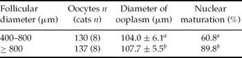
We collected 267 oocytes with normal morphology from eight cats (33.4 ± 13.9/female, mean ± SD). Oocytes derived from the ≥800 μm (large) follicle group showed a larger diameter of ooplasm and a higher maturation rate than those from the 400–800 μm (small) group (p < 0.05, Table 1). These results indicate that feline oocytes grow until the follicular diameter reaches 800 μm. Luteinizing hormone binding sites were detected in the granulosa cell layer of feline follicles that were ≥800 μm in diameter (Saint-Dizier et al., Reference Saint-Dizier, Malandain, Thoumire, Remy and Chastant-Maillard2007). The diameter of ooplasm seemed to have an effect on the maturational competence of feline oocytes, as has been shown in other species (Sato et al., Reference Sato, Matsuo and Miyamoto1990; Fair et al., Reference Fair, Hyttel and Greve1995; Arlotto et al., Reference Arlotto, Schwartz, First and Leibfried-Rutledge1996; Otoi et al., Reference Otoi, Yamamoto, Koyama, Tachikawa and Suzuki1997; Ledda et al., Reference Ledda, Bogliolo, Leoni and Naitana1999). In a previous study, it was demonstrated that there is a strong relationship between diameter and meiotic competence of cat oocytes (Otoi et al., Reference Otoi, Murakami, Ooka, Karja and Suzuki2001) although follicle diameter was not measured. Thus, the present results confirm earlier reports (Otoi et al., Reference Otoi, Murakami, Ooka, Karja and Suzuki2001; Saint-Dizier et al., Reference Saint-Dizier, Malandain, Thoumire, Remy and Chastant-Maillard2007) indicating that oocyte growth occurs and maturational competence improves as follicular diameter grows up to ≥800 μm in diameter.
Oocyte size and maturational ability in small (400–800 μm) vs. large (≥800 μm) follicles

a ,b Values with different superscripts in the same column differ significantly (p < 0.05).
Oocyte diameter values are mean ± standard deviation (SD). Data are pooled from eight cats.
As shown in Table 2, there was no difference in diameter of ooplasm and maturation rate of oocytes derived from follicles with the same size, regardless of cumulus cell morphology. Bovine oocytes with a multilayered cumulus cells showed higher rates of nuclear maturation, fertilization and development after IVP than denuded oocytes and oocytes with corona radiata alone (Xu et al., Reference Xu, Greve, Smith and Hyttel1986; Shioya et al., Reference Shioya, Kuwayama, Fukushima, Iwasaki and Hanada1988; Yang & Lu, Reference Yang and Lu1990; Madison et al., Reference Madison, Avery and Greve1992; Momozawa & Fukuda, Reference Momozawa and Fukuda1995; Stojkovic et al., Reference Stojkovic, Machado, Stojkovic, Zakhartchenko, Hutzler, Goncalves and Wolf2001). However, we could not detect an obvious difference of maturational competence between cat oocytes with different cumulus cell morphologies. In contrast, oocytes of similar diameter from large follicles having cumulus cells with intact membrane integrity (grade 1) showed a higher maturation rate (p < 0.05) than oocytes having poor cumulus integrity (grade 3) (Table 3). In addition, oocytes surrounded by cumulus cells with grade 1 integrity from small follicles were larger in diameter than oocytes enclosed by cumulus cells of grades 2 and 3 (p < 0.05). However, the diameter of oocytes and the nuclear maturation rate was lower than those of oocytes surrounded by cumulus cells with grade 1 integrity from large follicles. Taken together, these results indicate that live and functional cumulus cells support the growth and nuclear maturation of feline oocytes.
Effects of cumulus morphological categories on size and maturational ability of cat oocytes from small (400–800 μm) vs. large (≥800 μm) follicles

a ,b Values with different superscripts in the same column differ significantly (p < 0.05).
cCumulus category: A, oocytes tightly surrounded by at least three layers of cumulus cells; B, oocytes surrounded by less than three layers of cumulus cells; C, oocytes surrounded by only corona radiata cells. Oocyte diameter values are mean ± standard deviation (SD).
Effects of membrane integrity of cumulus cells on size and maturational ability of cat oocytes from small (400–800 μm) vs. large (≥800 μm) follicles

a ,b Values with different superscripts in the same column differ significantly (p < 0.05).
A ,B Values with different superscripts in the same row differ significantly (p < 0.05).
cGrade of oocytes: grade 1, one-quarter of cumulus cells stained by propidium iodide (PI); grade 2, one-half of cumulus cells stained by PI; grade 3, more than one-half of cumulus cells stained by PI. Values of oocyte diameter are means ± standard deviation (SD).
In large follicles, maturation rate of oocytes selected by morphological category A (92.0%, n = 75) was similar to that of oocytes selected by grade 1 of cumulus cell integrity (93.8%, n = 80). However, the oocytes surrounded by at least three layers of cumulus cells (category A) derived from large follicles were divided by their cumulus cell integrity into grade 1 (72%, 54/75) and grades 2 and 3 (28%, 21/75), and maturation rate of oocytes having cumulus cells with grade 1 integrity (96.3%, n = 54) was higher than that of oocytes having cumulus cells classified as grades 2 and 3 (81.0%, n = 21) (p < 0.05). This result also suggests that evaluating cumulus cell integrity concomitantly with cumulus morphology will improve IVP outcome in cats.
In conclusion, evaluation of membrane integrity of cumulus cells by propidium iodide staining can be used as a tool for predicting the maturational competence of oocytes.
Acknowledgements
This study was supported by a Grant-in-Aid for Scientific Research (no. 21780286) from the Ministry of Education, Science, Sports and Culture, Japan, to M. Nagano.