Introduction
The mammalian testis fulfils two essential functions in male reproduction: steroidogenesis and spermatogenesis. Steroidogenesis corresponds to the biosynthesis of sex steroid hormones whereas spermatogenesis is the complex cellular process that transforms spermatogonial cells into spermatozoa. Progression of spermatogenesis takes place in the seminiferous tubules, the functional units of the testis, and encompasses tightly coordinated mitosis, meiosis and cell differentiation events (Ref. Reference Hermo1). Mitotic divisions maintain the population of spermatogonial cells and differentiation of spermatogonia originates primary spermatocytes. The first meiotic division produces secondary spermatocytes, which undergo the second meiosis originating spermatids. Spermiogenesis is the cellular restructure process that transforms round spermatids in elongated spermatids and finally in spermatozoa (Ref. Reference Hess and de Franca2). Nonetheless, not all germ cells achieve maturity, and the spontaneous cell death by apoptosis is a common event in the testis, particularly, in the first waves of spermatogenesis (Ref. Reference Aitken3). On the other hand, deregulated apoptosis may be indicated in the aetiology of male infertility, since augmented rates of apoptosis were observed in the testes of subfertile and infertile men (e.g. Refs Reference Lin4, Reference Eguchi5, Reference Streichemberger6).
Development and maintenance of spermatogenesis depend on an assortment of hormonal messengers, which exert their actions by endocrine, paracrine, and autocrine signalling mechanisms. Androgens are widely recognised as the main regulators of male reproductive function, and androgenic actions are absolutely required for successful spermatogenesis (Ref. Reference Yeh7). Over the years, the dogma that spermatogenesis depends solely on androgens and gonadotropins has changed, and cumulative evidence has demonstrated the importance of oestrogens on the regulation of spermatogenic process. However, discordant reports about the role of oestrogens in male reproduction have been produced. For example, studies from mice and men with inability to synthesise oestrogens have described altered testis size, increased apoptosis of germ cells, oligozoospermia and/or reduced sperm motility, which indicated that these sex steroid hormones are required for successful spermatogenesis and male fertility (Refs Reference Carani8, Reference Herrmann9). On the contrary, it is known that exposure to oestrogens or oestrogen-mimicking compounds early in life, adversely affects spermatogenesis and hampers the reproductive potential of adult (Ref. Reference Phillips and Tanphaichitr10). The present review will discuss the current knowledge regarding the effects of oestrogens as survival or apoptosis-inducers in the testis, and summarise the molecular mechanisms driven apoptotic effects in male germ cells. This issue is of paramount importance for clinical management of male infertility because hyperoestrogenism has been a condition associated with cases of idiopathic infertility.
Apoptosis in spermatogenesis
Signalling mechanisms of apoptotic cell death
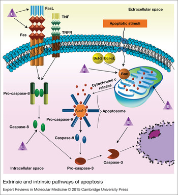
Apoptotic cell death is characterised by several hallmarks, such as, cell shrinkage, DNA fragmentation and externalisation of phosphatidylserine at cell membrane, and may be triggered by two distinct pathways (Ref. Reference Elmore11): the receptor-mediated (or extrinsic) and the mitochondrial (or intrinsic) (Fig. 1; Ref. Reference Lawen12). In both cases, cell-death depends on the specific proteases, the so-called caspases, which are the executioners of apoptosis. The caspases enzymes are synthesised as inactive zymogens (procaspases) becoming active in response to death stimuli (Ref. Reference McIlwain, Berger and Mak13). Initiator caspases (caspases-8 and -9) are directly activated by dimerisation and, in turn, cleave the effector caspases (caspases-3, -6 and -7) promoting their activation (Ref. Reference McIlwain, Berger and Mak13). The extrinsic pathway (Fig. 1) is initiated by activation of death receptors at cell membrane, namely the Fas (CD95/Apo-1) and the tumour necrosis factor receptor 1 (TNFR1), which induce the activation of procaspase-8 (Ref. Reference Lawen12). The intrinsic pathway of apoptosis (Fig. 1) could be activated by different stimuli, such as DNA damage, starvation, oxidative stress and autophagy. It is characterised by a decrease in the membrane potential of mitochondria and the release of cytochrome c, which interacts with dATP, cytosolic apoptotic protease activating factor 1 (Apaf-1) and procaspase-9, assembling the apoptosome complex (Ref. Reference Kaufmann and Gores14). Both extrinsic and intrinsic pathways of apoptosis converge at the activation of effector caspase-3 (Fig. 1), which has been considered an end-point of apoptotic process (Ref. Reference Elmore11).
Extrinsic and intrinsic pathways of apoptosis. Extracellular ligand binding (FasL or tumour necrosis factor, TNF) to death receptors (Fas and TNF receptor, TNFR) triggers the receptor-mediated (extrinsic) pathway resulting in the direct activation of initiator caspase-8. The mitochondrial (intrinsic) pathway is initiated in response to apoptotic stimuli leading to the activation of proapoptotic members of the Bcl-2 protein family, namely, Bax. The Bax protein is translocated to the mitochondria allowing the permeabilisation of mitochondrial membrane with consequent release of cytochrome c, which in turn, together with apoptotic protease activating factor 1 (Apaf-1), forms the apoptosome and activates caspase-9. Extrinsic and intrinsic pathways converge at the activation of the executioner caspase-3. Activation and inhibition are indicated by arrows and bar-headed arrows, respectively. Target sites of oestrogens (E) actions in the oestrogen-induced apoptosis of germ cells are highlighted by triangles.

Apoptosis of germ cells has been shown to play an important role in controlling sperm output in many species, and massive germ cell death occurs under physiological conditions during the first waves of spermatogenesis (constitutive apoptosis). This issue has been revised in recent reports (Refs Reference Aitken3, Reference Shaha, Tripathi and Mishra15) and will not be further explored here.
In the adult testis, apoptosis is a relevant mechanism for elimination of damaged germ cells avoiding passage of defects to the future generations (Ref. Reference Shaha, Tripathi and Mishra15). On the other hand, male germ cells strictly depend on the physical and biochemical support of Sertoli cells (SCs), the somatic cells within the seminiferous tubules, which nourish and sustain the developing germ cells (Ref. Reference Griswold16). However, SCs have a limited capacity for the number of germ cells they can support (Ref. Reference Sharpe17), and it has been accepted that they play a crucial role in determining germ cell fate. SCs secrete paracrine factors and establish cell-to-cell membrane contacts with adjacent germ cells, which promote germ cell survival or death maintaining the appropriate ratio of germ cell to SCs (Ref. Reference Mruk and Cheng18). The stem cell factor (SCF), a membrane-bound cytokine at the surface of SCs (Ref. Reference Rossi19), and its tyrosine kinase receptor the c-kit, present on the surface of adjacent germ cells (Ref. Reference Yoshinaga20), are the main mediators of survival communication between SCs and germ cells (Ref. Reference Yan, Suominen and Toppari21). Perturbations in the SCF/c-kit system by abolishment of SCF production (Ref. Reference Sato22), impairment of SCF binding to c-kit (Ref. Reference Lizama23) or disruption of c-kit signalling pathways (Ref. Reference Kissel24) have been linked to an increased apoptosis and reduced proliferation of germ cells. The death communication between SCs and germ cells is established by the Fas system being accepted that SCs express the Fas ligand (FasL) while germ cells mostly express the death receptor Fas (Refs Reference Lee25, Reference Pentikäinen, Erkkilä and Dunkel26, Reference Lee27).
Clinical findings of deregulated apoptosis in the testis of infertile men
Taking into account the delicate control of apoptosis in the germ line, it is not surprising that augmented rates of apoptosis have been identified in the testes of subfertile and infertile men (Refs Reference Lin4, Reference Eguchi5, Reference Streichemberger6, Reference Takagi28, Reference Lin29, Reference Stiblar-Martincic30, Reference Kandirali31, Reference Martincic32, Reference Kilic33, Reference Bozec34). Accordingly, altered expression patterns of a panoply of apoptosis-related genes have been described in human testes with defective spermatogenesis. The association of death receptors and the extrinsic pathway of apoptosis with male infertility have been suggested by several studies, which showed enhanced expression of FasL in cases of maturation arrest and Sertoli cell-only syndrome, the later characterised by the absence of germ cell in the seminiferous epithelium (Refs Reference Eguchi5, Reference Bozec34, Reference Kim35, Reference Almeida36, Reference Francavilla37, Reference Francavilla38). Cavalcanti et al. found an increased expression of tumour necrosis factor alpha (TNF-α) family member 10 (also known as TNF-α-related apoptosis inducing ligand, TRAIL) in the testes of subfertile men comparatively with potentially fertile (Ref. Reference Cavalcanti39).
As stated previously, caspases are the key elements and the executioners of apoptotic cell death. Increased expression of effector caspase-6 was found in the testes of subfertile men comparatively with potentially fertile (Ref. Reference Cavalcanti39). Also, the expression of effector caspase-3 was enhanced in cases of maturation arrest and Sertoli cell-only syndrome (Refs Reference Bozec34, Reference Kim35, Reference Almeida36). In addition, increased activity of caspase-9 and caspase-3 was found in spermatozoa of oligozoospermic and azoospermic patients comparatively with normozoospermic (Refs Reference Almeida40, Reference Zalata41), which is indicative of active apoptosis in spermatozoa of infertile patients. In fact, the occurrence of DNA fragmentation and the externalisation of phosphatidylserine, which are late apoptotic events indicating cell commitment to apoptosis, can be detected in spermatozoa of infertile men (Refs Reference Barroso, Morshedi and Oehninger42, Reference Weng43).
The novel apoptosis regulators such as Aven, survivin and regucalcin (RGN) have also displayed altered expression in human testis with defective spermatogenesis (Refs Reference Laurentino44, Reference Laurentino45, Reference Weikert46). Aven is an apoptosis inhibitor that acts by binding both Bcl-xL and Apaf-1 (Fig. 1), which enhances the antiapoptotic function of Bcl-xL and inhibits the assembling of a functional apoptosome complex, thus preventing caspase activation (Ref. Reference Chau47). Diminished expression of Aven was found in the testes of non-obstructive azoospermic men, which was correlated with the severity of spermatogenic defect and increased rates of apoptosis (Ref. Reference Laurentino44). In the case of survivin and RGN, both proteins with a dual role controlling apoptosis and cell cycle progression (Refs Reference Laurentino45, Reference Marques48, Reference Mita49), the balance of their expression levels should be determinant for maintenance of an appropriate germ cell number. Indeed, we have found increased expression of RGN in the testis of human infertile patients with hypospermatogenesis (Ref. Reference Laurentino45). Although the causes of idiopathic hypospermatogenesis may be diverse, accelerated apoptosis, rather than proliferative dysfunction, has been implicated in the decreased number of spermatogonia (Ref. Reference Takagi28). In a recent report, we have demonstrated that in vivo overexpression of RGN leads to suppression of thapsigargin- and actinomycin D-induced apoptosis in the testis through modulation of expression and activity of key regulators of apoptosis (Ref. Reference Correia50). This suggests that RGN may act as a protective molecule counteracting increased rates of apoptosis associated with infertility cases such as hypospermatogenesis. On the other hand, manipulation of RGN levels may represent a suitable mechanism for fertility preservation upon treatment of oncologic conditions.
Sex steroid hormones are the crucial orchestrators of the cellular and molecular events underlying normal spermatogenesis and their roles also rely on the control of germ cell survival and apoptosis. It has been shown that androgens withdrawal induces disruption of spermatogenesis with a severe reduction in the number of spermatocytes and spermatids in consequence of increased apoptosis (Refs Reference Woolveridge51, Reference Chang52). Whereas the action of androgens has been well documented and fully accepted as an essential requirement for the development and survival of testicular cells (Ref. Reference Wang53), in the case of oestrogens a dual behaviour has been assigned with independent reports describing their actions both as germ cell survival factors and as apoptosis inducers. The following topics of this review will focus on the role of oestrogens as regulators of testicular apoptosis.
Oestrogen receptors in testicular cells
The classical nuclear oestrogen receptors
Sex steroid hormones exert their effects mainly by the interaction with intracellular receptor proteins, which act as nuclear transcription factors regulating the expression of target genes in specific cell types (Refs Reference Mangelsdorf54, Reference Gronemeyer, Gustafsson and Laudet55). The steroid receptors belong to the nuclear receptor superfamily that includes, among others, oestrogen receptors (ERs), androgen receptor and progesterone receptor (Ref. Reference Mangelsdorf54). The ERs share with other members of the nuclear receptor superfamily a common arrangement of structural–functional domains (Ref. Reference Beato and Klug56), namely, the N-terminal region (Ref. Reference Mangelsdorf54), the DNA-binding domain (DBD) (Ref. Reference Klug and Schwabe57) and the ligand-binding domain (LBD) (Ref. Reference Mangelsdorf54).
In general, oestrogenic actions via nuclear ERs involve ligand-binding, dissociation of chaperones, receptor phosphorylation, receptor dimerisation, nuclear translocation, DNA-binding, interaction with cofactors and, consequently, modulation of transcription activity (Ref. Reference O'Donnell58). Alternatively, ERs also can modulate transcription via interfering with other promoter-bound transcription factors or via influencing a variety of intracellular signalling pathways (Ref. Reference Schultz, Petz and Nardulli59).
Two subtypes of nuclear ERs, ER alpha (ERα) and ER beta (ERβ), encoded by separate genes located on different chromosomes have been found along the evolutionary line of vertebrates (Refs Reference Mak, Ho and Callard60, Reference Krust61, Reference Kuiper62, Reference Mosselman, Polman and Dijkema63, Reference Socorro64). Even so, and depending on the species considered, ERα and ERβ proteins share 41–65% of overall amino acid identity with the highly conserved DBD reaching 96% of similarity (Ref. Reference Socorro64). The LBD also displays high conservation (57–60%), and both receptors bind the natural ligand 17β-estradiol (E2) (Ref. Reference Kuiper65), phytoestrogens and other natural or synthetic compounds (Ref. Reference Dutertre and Smith66). However, selective agonists and antagonists for ERα or ERβ have been identified helping to disclose their specific actions in a broad range of tissues (Ref. Reference McDonnell and Wardell67).
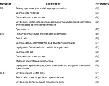
The biological function assigned to the ERs in a tissue or organ is not only dependent on ligand itself but also on the cellular localisation of receptors, as well as the balance between ERα and ERβ protein levels. Localisation of ERs on different cell types of the testis is species-specific and associated with the developmental stage of germ cells (Ref. Reference O'Donnell58). In rodents, ERα expression is more closely associated with Leydig cells and peritubular cells, whereas ERβ is found in multiple cell types such as, Leydig cells, peritubular cells, SCs, spermatogonia, spermatocytes and spermatids (Ref. Reference Carreau and Hess68). Similarly, in humans (Table 1), ERβ has been located in peritubular cells, Leydig cells, SCs, spermatogonia, spermatocytes, spermatids and spermatozoa (Refs Reference Pentikainen69, Reference Taylor and Al-Azzawi70, Reference Makinen71, Reference Saunders72, Reference Aquila73, Reference Lambard74, Reference Solakidi75, Reference Cavaco76). However, the cellular distribution of ERα in testicular cells remains controversial, and some investigators have even failed to detect this ER subtype in human testis (Refs Reference Makinen71, Reference Saunders72). The absence of consensus on the detection of ERα by distinct independent studies (Refs Reference Pentikainen69, Reference Taylor and Al-Azzawi70, Reference Makinen71, Reference Saunders72, Reference Aquila73, Reference Lambard74, Reference Solakidi75, Reference Durkee, Mueller and Zinaman77), together with the wide expression of ERβ in testicular cells (Refs Reference Pentikainen69, Reference Makinen71, Reference Saunders72, Reference Aquila73, Reference Lambard74, Reference Solakidi75), lead some authors to assume ERβ as the main mediator of oestrogenic actions in human testis. Recently, using testicular biopsies with normal and disrupted phenotypes of spermatogenesis, our research group have demonstrated that both ER subtypes are expressed in human testis (Ref. Reference Cavaco76). ERα was detected in Leydig cells, SCs, spermatogonia, spermatocytes, round spermatids and elongated spermatids/spermatozoa (Table 1), which highlighted for its relevant role in human spermatogenesis. The importance of ERα in human testicular physiology is also supported by studies reporting associations of ERα mutations and polymorphisms with low sperm counts, reduced sperm quality and male infertility (Refs Reference Smith78, Reference Galan79, Reference Guarducci80, Reference Meng, Mu and Wang81, Reference Safarinejad, Shafiei and Safarinejad82, Reference Lazaros83).
Localisation of ERα, ERβ and GPER proteins in human testis

The G-protein-coupled oestrogen receptor
Apart from the classical genomic effects, mediated by ERα and ERβ, actually, it is clear that oestrogens elicit rapid signalling events mediated through the G-protein-coupled receptor-30 [GPR30/GPER (Ref. Reference Prossnitz84)]. In rodents, the GPER was identified in mouse (Mus musculus) spermatogonia cell line, and in rat (Rattus norvegicus) SCs, pachytene spermatocytes and round spermatids (Refs Reference Sirianni85, Reference Chimento86, Reference Chimento87, Reference Lucas88). Moreover, there is an evidence of the functional activity of GPER in rat SCs (Refs Reference Chimento86, Reference Lucas88, Reference Royer89, Reference Loss, Jacobus and Wassermann90). As with nuclear ERs, also for GPER a lack of consensus exists regarding its localisation in human testis. Some authors exclusively identified GPER in Leydig and SCs (Ref. Reference Rago91) while others described a more widespread distribution both in somatic and germ cells indicating its localisation in SCs, spermatogonia and spermatocytes (Ref. Reference Chevalier92). Very recently, using immunohistochemistry together with gene expression analysis in isolated cell types or cellular fractions of human testicular biopsies, we have demonstrated that GPER is localised in somatic cells, Leydig and SCs, as well as in diploid germ cells, but it is absent in haploid germ cells (Ref. Reference Oliveira93). Nevertheless, further studies are needed to clarify the biological role of oestrogens actions in mammalian testis upon activation of GPER-mediated signalling pathways.
Overview of oestrogenic actions in the male
Role of oestrogens in sperm maturation and function
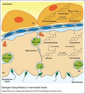
Besides being capable of responding to oestrogenic stimuli, the mammalian testes actively synthesises oestrogens. This process occurs through the aromatisation of androgenic precursors (namely, testosterone and androstenedione) by the activity of the cytochrome-c P450 aromatase enzyme (Fig. 2). It is actually established that E2, the most potent of oestrogens, is synthesised by rodent, sub-human primates and human testis, which has been demonstrated by studies characterising the presence of functional aromatase in testicular cells (Refs Reference Carreau and Hess68, Reference Devi94). The responsiveness to oestrogenic hormones, together with the fact that testicular cells can synthesise E2, has clearly supported the physiological role of oestrogens on testicular physiology. However, whether oestrogens have beneficial or detrimental effects on spermatogenesis has been a matter of discussion over the years.
Oestrogen biosynthesis in mammalian testis. Testosterone and androstenedione are converted, respectively, to 17β-estradiol and oestrone by the activity of aromatase enzyme. The 17β-hydroxysteroid dehydrogenase (17β-HSD) plays a role modulating the interconversion of androstenedione and testosterone, as well as, estrone and 17β-estradiol. In immature testis the primary source of 17β-estradiol seems to be the Sertoli cells (SC) whereas in the adult testis the main production is from Leydig cells (LC). However, germs cells also seem to be capable of synthesising oestrogens (Ref. Reference Carreau and Hess68). Legend: PTC, Peritubular cells.

Experimental data have been demonstrating that oestrogens exert a negative feedback over the hypothalamus–pituitary–testis axis, suppressing production of gonadotropin releasing hormone and luteinising hormone (LH) and thus, balancing the testosterone levels (Ref. Reference Chimento95). Oestrogens also can act directly on Leydig cells downregulating the expression of steroidogenic enzymes involved in testosterone biosynthesis and lowering testosterone production (Refs Reference Akingbemi96, Reference Strauss97, Reference Vaucher98).
In rodents, there are reports describing the direct effects of oestrogens inducing the onset of spermatogenesis (Refs Reference Pak, Lynch and Tsai99, Reference Gancarczyk100). Exposure to E2 accelerated the onset of spermatogenesis in the bank vole (Clethrionomys glareolus), while the antioestrogen ICI 182,780 blocked this effect (Ref. Reference Gancarczyk100). It was also showed that oestrogens may sustain spermatogenesis by promoting the proliferation of spermatogonia in a mechanism that seems to be mediated by the cross-talk between ERα and GPER (Refs Reference Sirianni85, Reference Li101, Reference Walczak-Jedrzejowska102). However, the ERβ-selective ligand 5α-androstane-3β,17β-diol also induced proliferation of spermatogonia in seminiferous tubules cultured ex vivo (Ref. Reference Wahlgren103). In the hypogonadal (hpg) mice, which are infertile because of the absence of gonadotropin production (Ref. Reference Cattanach104), oestrogens were able to restore spermatogenesis (Ref. Reference Ebling105). Interestingly, rather than a direct effect in the testis, this stimulatory effect of E2 in spermatogenesis has happened through stimulation of pituitary and elevation of follicle-stimulating hormone (FSH) levels (Refs Reference Ebling105, Reference Baines106).
In a rat model with depletion of all germ cell types after exposure to radiation, the administration of E2 has remarkably accelerated spermatogenic recovery by enhancing spermatogonial differentiation (Ref. Reference Porter107). In addition, it was shown that this oestrogen-mediated recovery of spermatogenesis is independent of the regulation of testosterone or gonadotropins production (Ref. Reference Porter107), and involves important changes in the expression of oestrogen-target genes (Ref. Reference Zhou108). Although this study has provided a valuable list of candidate genes, the identification of the molecular pathways underpinning the effect of oestrogens in the onset of spermatogenesis is still in the infancy.
Oestrogens also seem to be required for spermiogenesis, since treatment with aromatase inhibitors has strongly decreased the population of elongating/elongated spermatids in the testis of adult monkeys (Refs Reference Shetty109, Reference Shetty110). Accordingly, mice with disrupted aromatase CYP19 gene has displayed failure of spermiogenesis with a significant reduction in the number of round and elongated spermatids, and an abnormal development of acrosome (Refs Reference Robertson111, Reference Robertson112). These results were confirmed in rats administered subcutaneously with E2 but the observed increase in the number of elongating and elongated spermatids was proposed to occur in consequence of a failure in spermiation (Refs Reference D'Souza113, Reference Balasinor114). Recent work of Cacciola et al. has demonstrated that oestrogens play a role regulating chromatin remodelling in spermatids, particularly by preserving chromatin condensation and DNA integrity (Ref. Reference Cacciola115), and that this is involved in the nuclear size of epididymal spermatozoa (Ref. Reference Cacciola116).
Also, the function of E2 on capacitation, loss of acrosome and increased motility of spermatozoa in human ejaculated sperm has been reported (Ref. Reference Aquila73). Similarly, oestrogens have stimulated capacitation and acrosome reaction in mice (Ref. Reference Adeoya-Osiguwa117) and boar (Sus scrofa) (Ref. Reference Ded118) sperm.
Nevertheless, the most well-known effect of oestrogens in male reproductive function is probably the regulation of fluid absorption in the efferent ducts and rete testis (Ref. Reference Hess, Robaire and Hinton119). These are crucial events for male fertility by maintaining the appropriate osmolality and sperm concentration in the epididymis, as demonstrated by the elegant studies using the ERα knockout (αERKO) mice (Refs Reference Lee120, Reference Hess121, Reference Hess122).
Experimental and clinical evidence of oestrogens damaging effects for spermatogenesis
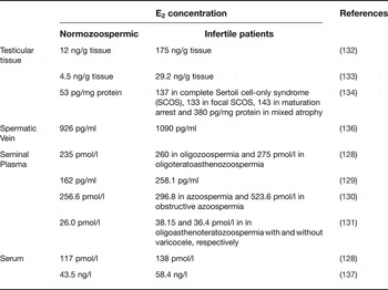
Contrasting with the beneficial actions of oestrogens in spermatogenesis and sperm maturation is the evidence of the deleterious effects of these hormones in male reproductive function. For example, it has been widely documented that foetal and post-natal exposure to oestrogens or oestrogen-like substances, that mimic endogenous oestrogens, cause adverse effects in male sexual differentiation (Refs Reference Toppari123, Reference Sultan124) and reproductive potential of the adult (Refs Reference Sweeney125, Reference Anway126, Reference Sweeney127). Noteworthy are the elevated concentrations of E2 (Table 2) detected in the semen (Refs Reference Luboshitzky128, Reference Bujan129, Reference Zhang130, Reference Zalata131), intratesticular fluid (Refs Reference Marie, Galeraud-Denis and Carreau132, Reference Levalle133, Reference Lardone134) and spermatic vein (Refs Reference Pasquier135, Reference Adamopoulos136) of idiopathic infertile men. Also, higher serum levels of E2 together with lower levels of testosterone were reported in men with severe impairment of spermatogenesis (Refs Reference Luboshitzky128, Reference Pavlovich137). This resulted in a decreased testosterone/E2 ratio, which was proposed to be the cause of male infertility since treatment with aromatase inhibitors corrected the endocrine defect and significantly improved the semen parameters in oligospermic patients (Refs Reference Pavlovich137, Reference Schlegel138). In this way, both experimental and clinical findings hinted for the possible involvement of oestrogens in the disruption of spermatogenesis and consequently, in male factor infertility.
Concentration of oestrogens in serum and reproductive tract of normozoospermic and infertile patients

Role of oestrogens controlling survival and death of testicular cells
The adverse effects of oestrogens on spermatogenesis are essentially linked to the induction of germ cell apoptosis. However, as described above, oestrogens seem to be able to restore spermatogenesis and play an important action promoting the proliferation of spermatogonia (Refs Reference Li101, Reference Walczak-Jedrzejowska102, Reference Ebling105, Reference Porter107). In fact, oestrogens are traditionally viewed as hormones that can regulate both proliferation and apoptosis in several tissues (Ref. Reference Lewis-Wambi and Jordan139), and this duality of action is also evident in the context of testicular cells.
Oestrogens as survival factors for male germ cells
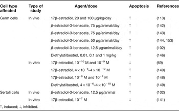
Oestrogens have been described as survival factors inhibiting the apoptosis of male germ cells (Table 3). E2 added to the culture medium of seminiferous tubules cultured ex vivo at 10−10 and 10−9 M concentrations was able to inhibit the death of germ cells (Ref. Reference Pentikainen69). Moreover, it was shown that E2 in the concentration range of 10−8–10−6 M activates the phosphatidylinositol-3-OH kinase/Akt survival pathway in human ejaculated spermatozoa (Ref. Reference Aquila73). Findings in the aromatase knockout mice support the ability of oestrogens to hold the survival of germ cells (Ref. Reference Robertson111). These mice, with a total absence of oestrogen biosynthesis, displayed progressive infertility by 14 weeks of age, which was consequence of impaired spermatogenesis at postmeiotic stages (Refs Reference Robertson111, Reference Robertson112). Depletion of round and elongated spermatids in the testis of aromatase knockout mice resulted from increased rates of apoptosis, and demonstrates the importance of oestrogens for the development and survival of male germ cells (Ref. Reference Robertson111). Also in the case of immature SCs, it has been shown that oestrogens improve their proliferation and inhibit apoptosis (Table 3) (Refs Reference Lucas140, Reference Simoes141), an effect that requires the translocation of both ERα and ERβ to the plasma membrane region with the consequent activation of epidermal growth factor receptor and the mitogen-activated protein kinase signalling pathway (Ref. Reference Lucas140). However, in vivo administration of β-estradiol-3-benzoate (estradiol benzoate) to adult male rats inhibited SCs maturation and increased their apoptosis (Ref. Reference Walczak-Jedrzejowska102).
Role of oestrogens controlling apoptosis of testicular cells

↑, induced; ↓, inhibited.
Oestrogens as apoptosis-inducers in germ cells
Remarkably, there is another scenario in the oestrogenic actions, which contrasts with the ability of oestrogens to maintain germ cell survival. It has been largely reported that E2 or oestrogens analogues have a profound impact in testicular physiology by enhancing the apoptotic rate of germ cells and, thus, decreasing sperm counts and fertility (Table 3). Estradiol benzoate, the synthetic ester of E2 has been widely used to study the apoptotic effects of oestrogens in mammalian testis. In vivo administration of estradiol benzoate to adult and newborn rats in doses ranging from 12.5 to 75 μg/animal markedly has increased the number of cells undergoing apoptosis for all germ cell types (Refs Reference Walczak-Jedrzejowska102, Reference Chaki142, Reference Kaushik143, Reference Blanco-Rodriguez and Martinez-Garcia144, Reference Blanco-Rodríguez and Martínez-García145). The germ cell loss was further confirmed by a reduction in testis weight (Refs Reference Walczak-Jedrzejowska102, Reference Kaushik143) and seems to occur mainly for spermatocytes and round spermatids (Refs Reference Chaki142, Reference Blanco-Rodriguez and Martinez-Garcia144).
Daily injections of E2 (20 or 100 μg/kg/day) to adult male rats for 10 days disrupted the pituitary–gonadal axis reducing serum gonadotropins and the intratesticular levels of testosterone (Ref. Reference D'Souza113). Both doses of E2 culminated in a significant increase of apoptosis in spermatids at stages VII and VIII, which correspond to the differentiation of round spermatids to elongated spermatids (Ref. Reference D'Souza113). Similar results were obtained through in vivo administration of the synthetic oestrogen diethylstilbestrol (DES). DES reduced testicular weight and spermatogenic cell loss in time- and dose-dependent manner (Ref. Reference Nair and Shaha146). Rat seminiferous tubules cultured ex vivo in the presence of 10−7 M E2 displayed increased rates of germ cell apoptosis (Ref. Reference Correia147), whereas a 10−9 M dose had no effect (unpublished results). Also, in vitro treatment of spermatogenic cells isolated from rat testis with 10−8 and 10−7 M of E2 induced the apoptotic cell death of spermatogonia, spermatocytes and spermatids (Ref. Reference Mishra and Shaha148). An 80% reduction in cell survival was observed 10 h after the initiation of E2 treatment (Ref. Reference Mishra and Shaha148).
Oestrogen-induced apoptosis was also observable in rat foetal testis cultured ex vivo in presence of E2 or DES. It was found as a deleterious effect on the gonocyte population in consequence of a dramatic increase of apoptosis (Ref. Reference Lassurguère149). The application of in vitro and ex vivo experimental approaches have demonstrated that oestrogens promote apoptosis of germ cells by direct actions in the testis, which do not depend on the interference of the hypothalamus–pituitary axis.
It is worth noting that oestrogenic effects inhibiting or promoting apoptosis may strictly depend on the hormonal dose used. This becomes particularly evident when comparing the same cell type and experimental approach. For example, 10−10 and 10−9 M of E2 promoted germ cell survival in vitro (Ref. Reference Pentikainen69), whereas 10−8 and 10−7 M doses induced the apoptotic death of germ cells (Refs Reference Correia147, Reference Mishra and Shaha148). Since 10−10 and 10−9 M resemble the range of physiological concentrations described in male (Ref. Reference Pentikainen69), it is liable to assume that the detrimental effects of oestrogens depend on the E2 concentrations increased above the normal physiological limits. Indeed, the study of Gancarczyk et al (Ref. Reference Gancarczyk100) has demonstrated a duality of effects for low and high doses of E2 in the testis of blank vole. A low dose of E2 (0.1 μg/g body weight) accelerated the advance of spermatogenesis showing a positive correlation with the number of spermatids. In contrast, males treated with a high dose of E2 (10 μg/g body weight) had disrupted testicular morphology, tubular atrophy and increased frequency of apoptosis (Ref. Reference Gancarczyk100). The elevated concentrations of E2 detected in the testis and reproductive fluids of idiopathic infertile patients (Table 2) (Refs Reference Marie, Galeraud-Denis and Carreau132, Reference Levalle133, Reference Lardone134) sustain the idea that oestrogens could be the cause underlying oligozoospermia and azoospermia by inducing spermatogenic cellular dysfunction due to increased rates of apoptosis.
Signalling pathways implicated in oestrogen-induced apoptosis of germ cell
Although the mechanisms by which oestrogens exert its apoptotic effects are not fully elucidated, it has been suggested that there is an involvement of the extrinsic (death receptor) pathway of apoptosis (Fig. 1) Mechanistic studies using several experimental approaches, such as, in vivo exposure, in vitro culture of seminiferous tubules or isolated spermatogenic cells have indicated that oestrogens activate the receptor-mediated pathway by upregulating the expression of both Fas and FasL (Refs Reference Nair and Shaha146, Reference Correia147, Reference Mishra and Shaha148). Increased expression of Fas system was responsible for initiating the apoptotic death of spermatogenic cells since it was followed by the activation of caspase-8 (Fig. 1) (Refs Reference Nair and Shaha146, Reference Mishra and Shaha148). Chaki et al. have also reported an increased activity of caspase-8 in the rat testis after administration of estradiol benzoate for 30 days (Ref. Reference Chaki142).
Other works have linked the apoptotic effects of oestrogens with the modulation of the intrinsic (mitochondrial) pathway of apoptosis. Bcl-2 protein family members are the key regulators of this pathway, and the interaction and relative abundance between proapoptotic and antiapoptotic members form a dynamic equilibrium that determines the threshold for apoptosis (Ref. Reference Oltval, Milliman and Korsmeyer150). It was shown that E2 or estradiol benzoate upregulated the expression of Bax in testicular cells (Refs Reference Chimento87, Reference Chaki142, Reference Correia147), which was accompanied by augmented protein ratio of Bax (proapoptotic)/Bcl-2 (antiapoptotic) and increased activity of caspase-3 (Ref. Reference Correia147). Treatment with oestrogens was also able to induce the hyperpolarisation of mitochondrial membrane (Ref. Reference Mishra and Shaha148) and the translocation of Bax from the cytosol to the mitochondria, which was followed by the release of cytochrome c and activation of caspase-9 and caspase-3 (Refs Reference Chaki142, Reference Nair and Shaha146). However, the use of anti-Fas antibody inhibited mitochondrial hyperpolarisation and E2-induced cell death, and the inhibition of caspase-8 prevented cell death, which indicates the major importance of the death-receptor pathway (Ref. Reference Mishra and Shaha148). Moreover, it was demonstrated that the mitochondrial hyperpolarisation is induced by the augmented generation of nitric oxide and superoxide, respectively, in consequence of increased expression of inducible nitric oxide synthase and E2 metabolism (Ref. Reference Mishra and Shaha148). Indeed, it has been widely accepted that oxidative stress is damaging for testicular function representing one of the main factors inducing the apoptotic cell death of male germ cells (Ref. Reference Turner and Lysiak151). Other study showed that estradiol benzoate treatment enhanced rates of apoptosis in rat testis, concomitantly with increased lipid peroxidation and reduced antioxidant defence, because of decreased enzymatic activity of superoxide dismutase and catalase (Ref. Reference Chaki142).
Apart from the effects on induction of apoptosis there are reports describing the anti-proliferative actions of E2 and the underpinning signalling mechanisms. E2 administration reduced the germ cell proliferation in rat seminiferous tubules cultured ex vivo with downregulated expression of the tyrosine kinase receptor c-kit (Ref. Reference Correia147). c-kit receptor and its ligand, the SCF, are recognised as the main mediators of survival communication between SCs and germ cells and their actions protect germ cells from apoptosis (Refs Reference Yan, Suominen and Toppari21, Reference Rossi152). Therefore, it is expected that disruption of the SCF/c-kit system by oestrogens deeply affects the spermatogenic output. Also, spermatocytes isolated from rat testis displayed diminished proliferation in response to E2 treatment (Ref. Reference Chimento86). This effect occurred by the activation of the EGFR/ERK/c-Jun pathway with the downstream down-regulation of the expression of cell cycle regulators cyclin A1 and B1 (Ref. Reference Chimento86).
Conclusion
Besides the ability responding to endocrine oestrogenic stimuli, the mammalian testis can also synthesise oestrogens, and the role of these sex steroid hormones in male reproduction has gained increasing interest in the last decade. Our knowledge of the oestrogenic actions in male reproduction has highlighted the important role of oestrogens in the control of spermatogenesis. Although classically known as mitogenic agents, it was shown that oestrogens can regulate both the survival and death of testicular cells. However, the literature contains controversial reports on the beneficial or detrimental effects of oestrogens for spermatogenesis jumping from ‘heaven to hell’. Presently, a substantial amount of data has been generated pointing oestrogens as apoptosis-inducers of male germ cells and consequently as deleterious agents for male fertility. However, the survival or apoptotic actions of oestrogens seem to be related with the hormone dosage used in the experimental assays. At doses above the physiological concentrations, oestrogens are no longer survival agents but instead they become powerful apoptotic triggers inducing the apoptosis of germ cells. Moreover, the apoptotic effects of oestrogens are consistent with the clinical findings in male infertile patients that display decreased ratios of testosterone to E2, because of elevated concentrations of E2. In this way, the role of oestrogens as apoptosis inducers, supported by a huge amount of experimental data, provided a rationale to explain the successful use of aromatase inhibitors to treat male infertility. Finally, disclosing the complexity of oestrogens signalling mechanisms in the induction of apoptosis of germ cells will provide new clues to understand the aetiology of spermatogenic disorders and to identify new therapeutic targets for male infertility.
Financial support
This work was partially supported by Portuguese Foundation for Science and Technology (FCT) under Program COMPETE (PEst-C/SAU/UI0709/2011) and fellowships to Correia S (SFRH/BD/60945/2009).
Conflicts of interest
None.