Introduction
Ocular diseases, including keratoconus (KC), diabetic retinopathy, glaucoma and age-related macular degeneration (AMD) have multifactorial and complex pathophysiologies, which include inflammatory or angiogenic aspects. Therefore, protein signatures of health and disease can be detected in the major eye-derived humours (tear fluid, aqueous humour or vitreous humour), which may reveal critical information about the state of eye health. While enzyme-linked immunosorbent assay (ELISA) (considered the gold standard for protein detection) is certainly adequate for measurement of individual proteins in these fluids, a complete understanding of ocular pathologies is unlikely to arise from studying a single protein. Moreover, the utility of ELISA is severely limited by the low fluid volumes, which are typically collected from the eye. Advancing our understanding of eye disease requires assaying many proteins in parallel, thus maximising the information generated from the major eye-derived fluids. Multiplex immunoassays are an emerging proteomic technique that has been used for the detection of key proteins involved in both homeostasis and disease states of the eye. Multiplex immunoassays are available on two major platforms: (1) antibody arrays (capture antibodies spotted on planar solid supports such as membranes, glass slides or microtitre plates) and (2) bead-based assays (capture antibodies coupled to fluorescent-labelled microbeads). Both techniques are relatively simple to perform and have been employed to efficiently capture protein expression profiles from a variety of unique biological fluids, including cerebrospinal fluid (Refs Reference Broux1, Reference Kaplin2), saliva (Refs Reference Ichikawa3, Reference Tatarakis4), colostrum (Ref. Reference Kverka5) and bronchoalveolar lavage (Refs Reference Benabid6, Reference Honko and Mizel7). Moreover, multiplex immunoassay technologies have proven indispensable in numerous instances of disease biomarker identification for diverse disease processes, including asthma, renal disease, neurological dysfunction, immunologic disorders and others (Ref. Reference Wilson8).
Other proteomic techniques have been applied to the characterisation of ocular fluids and to the search for disease biomarkers therein. These include two-dimensional (2D) gel electrophoresis coupled with various methods of single or tandem mass spectrometry (MS). While these methods have revealed a wealth of information in the characterisation of tears, aqueous humour or vitreous humour, the sensitivity limitations of MS have hampered routine detection of many low-abundance biomarkers which may reveal critical information about disease status. With this and other limitations, multiplex immunoassay platforms are an excellent and necessary complement to exploratory MS techniques, as demonstrated in several ocular research studies employing both methods with good success (Refs Reference Chowdhury9, Reference Schwartzman10).
In this review, we present studies that demonstrate the utility of multiplex immunoassay technologies in the identification of cytokine expression patterns in vitreous humour, aqueous humour and tears. We also describe how the data generated contributed to a description of the molecular basis of a variety of ocular pathologies. Finally, we discuss the contribution of genomic studies to ocular disease characterisation and how these data may complement the findings of proteomic approaches.
Protein signatures in tear fluid
The tear film is an aqueous layer containing a highly complex mixture of as many as 1543 unique proteins, peptides, lipids and electrolytes and coats the ocular surface to provide an optically smooth interface with the atmosphere (Ref. Reference Zhou11). The constituents of tear fluid are produced by multiple structures and cell types within the eye, including ocular surface epithelial cells, goblet cells, resident immune cells, lachrymal glands, Meibomian glands and even serum exudates (Ref. Reference Tiffany12). The most abundant tear proteins include lysozyme, secretory IgA, lactoferrin and lipocalin (Ref. Reference Zhou and Beuerman13), which are present at concentrations as high as 1–2 mg/ml, and contribute to the antimicrobial functions of the tear film. However, the tear proteome contains a multitude of other components such as cytokines, chemokines, growth factors, angiogenic factors, and proteases, all of which are bioactive at much lower concentrations (ng/ml to pg/ml), and which regulate a wide variety of homeostatic processes (Refs Reference Fung, Morris and Duncan14, Reference Sullivan, Dartt and Meneray15). As such, tears are a rich source of potential biomarkers, which may be used to diagnose or determine the stage of ocular disease, elucidate disease mechanisms and assess treatments. Tears are also an easily accessible body fluid, which can be collected by minimally invasive methods (typically using capillary tubes or Schirmer filter paper strips), making them particularly inexpensive and amenable to population screening (Ref. Reference Pieragostino16).
Interestingly, the protein profiles of open-eye (OTF) and closed eye (CTF) tear fluids have been compared by Sack and others (Ref. Reference Sack17) using semi-quantitative membrane-based antibody arrays, and were found to be profoundly different in their levels of several cytokines and angiogenic factors. Of the 79 cytokines screened in pooled tears from six healthy donors, 39 targets were detectable, with 11 exhibiting strong signals in both OTF and CTF. Amongst these 11, interleukin-8 (IL-8), tissue inhibitor of metalloproteinase (TIMP)-1, TIMP-2, epidermal growth factor (EGF) and monocyte chemoattractant protein-1 (MCP-1) were corroborated by previous studies (Refs Reference Goren18, Reference Thakur, Willcox and Stapleton19), while angiogenin, growth-related oncogene (GRO) and [interferon γ (IFN-γ)-induced protein 10 (IP-10), CXCL-10] had not been previously reported in tear fluid. The presence of angiogenin, a potent pro-angiogenic factor, was especially interesting given the avascular nature of the cornea. However, angiogenins have been previously reported to have microbicidal activity within the epithelial surfaces of the gut, a function which may serve to influence the resident microbial landscape (Ref. Reference Hooper20). This suggests that angiogenin may contribute a protective rather than a vasculogenic function in the context of healthy tears.
Importantly, the array data also revealed that the majority of the detected tear cytokines were present at significantly higher levels in CTF compared with OTF. This increase is partially attributable to the accumulation of cytokines as a result of reduced tear flow after eye closure. Additionally, a temporary state of sub-clinical inflammation is associated with eye closure, characterised by recruitment of PMN cells and conversion of complement (Refs Reference Thakur, Willcox and Stapleton19, Reference Sack21).
Ocular allergy
Sack's group explored the pathogenesis of chronic ocular allergies (CA) in a subsequent study using CTF from normal and CA patients (Ref. Reference Sack22). After analysis of tears by both membrane-based antibody arrays and microplate-based arrays, the authors reported that the difference in cytokine expression between normal donors and active CA patients was far more pronounced for CTF samples than for OTF samples. This study was the first to validate the potential utility of CTF for detection of inflammatory mediators at the ocular surface. Collectively, the findings of Sack et al. (Refs Reference Sack17, Reference Sack22) have shed light on the functional differences between CTF and OTF, and revealed molecular signatures which could not be captured by MS data from previous studies (Ref. Reference Mulvenna23).
Contemporaneously, the work of Shoji et al. (Ref. Reference Shoji, Inada and Sawa24) explored the differential cytokine expression patterns in tears of vernal keratoconjunctivitis (VKC, a seasonal allergic condition) and giant papillary conjunctivitis (GPC) patients. Because VKC and GPC have similar clinical manifestations (large papillae on the upper tarsal conjunctiva) and histopathological characteristics (infiltration of the epithelium by basophils, eosinophils and mast cells), the two conditions are thought to belong to the same clinical spectrum. Employing a membrane antibody array of 40 inflammatory cytokines to analyse normal and pathological samples, they observed that IL-6sR (IL-6 soluble receptor) increased significantly in both the GPC and VKC groups. This finding agreed with previous studies linking allergic rhinitis to increased expression of IL-6sR in nasal fluid (Refs Reference Kokuludag25, Reference Benson26). The authors speculated that the Th2 cell regulatory function of IL-6sR may have a role in the formation of the conjunctival papillae common to both diseases. Also noteworthy was a statistically significant increase in eotaxin-2 in the VKC group, which was presumed to be related to the eosinophilic inflammation also seen.
Keratoconus
KC) is a degenerative ocular disease, wherein progressive corneal thinning gives rise to a cone-shaped cornea instead of the normal spherical shape. Central corneal scarring is also characteristic of the condition, leading to a loss of transparency of the cornea. This distortion results in a debilitating level of visual impairment which must be corrected by gas-permeable (GP) rigid contact lenses, or in the most severe cases, by penetrating keratoplasty (corneal transplant). While the pathophysiology of KC has been extensively described, our understanding of its aetiology remains frustratingly incomplete.
KC has been classically defined as a noninflammatory condition, but a growing body of molecular studies has called this notion into question. The latest studies suggest that defects in the stromal wound repair cascade (Ref. Reference Cheung, McGhee and Sherwin27) and possibly oxidative stress (Ref. Reference Kenney28), are implicated in the disease. In a study by Pannebaker et al., the mechanism of KC was explored by analysing the protein profiles of tear samples from keratoconic patients (with or without GP contact lenses) and healthy controls with GP lenses (Ref. Reference Pannebaker, Chandler and Nichols29). Cytokine analysis of the tear fluids, which was performed using a glass slide-based antibody array of 40 cytokines, detected a significant increase in matrix metalloproteinase-1 (MMP-1) in the 2 keratoconic groups compared with controls. MMP-1 had been previously been found to be upregulated in keratoconic corneal epithelial cells (Ref. Reference Seppala30), but this was the first time elevated MMP-1 had been detected in keratoconic tear fluids. MMP-1 is a proteolytic enzyme with specific collagenase activity. Given that the cornea is 70% collagen by weight (Ref. Reference Collier31), and that MMP-1 should have minimal expression in healthy tissues, the overexpression of MMP-1 in KC hints at a dysfunctional wound repair mechanism. Along with these findings, it is important to note that the characteristic corneal distortion and thinning in KC is in large part because of a damaged extracellular matrix (ECM) and a decrease in types I and IV collagen (Refs Reference Collier31, Reference Abalain32). This putative defect in the corneal wound healing cascade was later explored by Cheung et al., through examination of cytokine profiles of corneal stromal cells (Ref. Reference Cheung, McGhee and Sherwin33). In this study, corneal biopsies were collected from keratoconic patients and nonkeratoconic controls following penetrating keratoplasty. After excision, an ex vivo post-surgical incision wound (SI) was applied to the corneal tissue in order to examine the reparative responses to new injury. After isolating stromal cells from each cornea, lysates were prepared and analysed using a nitrocellulose membrane-based antibody array. From the 120 proteins screened for in the array, two important trends were noted. First, both keratoconic corneas and wounded control corneas exhibited increased levels of interleukin-1α (IL-1α), insulin-like growth factor 1 (IGF-1), tumour necrosis factor-α (TNF-α) and transforming growth factor-β1 (TGF-β1) compared with unwounded controls. Secondly, compared with wounded controls, the wounded keratoconic cells expressed much lower levels of IL-1α, IGF-1, fibroblast growth factor (FGF)-2, EGF, TGF-β1 and platelet-derived growth factor (PDGF). Collectively, these observations suggest that the expression profile of keratoconic corneas mimics that of injured normal corneas, and further, that keratoconic corneas exhibit a blunted reparative response to wounding. Taken together, these antibody array-based studies have helped reshape our understanding of the pathological basis for KC, pointing to an underlying inflammatory mechanism.
Protein signatures in vitreous
Vitreous humour, an acellular, transparent gel that fills the space between the lens and the retina, accounts for 80% of the volume of the eyeball. Permanently formed at birth, the vitreous humour undergoes a very slow rate of metabolic exchange with the systemic circulation, changing very little over time. The main functions of the vitreous are to transmit light to the retina and to maintain its rounded shape by exerting pressure to keep it tightly pressed against the choroid. Vitreous fluid must be obtained by more invasive procedures compared with tear fluid; however, it is a rich source of biomarkers relevant to retinochoroidal diseases. Intraocular tissues contain a variety of cell types such as RPE (retinal pigment epithelium) cells, pericytes, endothelial cells and Müller cells which secrete angiogenic factors and cytokines into the vitreous fluid. The expression levels of these markers are altered during injury or hypoxia (Ref. Reference Semeraro34). Multiplex immunoassay-based profiling of vitreous humour has been extensively used to describe various ocular pathologies, enabling more global cytokine changes to be evaluated within small sample volumes (Refs Reference Maier35, Reference Maier36, Reference Nagata37, Reference Sato38).
Diabetic retinopathy (DR)
DR, one of the leading causes of blindness worldwide, is caused by pathological angiogenesis in the retina leading to damage of its microvasculature (Ref. Reference Aiello39). The condition is strongly correlated with chronic hyperglycaemia, which may induce vascular damage through a number of possible mechanisms, including alterations of PKC (protein kinase C) signalling, oxidative stress or increases in advanced glycation end products (Refs Reference Aronson40, Reference Kowluru41, Reference Kowluru, Tang and Kern42). In the early stages of the disease, known as nonproliferative diabetic retinopathy (NPDR), the patient typically experiences little or no change in vision but microvascular abnormalities can be detected in the retina. As the disease progresses, upregulation of angiogenic factors in the vitreous leads to neovascularisation of the retina. This destructive phase, called proliferative diabetic retinopathy (PDR), is characterised by vision-impairing vitreous haemorrhages that ultimately destroy retinal architecture.
With the aid of multiplex immunoassay techniques, a considerable body of evidence has been built implicating specific vitreal proinflammatory and angiogenic factors in the pathogenesis of DR. Multiple studies have correlated PDR with increases in vitreal MCP-1 (Refs Reference Capeans43, Reference Elner44, Reference Hernandez45, Reference Mitamura46), IL-8 (Refs Reference Elner44, Reference Hernandez45, Reference Canataroglu47, Reference Yuuki48) and vascular endothelial growth factor (VEGF) (Refs Reference Aiello49, Reference Wang50) using ELISA or radioimmunoassay. To maximise the information generated from precious vitreous humour specimens, Banerjee et al. expanded the query to 19 cytokines, examining patients with PDR and several other vitreoretinal disorders using a cytometric bead assay (Ref. Reference Banerjee51). From this screen, increases in VEGF, MCP-1, macrophage inflammatory protein-1β (MIP-1β), IL-6, IL-8, IL-12 and regulated on activation, normal T cell expressed and secreted (RANTES) were observed in PDR (n = 10) compared with nonproliferative controls. This agreed with a previous multiplex study, which noted an increase in VEGF, MCP-1 and angiogenin in the vitreous of diabetic patients (Ref. Reference Maier35). The data further revealed the expression patterns in uveitis (an inflammatory disorder), which were distinct from PVR (proliferative vitreoretinopathy) and PDR patients (Ref. Reference Banerjee51), suggesting that vitreal cytokine signatures can distinguish between various types of retinopathies.
A later study utilised a broader screening approach to identify diabetic molecular profiles in the eye (Ref. Reference Schwartzman10). The study began with a large antibody array panel of 79 targets, which was employed to detect bioactive proteins in cadaver vitreous humour. From this preliminary screen, 12 proteins exhibited detectable signals: GRO, IL-8, IL-6, IL-1α, IL-1β, MIP-1α [colony-stimulating factor (SCF)], MCP-1, angiogenin, leptin, IP-10, TIMP-1 and TIMP-2. Subsequently, a smaller array focused on this detectable cytokine panel was used to identify expression pattern differences between NPDR, PDR, nondiabetic retinal detachment (RD) and control samples. The array revealed several important trends. As observed in previous studies, VEGF expression levels were increased in NPDR and PDR vitreous (Refs Reference Aiello49, Reference Aiello52, Reference Itakura53, Reference Murata54), and furthermore, this increase was diabetes-specific (i.e. not increased in RD patients). The array also revealed for the first time the presence of vitreal vascular endothelial growth factor receptor-2 (VEGFR-2) in all patient groups. The VEGFR-2 levels were significantly lower in NPDR and PDR samples compared to RD and control, resulting in a significantly increased ratio of VEGF/VEGF-R2 expression (P < 0.05). Since soluble receptors are known to act as inhibitors of their respective ligands, it was postulated that the relative decrease may further contribute to the angiogenic phenotype of the retina in DR. Additionally, LC/MS/MS-based analysis of arachidonic acid-derived eicosanoids in the vitreous revealed a 5-fold, diabetes-specific increase in the proinflammatory metabolite 5-HETE (5-hydroxyicosatetraenoic acid). Taken together, the data suggest diabetic vitreous contains an overall proinflammatory molecular profile.
The antibody array approach to analysing vitreous humour has revealed that not only the expression levels, but also the tyrosine phosphorylation states of various cytokines can be important indicators of disease states (Ref. Reference Reverter55). In one study, a membrane-based array detecting 42 soluble factors was adapted for phosphorylation profiling by employing an antiphosphotyrosine detection antibody coupled with cytokine-specific capture antibodies. From the vitreous of diabetic retinopathy patients, the array showed a striking decrease in the phosphorylation status of several inflammatory and angiogenic proteins, including IL-1α and IL-1β (50% decrease) as well as epithelial-derived neutrophil-activating protein (ENA)-78, granulocyte colony-stimulating factor (GCSF), granulocyte-macrophage colony-stimulating factor (GM-SCF), IL-5 -6, -7, -8, -10, -12, monokine induced by IFN-γ protein (MIG), MIP-1γ and RANTES (20–40% decrease) compared with healthy controls. This altered phosphorylation signature was attributed to two possible mechanisms: an excess of peroxynitrates in the diabetic milieu of the eye, or dysregulation of phospho-dephosphorylation mechanisms because of alterations in insulin signalling through Akt, GSK-3 (glycogen synthase kinase 3) or other tyrosine kinases.
Protein signatures in aqueous humour
The aqueous humour is a transparent, watery fluid located within the anterior and posterior chambers of the eye (in the space between the lens and the cornea), and contains a complex mixture of organic solutes, electrolytes, cytokines and growth factors. This fluid is in direct contact with the nonvascularised tissues of the cornea, lens and trabecular meshwork (TM), bathing them in nutrients and maintaining homeostasis of the anterior segment. Unlike the vitreous, which undergoes very little fluid exchange with the circulatory system, aqueous humour is regularly drained and replenished through the ciliary body. During glaucoma or inflammatory ocular conditions, the protein composition of aqueous humour can change significantly, providing a source of potential diagnostic biomarkers.
To gain insight into the soluble factors involved in maintaining homeostasis of the anterior segment, the proteome of aqueous humour has been explored using a combination of high-density antibody arrays, quantitative antibody arrays and nanoflow liquid chromatography electrospray ionisation tandem mass spectrometry (nano-ESI-LC/MS/MS) (Ref. Reference Chowdhury9). This combined approach served to detect aqueous humour targets in both high-abundance (enzymatic, catalytic and structural proteins) and low abundance (cytokines, growth factors and soluble receptors). Of the 676 proteins identified in the study, 328 were identified by a high-density biotin label-based antibody array containing a panel of 507 antibodies. Interestingly, only 10 of these 328 were detectable by nano-ESI-LC/MS/MS. These low-abundance factors included members of the TGF, TNF, FGF and IL families along with their receptors. Collectively, this work continues to serve as a reference for the discovery of biomarkers or therapies for pathological conditions of the anterior segment.
Uveitis
Uveitis is broadly defined as inflammation of the inner eye tissues as a result of infection, injury or autoimmune disease. The term ‘uveitis’ encompasses a broad and heterogenous spectrum of clinical phenotypes that is characterised by infiltration of leukocytes (CD4+ T cells) into the intraocular cavity, aberrant secretion of cytokines, all of which leads ultimately to destruction of eye tissues. Although a relatively uncommon condition, uveitis a significant contributor to visual impairment, accounting for 10% of blindness worldwide (Ref. Reference Wakefield and Chang56). The causes are numerous but poorly understood, and include infection, injury, exposure to toxins or autoimmune responses. The disease may be classified by the anatomical location of the introaocular inflammation (posterior, anterior or panuveitis); or by general aetiology (infective, noninfective or idiopathic). Infective uveitis may occur as a complication of systemic infection, as in ocular toxoplasmosis (Ref. Reference Bodaghi57), ocular tuberculosis (Ref. Reference Gupta, Gupta and Rao58) or Ebola-associated uveitis (Refs Reference Hunt and Knott59, Reference Varkey60). Noninfective uveitis is thought to have an autoimmune component, such as in association with Behçet's disease, sarcoidosis or Reiter's syndrome. However, the clinical entities are diverse and exact aetiologies are currently unclear.
Under normal circumstances, the eye is protected from infiltrating leukocytes by an efficient blood–retinal barrier. In addition, the intraocular microenvironment contains immunoinhibitory molecules, including TGF-β2, MIF (macrophage migration inhibitory factor), Fas ligand, α-MSH (α-melanocyte-stimulating hormone), CGRP (calcitonin gene-related peptide) and CD200 receptor, all of which curb the tissue destructive activities of inflammatory cells (Ref. Reference Perez and Caspi61). Together, these properties contribute to immune privilege in the eye. During uveal inflammation however, inflammatory cytokine levels in ocular tissues and fluids increase significantly, drastically altering immune cell trafficking within the ocular chambers and the affected tissue. Considerable research efforts are currently focusing on the mechanisms of sustained intraocular inflammation with the intent of identifying key pathogenic molecules upon which to focus new therapies. Given the central role of cytokines in inflammation, multiplex immunoassays have proven valuable for advancing our understanding of the disease.
In studies which compared the aqueous humour with the serum of uveitic patients, it was repeatedly observed that the cytokine profiles of the two fluids are quite disparate. For example, Valentincic et al. reported that TNF-α was increased in the serum of idiopathic uveitis patients compared with controls, whilst IL-8 and IL-6 (among other cytokines) were significantly increased in the AH; no overlap between the two fluids was observed (Ref. Reference Valentincic62). More recently, it was reported that out of 15 cytokines profiled, only IL-6 was significantly elevated in the serum of uveitis patients versus control, while the aqueous humour showed significant increases in 11 cytokines, including IL-17A and IFN-γ. The aqueous humour expression patterns thus proved more informative, and further suggested that subjects with idiopathic acute anterior uveitis (AAU) exhibited a predominantly Th17-driven immune response, whereas a Th1-dominant immune response was characteristic of HLA-B27-associated AAU patients (Ref. Reference Chen63). Not surprisingly, the molecular composition of aqueous humour provides a more detailed depiction of the ocular inflammatory response than that of peripheral blood. This highlights the importance of characterising the ocular microenvironment in the context of active uveitis.
Though the molecular aetiologies of the various uveitic entities are cryptic, our current understanding of the pathological processes involved has advanced considerably in recent years. Much of the progress is attributable to animal models of uveitis and clinical trials in which infiltrating cells and cytokine profiles were examined. The studies using these approaches have contributed valuable insights not only into the pathogenesis of uveitis, but also have pointed us towards key therapeutic targets and discriminatory biomarkers. For example, endotoxin-induced uveitis (EIU), which is induced by systemic or local injection of bacterial lipopolysaccharide in rats or mice, is a widely used model for noninfective autoinflammatory uveitis. Using this model, a study examined the aqueous humour of EIU rats in order to assess the protective effects of guggulsterone, an anti-inflammatory plant sterol. Here, administration of guggulsterone to rats decreased the numbers of infiltrating cells in the AH. Cytokine profiles of the aqueous humour further revealed that 14 cytokines were significantly upregulated in EIU, and significantly suppressed with guggulsterone pre-treatment. The anti-inflammatory action of guggulsterone was shown to occur through inhibition of iNOS and blockade of [nuclear factor kappa-B (NF-κB), a transcription factor and master regulator of innate immune responses] translocation to the nuclei (Ref. Reference Kalariya64). This study provides rationale for the use of systemically administered anti-inflammatory compounds to attenuate uveitis.
The prevailing thought is that autoimmune uveitis is largely CD4+ T cell driven. Early studies have established the importance of Th1 cells in the development of uveitis; however, more recently, Th17 cells (a newly described T-helper cell lineage) have been identified as pivotal players as well (Ref. Reference Takeda, Sonoda and Ishibashi65). The evidence for this paradigm has been collected largely from studies in animal models, many of which utilised cytokine profiling and analysis of infiltrating lymphocytes (Refs Reference Lee66, Reference Ooi67). A 2005 clinical study of idiopathic uveitis patients reported that their aqueous humour protein expression profiles were dominated by Th1 cytokines, while TGF-β2 was reduced compared with noninflammatory controls. Furthermore, using random forest and cluster analysis of the bead array data, six cytokines were identified (IL-6, IL-8, MCP-1, IL-13, IL-2 and TNF-α) which could completely discriminate idiopathic uveitis patients from controls (Ref. Reference Curnow68). It was later confirmed that IL-6 and IL-8 in aqueous humour were markers for intermediate uveitis (and for uveitis in general) (Ref. Reference Valentincic62). In another study with chronic refractory uveitis patients, IL-6 and IL-23 (both Th17 cytokines) were identified as contributors to ocular inflammation via Th17 differentiation. This group further explored the role of these two cytokines in the experimental autoimmune uveitis (EAU) mouse model, and observed that IL-6 or IL-23-deficient mice exhibited diminished uveitic disease symptoms. Administration of a systemic blocking antibody against the IL-6 receptor also ameliorated EAU (Ref. Reference Yoshimura69). Another study analysed aqueous humour specimens from viral and toxoplasmic uveitis patients, and revealed Th1- and Th17-weighted cytokine profiles that included IL-6, MIP-1β and IFN-γ (Ref. Reference Lahmar70). Collectively, these and other such studies suggest that in uveitis, Th2-like responses are polarised towards Th1 and Th17, while disease resistance is characterised by polarisation to a Th2 pathway. This deeper understanding of the pathogenic mechanism of uveitis has illuminated new therapeutic strategies, in particular, biologic agents targeted against cytokines. These include biologics targeting IL-6, TNF-α, IFNs or cell surface molecules. Th17 cytokine-targeted therapies (including blockers of IL-6, IL-23 and TNF-α) have been successfully utilised for treating noninfective uveitis as well as a number of other autoimmune diseases such as Crohn's disease, rheumatoid arthritis and psoriasis (Ref. Reference Takeda, Sonoda and Ishibashi65).
Glaucoma
Glaucoma is a general term for a group of optic neuropathies in which progressive degeneration of retinal ganglion cells (RGCs) leads to optic nerve damage, ultimately resulting in vision loss. Glaucoma is frequently associated with an increase in intraocular pressure (IOP), a condition resulting from improper drainage of the aqueous humour through the TM. The mechanisms leading to RGC death and glaucoma are a subject of intense investigation. Current views are that the neurodegenerative processes involved are multifactorial and include both genetic and age-related susceptibility factors, along with secondary inflammatory or autoimmune responses including glial cell activation (Refs Reference Tezel71, Reference Tezel72).
Seeking to explore the inflammatory aspect of glaucoma pathogenesis, Kuchtey et al. employed a bead-based 12-plex cytokine assay to test aqueous humour of patients with primary open-angle glaucoma (POAG) (Ref. Reference Kuchtey73). They observed that IL-8 was significantly elevated in the aqueous humour of POAG patients compared with nonglaucoma controls and also that the concentration of IL-8 correlated with the severity of visual field defect. Immunohistochemical staining of whole eyes revealed that IL-8 is expressed in the optic nerve and the inner retina. Because IL-8 is a proinflammatory cytokine with demonstrated neurotoxic effects (Ref. Reference Thirumangalakudi74), this finding supported their hypothesis that neuroinflammatory processes are activated in glaucoma and may underlie disease symptoms.
Although human aqueous humour samples may be readily collected during routine cataract surgeries, these specimens are generally in limited supply, making studies which associate protein changes with disease state very difficult to execute. Therefore, many researchers rely on animal models to study diseases of the anterior segment. One study (Ref. Reference Birke75) investigated the pathogenesis of glaucoma by using the D2J mouse strain which develops optic neuropathy and glaucomatous eye phenotype with age (Ref. Reference Chang76). Using a mouse membrane-based array, they tested aqueous humour of D2J mice aged 2, 7 and 10 months, and found a statistically significant increase in osteopontin (OPN) in the 10-month-old DJ2 mice only, suggesting that OPN is an age-dependent marker. Moreover, elevated OPN correlated with the optic nerve degeneration phenotype (DJ2+ mice). These observations corroborate the work of Chowdhury et al. (Ref. Reference Chowdhury9), who observed detectable levels of OPN in their proteomic screen of aqueous humour. Interestingly, elevated IOP, which is a widely recognised risk factor for glaucoma, was also measured in the mice, but did not correlate with OPN concentrations. They concluded that dysregulation of OPN could be a key factor in the pathological processes involved in optic nerve degeneration in the D2J mouse model, and suggested that OPN may be a biomarker relevant to human glaucoma. Based on this work, Neumann et al. subsequently characterised OPN as a TGF-β2-activated factor that regulates the metabolic activity of astrocytes, and may have a cytoprotective effect in response to TGF-β2-mediated astrocyte damage (Ref. Reference Neumann77). Similarly, OPN was also found to have a neuroprotective function in cultured photoreceptor cells (Ref. Reference Del Rio78).
Age-related macular degeneration
AMD is degeneration of the macula lutea (a 5.5 mm area of the retina responsible for high resolution, central vision) and is a leading cause of irreversible vision loss in the >50 year age group. AMD is classified as two types: exudative, which is characterised by choroidal neovascularisation and nonexudative, which involves thinning of the macular tissue. The same 507-target biotin label-based array used in Birke's study (Ref. Reference Birke75) has also been used to explore the pathophysiology of AMD (Ref. Reference Cha79). Conducting a comparative analysis of aqueous humour from patients with exudative AMD and normal controls, this study uncovered increased expression of insulin-like growth factor binding protein (IGFBP)-2, IGFBP-6, and insulin-like growth factor binding protein-related protein-1 (IGFBPrp1) in the exudative AMD group. Since the IGFBPs are known to regulate the function of IGF-1, they followed up on this finding by testing IGF-1 levels by ELISA, and found that it was increased as well in AMD patients. The possible involvement of the IGF axis in the pathological processes of AMD was a novel finding, and in good agreement with previous evidence that IGF-1 could induce choriocapillary endothelial cells to proliferate (Ref. Reference Spraul80).
Diabetic retinopathy
Cheung and others collected cytokine profiles from aqueous humour of diabetic patients in an effort to explore biomarkers for the early stages of DR (Ref. Reference Cheung81). This study included three groups of subjects: diabetic patients without retinopathy, diabetic patients with DR, and nondiabetic controls. Interestingly, analysis of the aqueous humour by a 22-cytokine cytometric bead assay revealed that the cytokine signatures of diabetic patients with DR closely mirrored that of diabetic patients without DR. In particular, VEGF and IL-6 levels were significantly higher in diabetic subjects compared to controls, while IL-12, TNF-α, IFN-α and GM-CSF levels were reduced. Thus, even without detectable retinopathy, the diabetic eyes displayed a cytokine profile, which was markedly similar to that of patients with DR, and which was also statistically distinct from the nondiabetic controls. It was concluded that these cytokines are potential biomarkers for the early detection or prognosis of DR.
Genome-wide association studies (GWAS) for the discovery of ocular disease risk variants
With data from the Human Genome Project and the International HapMap Project now accessible in computerised databases, researchers can rapidly and accurately screen whole genomes for genetic variations associated with disease onset. GWAS are one such high-throughput approach, which serves to identify single-nucleotide polymorphisms (SNPs) and associate them with disease progression. Indeed, compared with diseases in other tissues, GWAS approaches have been particularly fruitful for the discovery of novel factors associated with the pathogenesis of ocular disease (Ref. Reference Altshuler, Daly and Lander82). Additionally, GWAS samples can be obtained from cells collected by minimally invasive methods (such as blood samples or cheek swabs), allowing for much larger population studies (including healthy controls) than can be accomplished with intraocular fluids or biopsy specimens.
Two reports that were published together in Nature Genetics in 2010 reported the use of GWAS to identify Behçet's disease susceptibility loci. One study discovered 54 SNPs at 35 discrete loci that exhibited significant correlation with Behçet's disease. The strongest correlative association was found to be a 47-kb locus between IL23R, which encodes IL-23 receptor, and IL12RB2, which encodes IL-12 receptor β2. The second most significant was a region between SNP within the IL10 gene (encoding IL-10) (Ref. Reference Mizuki83). The other study also identified associations of IL10 and IL23R – IL12RB2 variants with Behçet's disease, but from a different population (Ref. Reference Remmers84). A subsequent meta-analysis of the data revealed that the significant associations were replicated across the two studies (Ref. Reference Kirino85). In addition, association of IL10 and IL23R – IL12RB2 variants with Behçet's disease were also identified in an Iranian population (Ref. Reference Xavier86). Interestingly, risk variants for these genes have also been identified in other chronic inflammatory diseases, including psoriasis (IL23R), and inflammatory bowel diseases (IL23R and IL10), suggesting perhaps common aetiologies with noninfective uveitis. In fact, dysregulation of the IL-23/Th17 signalling axis has indeed been described in all three diseases (Refs Reference Chen63, Reference Abraham and Cho87, Reference Di Cesare, Di Meglio and Nestle88, Reference Merida89).
In addition to Behçet's disease, GWAS has been used to identify factors associated with AMD. A genetic link to AMD had been proposed for decades mainly on the basis of discoveries made in familial aggregation studies. Seminal GWAS-based studies discovered AMD risk associated with variant alleles of complement factor H (CFH) in various populations throughout the world (Refs Reference Klein90, Reference Edwards91, Reference Haines92). Another locus linked to AMD susceptibility is the age-related maculopathy 2 gene (ARMS2) at SNP rs10490924 (Ref. Reference Rivera93), and a closely associated gene, high-temperature requirement A1 (HTRA1), was found to be associated with neovascular AMD (Ref. Reference Dewan94). Subsequent meta-analysis revealed that both ARMS2 and HTRA1 may contribute towards AMD independently with an amplified collective influence (Ref. Reference Tong95). In 2007, the AMD Gene Consortium identified another 19 susceptibility loci associated with the development of AMD (Ref. Reference Fritsche96). This study involved a collaborative GWAS examination of 17 100 advanced AMD patients and >60 000 healthy controls. Pathways found to be enriched included those involved in lipid metabolism, ECM remodelling, and angiogenesis. Seven loci were identified with particularly significant genetic risk score, near the genes COL8A1-FILIP1L, IER3-DDR1, SLC16A8, TGFBR1, RAD51B, ADAMTS9 and B3GALTL. These findings opened new avenues of inquiry for therapeutic targets of AMD. In addition, a study involving a Japanese population with exudate AMD identified TNFRSF10A as novel susceptibility locus (Ref. Reference Arakawa97). Together, these landmark GWAS studies for the discovery of AMD risk variants have facilitated a comprehensive explanation of the hereditability of this condition.
Prior to GWAS, mutations in myocilin (MYOC), optineurin (OPTN) and WD repeat domain 36 (WDR36) were by and large, the only genes known to be associated with increased risk of POAG. However, a GWAS within a Japanese cohort discovered six polymorphisms adjacent to the ZP4 and PLXDC2 genes (Ref. Reference Nakano98), although these variants were not replicated in a subsequent study in a cohort in India (Ref. Reference Rao, Kaur and Chakrabarti99). In a much larger GWAS, Thorleifsson et al. reported two SNPs with linkage to caveolin-1 and 2 (CAV1 and CAV2), which are highly expressed throughout ocular tissues (Ref. Reference Thorleifsson100). Caveolin-1 is an integral membrane protein which is known to interact with the TGF-β type 1 receptor, imparting important regulatory effects on TGF-β signalling. This finding, together with the discovery of the protein biomarker OPN (Refs Reference Birke75, Reference Neumann77), implicate TGF-β signalling as a key pathogenic pathway in glaucoma. In a similar study, an additional two regions harbouring risk variants were identified adjacent to TMCO1 and CDKN2B, both of which are thought to function in apoptosis within the eye (Ref. Reference Burdon101).
GWAS has also been employed to identify markers of corneal ocular diseases. For example, GWAS identified risk variants for Fuchs' corneal endothelial dystrophy associated with transcription factor 4 (TCF4), which is involved in cellular growth and differentiation (Refs Reference Baratz102, Reference Li103, Reference Thalamuthu104). In addition, risk variants were identified by GWAS for KC at RAB3GAP1 and on the promoter region of HGF (Refs Reference Li105, Reference Burdon106).
Without doubt, GWAS has identified a plethora of genetic variants that predispose patients to ocular diseases, providing insights into their pathogenesis and the potential for early therapeutic intervention. However, finding a risk variant is only the beginning of the full description of disease aetiology. Genetic association initiates the formulation of a hypothesis for further examination. But for each disease condition, the extent to which environmental factors in combination with risk allele variants contribute to disease development and presentation must be determined. Do all patients with the risk allele develop the particular associated ocular condition? Conversely, do all patients with the disease carry the variant allele? If the answers to both of these questions are negative, then additional contributing environmental influences for disease development remain to be discovered.
In comparing the molecular signatures discovered from multiplex immunoassays, we see some, but not extensive overlap in the biological pathways identified by GWAS. For example, inflammatory genes of the interleukin families were identified by both approaches, as well as growth and differentiation factors. Identification of any single common factor between the two methods, however, is relatively rare; this is likely because of the nature of the interrogation. GWAS is an open-ended unbiased approach, whereas multiplex immunoassay involves a defined and pre-selected candidate protein list. In addition, genetic risk variants and proteins function at different stages of disease progression. Clearly, there is a need to amalgamate both genomics and proteomics in a coordinated strategy to identify both diseases aetiologies and disease progression. Collecting protein signatures in the ocular fluids of a full GWAS cohort would yield invaluable information for testing hypotheses rationalised on genetic risk variant data.
Conclusion
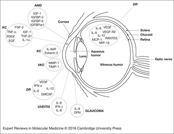
Perfect functioning of the eye involves light passage through all ocular fluids, including the tear film, the aqueous humour and the vitreous humour, in order to permit focusing of images onto the retina. Owing to their close contact with the vascular structures in the eye (epithelial surfaces, ciliary body, TM, retina, choroid, macula and so on), these fluids contain molecular signatures that are uniquely representative of local homeostatic or pathological processes within the immune privileged microenvironment of the eye. For this reason, the ocular fluids are the preferred specimens for studying the pathophysiology and molecular signatures of complex eye diseases. Indeed, while circulating biomarkers have been reported for DR and AMD, they have thus far demonstrated limited utility (Refs Reference Gu107, Reference Nguyen108). Many of the signalling pathways involved in their pathophysiology modulate activity of cytokines, chemokines, growth factors and/or soluble receptors which are relatively low in abundance within the eye (ng/ml to pg/ml) and challenging to detect by MS. Multiplex immunoassay techniques, including antibody arrays and cytometric bead assays, have identified associations between disease states and expression patterns of intraocular proteins, maximising the data generated from low sample volumes, all while providing comparable sensitivity and reproducibility to ELISA. A summary of the key disease-associated proteins identified in each ocular fluid is depicted in Figure 1.
Diseases associated with aberrant cytokine expression in various structures of the eye. Cytokines are positioned with the fluid or tissue type within which they were observed to be differentially expressed in the indicated disease, as described in Table 1. AMD = age-related macular degeneration; DR = diabetic retinopathy; KC = keratoconus; VKC = vernal keratoconjunctivitis.

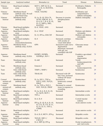
Application of antibody arrays to tear fluid analysis has revealed a dysregulated wound repair response in the corneas of keratoconic patients, contributing new insight to the pathophysiology of the disease. Multiplex immunoassay-based analysis of vitreous humour has facilitated the characterisation of angiogenic and inflammatory processes of DR, and even revealed cytokine signatures which are distinct for various types of retinopathy. Analysis of aqueous humour by antibody arrays and cytometric bead assays led to the discovery of OPN as a cytoprotective molecule in optic neuropathy, and a potential biomarker for glaucoma in humans. Additionally, many studies analysing infiltrating cells and intraocular cytokine profiles in uveitic animal models have pointed to the activation of the Th1 and Th17 immune response. Collectively, these findings have provided a greater understanding of the intraocular microenvironment in which ocular pathologies occur, insights into their underlying pathogenic mechanisms, and many potential biomarkers for early detection or classification of ocular diseases (Table 1). However, despite these advancements, the ocular diseases described here still have unclear aetiologies and clinically useful biomarkers are few and far between, indicating that current studies have only scratched the surface of their molecular bases. These significant gaps in knowledge can be addressed by further studies profiling the expression of bioactive proteins in the chambers of the eye. Before the advent of multiplex immunoassays, this task could only be accomplished by MS. While MS is represents an excellent tool for unbiased screening of molecular markers, these methods are costly, laborious and insufficiently sensitive for the routine detection of low-abundance markers (Ref. Reference Chowdhury9). Multiplex immunoassays, while limited to the repertoire of antibodies contained within the panel, are rapid and efficient to perform and can detect most proteins at pg/ml levels. As such, the two methods complement one another and integration of their respective data sets have proven useful for characterising intraocular environment of the diabetic eye (Ref. Reference Schwartzman10).
Disease-specific cytokine and growth factor expression trends in ocular fluids or tissues

In addition to contributing pathophysiological insights and revealing crucial but unexpected expression of key factors, the data from multiplex immunoassays have laid essential groundwork for new therapeutic strategies for the treatment of inflammatory ocular diseases. The collective work of multiple studies have firmly established Th1 and Th17 cell activities as pathophysiological hallmarks of uveitis, and have led to clinical trials with numerous biological agents targeting cytokine signalling. In many instances, these therapeutics have been employed with success. For example, tocilizumab, a humanised monoclonal antibody against the interleukin-6 receptor (IL-6R), was originally developed for rheumatoid arthritis, but showed efficacy in the treatment of refractory uveitis associated with Behçet's disease (Ref. Reference Hirano109) and refractory uveitis-related cystoid macular oedema (Ref. Reference Adan110). Infliximab, another therapeutic antibody developed for treatment of autoimmune diseases, specifically targets TNF-α. A pilot study reported that vitritis, macular oedema, and vasculitis were all resolved in 90% of patients treated with infliximab (Ref. Reference Sfikakis111). Similarly, the TNF-α-inhibiting agent adalimumab, has been used to treat refractory uveitis (Refs Reference Bravo-Ljubetic112, Reference Simonini113), while gevokizumab, a monoclonal antibody which binds with high affinity to IL-1β, reportedly reduced intraocular inflammation of Behçet's disease (Ref. Reference Gul114). These few examples are proof of principle that cytokine blockade may be an effective tool for the treatment of uveitis and other inflammatory or autoimmune ocular diseases. This also suggests that other therapeutic targets identified in antibody-based screens may eventually lead to the development and implementation of specific, high-affinity agents to treat ocular disease. This advancement will depend however, upon the extensive validation of such targets through the execution of numerous high-quality studies, both in animals and human subjects. This approach holds promise for the future advancement of precision medicine.
The selection of an array panel for querying protein expression in ocular specimens is often guided by the researcher's intended application. If the goal is to characterise specific pathologies of a known disease or disease model, then a focused panel of 20–200 targets may be appropriate, containing a mixture of known biomarkers and proteins relevant to select disease processes. In cases of dry eye disease, glaucoma or uveitis, where inflammation or T-cell activation is expected, it is appropriate to screen the IL families, particularly IL-6, IL-8 and markers of the Th1/Th2 and Th17 response. The TNF family cytokines and their receptors should also be included, as well as other chemotactic chemokines and adhesion molecules (MCP-1, RANTES, eotaxin-1, fractalkine, the MIPs and others). Additionally, the extensive crosstalk between the TGF-β signalling axis and classical inflammatory cascades necessitates probing for any ligands of the TGF-β superfamily including activin, nodal, anti-müllerian hormone, the bone morphogenic proteins (BMPs) and GDFs (growth and differentiation factors). OPN should also be included for its involvement in inflammation and TGF-β signalling (Ref. Reference Neumann77). In the context of DR or AMD, it is critically important to query the angiogenic pathways as well as inflammation, and to build into the panel all members of the VEGF, IGF, FGF, EGF, HGF and TGF-β family proteins and their receptors. These targets are also highly relevant for KC, corneal ulcer and other vitreoretinopathies. To complement this panel, molecules involved in maintenance and turnover of the ECM should also be explored to allow a comprehensive inquiry of dysfunctional wound healing and vasculogenesis. This panel should thus include all available MMPs, TIMPs, integrins, fibronectin, collagens and laminins. If tissue biopsies or cell lysates are being examined, then cytoplasmic markers may be included in the assay, particularly those involved in the apoptotic machinery [caspases, cytochrome-C, bcl-2, bax, survivin, the inhibitors of apoptosis (IAPs) and others]. Additionally, the activation of other key cellular signalling pathways, such as the signal transducers and activators of transcription (STATs), MAP kinases, Akt, Notch, and the NF-κB, can be probed as well using phosphorylation-specific antibody arrays. All of the aforementioned multiplex panels are commercially available on one or more detection platforms. It is worth noting that if the goal is to screen for drug targets, prognostic biomarkers, novel mechanisms, or to characterise an unknown disease state, then the ideal panel would be large (>500 targets) and comprehensive (containing markers of a variety of disease processes).
As we described, the core processes underlying many ocular diseases involve inflammation, pathological neovascularisation, or dysregulated wound repair, with VEGF, IGF-1 and TGF-β routinely implicated as key players by multiple studies. The pleiotropic nature of these molecules necessitates further inquiry to gain a full understanding of their contribution to the disease process. VEGF, a key regulator of vasculogenesis and angiogenesis, exerts potent mitogenic and pro-survival effects on endothelial cells (Ref. Reference Ferrara, Gerber and LeCouter115). Previously known as vascular permeability factor, VEGF is also known to promote vascular leakage, an activity which contributes to inflammation or other pathological processes in the eye (Ref. Reference Ferrara, Gerber and LeCouter115). Expression of VEGF is induced by numerous growth factors and inflammatory cytokines, including the TGF-βs, TGF-α, IGF-1, FGF, IL-1α and IL-6. IGF-1, a key factor in retinal vascularisation (Ref. Reference Hellström116), has been recently shown to not only enhance VEGF-dependent signalling, but also to stabilise newly formed retinal vessels in vitro and ex vivo (Ref. Reference Jacobo and Kazlauskas117).
The critical role of VEGF signalling in the pathological angiogenesis of AMD and diabetic retinopathy is widely recognised (Ref. Reference Rajappa, Saxena and Kaur118), so much so that several anti-VEGF therapies have been investigated in clinical trials and have proven effective in preventing vision loss (Ref. Reference Sene, Chin-Yee and Apte119). However, because of the critical role of VEGF in vascular homeostasis and as a neuroprotective agent, prolonged VEGF inhibition can in some cases result in cell death and permanent vision loss. Thus, there is an urgent need for alternative therapies. Additionally, because angiogenesis is a complex and highly coordinated process involving many signalling molecules downstream of VEGF, a fuller understanding of the biochemistry of intraocular angiogenesis will greatly aid the design of treatments that complement or replace anti-VEGF therapy. Currently, several non-VEGF inhibitors targeting downstream or alternate pathways are under investigation for the treatment of AMD, including inhibitors of Src kinases (Ref. Reference Doukas120), mTOR (mammalian target of rapamycin) (Ref. Reference Dalal121) and PDGF (Ref. Reference Kaiser122).
There is evidence to suggest that TGF-β1 and TGF-β2 have causative roles in the development of elevated IOP, a condition which increases the risk of the onset of glaucoma. A number of mechanisms have been postulated for the role of TGF-β in this process, including remodelling of the optic nerve head or alterations to the architecture of the TM by apoptosis or ECM remodelling, which is thought to diminish aqueous humour outflow (Ref. Reference Prendes123). The TGF-βs have dynamic biological functions, which modulate a broad range of cellular processes, including but not limited to differentiation, proliferation, apoptosis and synthesis of ECM proteins. This diversity of function necessitates tight regulation, which is accomplished by an array of inhibitory molecules such as the latency-associated protein and the structurally related BMPs (Ref. Reference Wordinger, Sharma and Clark124). A comprehensive understanding of the role of TGF-β in glaucoma and of its potential as a therapeutic target can only be achieved following the examination of other proteins which regulate its function and the downstream target proteins which it activates. The utility of the antibody array towards this goal has been demonstrated with the identification of OPN as an age-dependent cytokine biomarker (Ref. Reference Birke75) whose activity is regulated by TGF-β2 (Ref. Reference Neumann77).
Though multiplex immunoassays have the capacity to process ocular fluid samples efficiently and in a high throughput manner, the availability of sufficient numbers of high quality clinical samples presents a significant barrier to biomarker and therapeutic target identification. Additionally, the invasive nature of aqueous humour and vitreous humour collection poses a considerable challenge in the design of longitudinal studies following disease progression in the same patient over time. For some rarer ocular diseases, such as KC, sample availability is yet another limitation. Thus, many of the cellular proteins and metabolites that have been reported to be associated with KC are those manifested during mid to late onset of disease and represent signalling pathways active during advanced disease rather than in the underlying initial pathogenesis. Indeed, by late disease stages, cytokine signalling networks will have already been activated for considerable periods and perhaps have undergone several cycles of pathway activation and transcriptional responses, as well as positive and negative feedback loops. For these reasons, little progress has been made towards understanding the underlying aetiologies or identifying discriminatory biomarkers for KC or for DR. The considerable progress that has been achieved in understanding the pathophysiology of uveitis was largely attributable to animal studies. Thus, it is expected that future studies of KC and other complex ocular diseases would be greatly aided by the establishment of a robust animal model, as well as by consistent and standardised biobanking procedures for the storage of human specimens.
A typical multiplex immunoassay detects between 10 and 40 targets, although planar antibody arrays can be designed to detect a thousand or more. The finite number of antigens within each panel represents a technological limitation to the discovery process because of the restricted size of the biomarker candidate pool. However, it can be argued that, because the antibody panel is pre-selected based on known relevance of the antigens to key pathogenic pathways, that the panel is more likely to yield a therapeutic target or a clinically useful biomarker than an open-ended, unbiased approach such as MS or GWAS. Cytokines control inflammation, apoptosis, stress response, angiogenesis and tissue remodelling, and thus represent nodes at which these key pathways connect.
Nevertheless, a comprehensive, fully inclusive inquiry of biomarker candidates necessitates interrogation of data from multiple high-content data collection platforms. Advancements in the sensitivity of proteomic, genomic, and metabolomic techniques have increased the potential for the identification of diagnostic and prognostic biomarkers. Integration of these large data sets, a daunting task in itself, is now possible with the continually improving computational tools within the emerging field of network biology (Ref. Reference Barabasi and Oltvai125). This integrative approach has the potential to reveal more in depth, comprehensive insights into disease pathogenesis, catalysing the rational design of therapeutic strategies.
It is clear that the signalling events contributing to homeostasis of the intraocular landscape are highly complex, involving interplay of numerous pleiotropic cytokines, growth factors, angiogenic modulators and receptors. The dysregulation of any of these proteins has highly contextual implications, which contribute uniquely to the intraocular environment. One thing is certain: observing one or a few markers at a time is woefully insufficient for characterising the key factors in ocular disease. Likewise, single biomarkers lack the discriminatory power to classify aetiologically complex and multifactorial disorders such as the ones described in this review. Instead, a panel of biomarkers, ideally from multiple ocular fluids and even peripheral blood, is ultimately necessary and will precede the development our landmark objective: a low-cost, powerful diagnostic biomarker panel. It is also clear that, because diseases arise from the dysregulation of protein functions or interactions, any novel diagnostic/prognostic biomarkers will likely arise from proteomics rather than genomics approaches. Moreover, our understanding of the pathologic dysregulation of these signalling events and the potential of therapeutic interventions will be significantly advanced by molecular studies, which examine multiple proteins in parallel.
Multiplex immunoassay technologies represent an efficient and sensitive method for uncovering biomarkers, assessing drug efficacy, and unlocking molecular mechanisms of disease while consuming low sample volumes. However, despite the advantages offered by these platforms in terms of miniaturisation and high content data output, challenges remain for the routine detection of very low-abundance markers which are bioactive at sub-picogramme-per-millilitre levels. The sensitivity of antibodies using standard ELISA methodologies rarely surpasses low pg/ml concentrations. The fact that many antigens escape detection in multiplex ELISA screens highlights an important limitation in the technology, and one which has been noted by several researchers (Refs Reference Sack17, Reference Copeland, Siddiqui and Remick126, Reference Ingvarsson127). Accordingly, new technological advancements are emerging with the intent of improving these limitations. One of the more promising new technologies is the so-called single-molecule ELISA, which is based upon the isolation of individual proteins on antibody-conjugated paramagnetic beads, which are individually trapped in femtoliter-sized wells. The fluorescent signal on each bead produces a ‘digital’ readout, allowing single immunocomplexes to be detected (Refs Reference Chang128, Reference Rissin129). In some cases, this approach has achieved attomolar sensitivity (Ref. Reference Rissin129). Single-molecule ELISA theoretically allows for multiplexing up to 10 antibodies (Refs Reference Fischer130, Reference Rissin131) and requires a similar reaction volume to standard ELISAs, microplate-based or glass chip-based arrays (100 µl). Although this platform has not yet been applied in ocular disease studies with eye-derived fluids, the technology has the potential to unlock new low-abundance biomarkers, as well as a wealth of information on previously cryptic cytokine dynamics.
Acknowledgements
We would like to express our thanks for the support of RayBiotech: the RayBiotech Innovative Research Fund, Guangdong Innovative Research Team Programme (grant no. 201001s0104659419), Foundation of Enterprise University Research Institute Cooperation Of Guangdong Province and Ministry of Education of China (grant no. 2012B090600021), Special Programme for the Development of Technology Business Incubators in Guangzhou (grant no. 2013J4200016), Foundation of Enterprise University Research Institute Cooperation Of Guangdong Province and Ministry of Education of China (grant no. 2012B091000145), National High Technology Research and Development Program 863 (grant no. 2014AA020905), ‘Five-Twelfth’ National Science and Technology Support Programme (2012EP001000), UK-China (Guangzhou) Healthtech Open Innovation (grant no. 2014Q-P037), Guangdong Provincial Science and Technology SME Technology Innovation Fund Programme (grant no. 2015A010101492).
Conflict of interest
All of the authors of this paper are employees of RayBiotech, Inc., a company producing commercial antibody arrays which were used in some cited references.