Introduction
Pantolestidae is a Paleogene Holarctic family of semi-aquatic mammals of uncertain phylogenetic affinity (Rose and Koenigswald, Reference Rose and von Koenigswald2005; Gunnell et al., Reference Gunnell, Bown, Bloch and Boyer2008). They have been placed in the Insectivora by Winge (Reference Winge1941), Simpson (Reference Simpson1945), and Romer (Reference Romer1966), who assigned it to suborder Proteutheria. Bown and Schankler (Reference Bown and Schankler1982) allocated Pantolestidae to the order Proteutheria, separate from Insectivora. Gunnell et al. (Reference Gunnell, Bown, Bloch and Boyer2008) also included Pantolestidae in “Proteutheria.” In contrast, McKenna (Reference McKenna1975) and McKenna and Bell (Reference McKenna and Bell1997) allocated the family to the suborder Pantolesta in the order Cimolesta. Carroll (Reference Carroll1988) recognized an order Pantolesta, unassigned within Eutheria, whereas Rose (Reference Rose2006) also recognized order Pantolesta but included it in the mirorder Cimolesta, following McKenna. For convenience, we adopt the latter classification here, but note that referral to Cimolesta may be erroneous and deserves careful analysis that is beyond the scope of the present study. A close relationship among pantolestids, palaeanodonts, and leptictids has been previously suggested (Szalay, Reference Szalay1977; Rose and Lucas, Reference Rose and Lucas2000; Hooker, Reference Hooker2013) and was supported by a recent comprehensive phylogenetic analysis of Late Cretaceous–Paleogene mammals based on craniodental and postcranial characters (Zack, Reference Zack2009).
The systematics of the Eocene Pantolestidae have been problematic since the discovery and initial description of fossils belonging to this group in the late 19th and early 20th centuries (Cope, Reference Cope1872; Marsh, Reference Marsh1872; Matthew, Reference Matthew1901), owing in part to the fragmentary nature and poor preservation of many of the early holotypes and referred specimens. The first member of the group to be described was Pantolestes longicaudus from the middle Eocene (Bridgerian) of the Bridger Basin, Wyoming (Cope, Reference Cope1872). Despite his observation that “[t]he molars in the only specimen are so worn as to preclude exact description” (Cope, Reference Cope1872, p. 467), Cope proceeded to name a new genus and species based only on the single, heavily worn specimen. He did not specifically mention the broader affinities of Pantolestes in his initial discussion, but noted a resemblance to the Bridgerian primate Notharctus. In 1873, Cope formally included P. longicaudus in the “Mesodonta” together with such genera as Omomys (Primates), Microsyops (Plesiadapiformes) and Hyopsodus (“Condylarthra”), and later placed this taxon within the Artiodactyla based upon its resemblance to an erroneous referral of a specimen of Diacodexis to Pantolestes (Cope, Reference Cope1873; Reference Cope1884; Matthew, Reference Matthew1909; Rose and Koenigswald, Reference Rose and von Koenigswald2005). In the same year as Cope’s description of Pantolestes, Marsh named two insectivore genera that he considered to be closely related to each other, Anisacodon and Passalacodon (Marsh, Reference Marsh1872). Both genera are now considered synonyms of Pantolestes but were not compared to Pantolestes at the time.
In 1881 Cope named Ictops didelphoides from the early Eocene (Wasatchian) of the Wind River Basin, Wyoming, and considered it to be closely related to leptictids, which he placed within the Creodonta. Twenty years later, Matthew (Reference Matthew1901) named the new genus and species Palaeosinopa veterrima, from the Wasatchian of the Bighorn Basin, Wyoming, and transferred I. didelphoides to this genus, agreeing with Cope (Reference Cope1881, Reference Cope1884) that Palaeosinopa was closely related to the Leptictidae and belonged in the Creodonta. Thus, at the beginning of the twentieth century, the relationship between Pantolestes and Palaeosinopa was not yet recognized: Pantolestes was considered by most to be an artiodactyl (but see Matthew (Reference Matthew1899) who initially thought it belonged in Primates or Creodonta), Anisacodon and Passalacodon remained among the insectivores, and Palaeosinopa (including P. veterrima and P. didelphoides) was considered to be a hyaenodontid creodont (Matthew, Reference Matthew1901). Because the holotypes of these species were initially considered to belong to very different groups of mammals, they were apparently not compared to each other.
The discovery of additional cranial, dental, and especially associated skeletal material of Pantolestes indicated to Matthew (Reference Matthew1909) that Palaeosinopa and Pantolestes were closely related, and that Anisacodon and Passalacodon were junior synonyms of Pantolestes. To recognize this, Matthew (Reference Matthew1909, p. 522) placed both genera within the Pantolestidae, noting: “Pantolestes is neither Artiodactyl, Primate nor Creodont, but an Insectivore of very peculiar and archaic type.” Pantolestids retained this taxonomic assignment for much of the last century.
Matthew (in Matthew and Granger, Reference Matthew and Granger1918) recognized three species of Palaeosinopa: P. veterrima, P. didelphoides, and P. lutreola, distinguished from each other mainly by size. Palaeosinopa didelphoides was further differentiated from P. veterrima by its “vestigial” paraconid and narrower molars, with P. veterrima being characterized as having a “robust” paraconid and wider molars (Matthew, Reference Matthew1901; Matthew and Granger, Reference Matthew and Granger1918). These distinctions are complicated by the tendency for pantolestid specimens (including some of the holotypes) to exhibit teeth that are so heavily worn that it is often difficult to discern the presence of a paraconid, not to mention its relative development (Rose and Koenigswald, Reference Rose and von Koenigswald2005). More recent researchers have generally followed the division of early Eocene Palaeosinopa into three species based mainly on size, noting where specimens are ambiguous (McKenna, Reference McKenna1960; Gazin, Reference Gazin1962; Delson, Reference Delson1971; Guthrie, Reference Guthrie1971). In their review of Willwood Formation insectivores, Bown and Schankler (Reference Bown and Schankler1982) included many of these ambiguous specimens in a new species, Palaeosinopa incerta. Their allocation of specimens to this new species relied primarily on scatter plots and statistical tests of dental dimensions rather than a detailed examination of morphology (Bown and Schankler, Reference Bown and Schankler1982:21).
In general, very few descriptive dental features have been used to distinguish among species of Palaeosinopa, and those characters that are used tend to be vague, such as a “robust” versus “vestigial” paraconid. Consequently, most authors have fallen back on the use of size to classify Palaeosinopa into “small,” “medium,” and “large” species based on gaps in size distribution. This rather imprecise approach is problematic when dealing with small samples as it can lead to apparent discontinuities in a truly continuous distribution resulting in erroneous identification of multiple groups. Conversely, the presence of large samples combined with geographic and/or time averaging can obscure true gaps in size distributions, making it more difficult to identify multiple species (Plavcan and Cope, Reference Plavcan and Cope2001).
Pantolestids have historically comprised a small proportion of fossil assemblages. Matthew had roughly 20 specimens of Palaeosinopa for comparison in the early 1900s (Matthew, Reference Matthew1901; Reference Matthew1909), whereas the most recent summary by Bown and Schankler (Reference Bown and Schankler1982) listed just over 80 specimens of early Eocene pantolestids (mostly from the Bighorn Basin), with sample sizes for individual tooth loci being much smaller. These samples are relatively small for Willwood Formation mammals, especially considering the three million year timespan of the formation, and may have led to an inaccurate representation of the species-level diversity of Palaeosinopa in the Willwood Formation.
Ongoing fieldwork in North America and Europe over the past century has substantially increased the sample of pantolestid dental and postcranial remains. The Wasatchian Bighorn Basin sample used in the present study is nearly three times the size of Bown and Schankler’s (Reference Bown and Schankler1982) sample, and by far the largest sample of pantolestids known from anywhere. In addition, relatively complete skeletons are now known of late Wasatchian Palaeosinopa from the Fossil Butte Member of the Green River Formation, Wyoming (Rose and Koenigswald, Reference Rose and von Koenigswald2005; Rose et al., Reference Rose, Dunn and Grande2014), and middle Eocene Buxolestes from Messel, Germany (Koenigswald, Reference Koenigswald, von1980; Pfretzschner, Reference Pfretzschner1993; Reference Pfretzschner1999), in addition to the Bridgerian species Pantolestes natans described by Matthew in 1909. Additional more fragmentary postcranial remains of North American Wasatchian and Bridgerian pantolestids have also been reported (Matthew, Reference Matthew1909; Rose and Koenigswald, Reference Rose and von Koenigswald2005). Based on these specimens, it appears that pantolestids were fairly conservative in postcranial morphology through time and space, and represent a semi-aquatic otter-like radiation of primitive eutherian mammals, confirming the initial ecological conclusions of Matthew (Matthew, Reference Matthew1909; Koenigswald, Reference Koenigswald, von1980; Bown and Schankler, Reference Bown and Schankler1982; Pfretzschner, Reference Pfretzschner1993; Reference Pfretzschner1999; Rose and Koenigswald, Reference Rose and von Koenigswald2005; Gunnell et al., Reference Gunnell, Bown, Bloch and Boyer2008).
In this paper we reassess species-level diversity and evolution of Palaeosinopa in the Willwood Formation of the Bighorn Basin based on the larger fossil samples now available, describe a newly discovered skeleton of Palaeosinopa veterrima from the Bighorn Basin, which is the oldest relatively complete pantolestid specimen known, and report on other new postcrania.
Sample and methods
The sample used for this study consists of 259 specimens of Palaeosinopa from North America, including the Bighorn Basin (227), Wind River Basin (25), and Powder River Basin (7) in Wyoming. All specimens were measured either under a microscope with an optical reticle or with dial calipers and were rounded to the nearest 0.05 mm. Molar areas were calculated using length and trigonid width unless otherwise noted. Casts were measured when the original specimens were not available. Effort was taken to take measurements consistently, although the advanced state of wear of some specimens makes measurements difficult and may result in larger measurement error. A few specimens used in previous works concerning Palaeosinopa could not be located at the time of study. Measurements for these specimens were provided by D.M. Schankler (personal communication). Our measurements were found to be equivalent within 0.1 mm based on comparison of specimens measured by both R.H.D. and D.M. Schankler. Statistics were performed using R (R Core Team, 2013).
Specimens from the central Bighorn Basin were assigned stratigraphic position based on the Fifteen Mile Creek composite section and associated sections (Bown et al., Reference Bown, Rose, Simons and Wing1994a). There is a stratigraphic discrepancy of approximately 70 m between the Elk Creek composite section (EC) of Schankler and Wing (Schankler, Reference Schankler1980) and the Fifteen Mile Creek composite section (FMC) of Bown and Kraus (Reference Bown and Kraus1993; Bown et al., Reference Bown, Rose, Simons and Wing1994a) above 570 m. Bown et al. (Reference Bown, Rose, Simons and Wing1994a) generated a new master section adjusting the meter levels of localities in the EC composite section to fit within the FMC, although this has subsequently been described as problematic (Clyde et al., Reference Clyde, Hamzi, Finarelli, Wing, Schankler and Chew2007; Chew, Reference Chew2009). This discrepancy is not a concern for our sample as only one specimen (USNM 510992) comes from above 570 m, and the locality (D–1647) was originally tied to the FMC composite section (Bown et al., Reference Bown, Rose, Simons and Wing1994a). The stratigraphic limits of faunal zones in the central Bighorn Basin follow Chew (Reference Chew2009, pers. comm.) and the corresponding time scale estimated based on sedimentation rates given therein. Specimens from the northern Bighorn Basin (including the Clark’s Fork and McCullough Peaks areas) were assigned to a faunal zone following the University of Michigan catalog. Where meter levels were available for individual localities (G. Gunnell, pers. comm.) these were correlated with central Bighorn Basin stratigraphy following Bown et al. (Reference Bown, Rose, Simons and Wing1994a), Chew (Reference Chew2005), and Clyde (Reference Clyde1997). A complete list of specimens, with their associated data can be found in Supplemental Data 1.
Repository information.
AMNH, Department of Paleontology, American Museum of Natural History, New York, NY; CM, Section of Vertebrate Paleontology, Carnegie Museum of Natural History, Pittsburgh, PA; UM, Museum of Paleontology, University of Michigan, Ann Arbor, MI; USGS, United States Geological Survey, Denver, collection now housed at USNM; USNM, Department of Paleobiology, United States National Museum of Natural History, Smithsonian Institution, Washington, D.C.; YPM, Peabody Museum of Natural History, Yale University, New Haven, CT; YPM-PU, Princeton University collection at Peabody Museum of Natural History, Yale University, New Haven, CT.
Systematic paleontology
Mirorder Cimolesta McKenna, Reference McKenna1975
Order Pantolesta McKenna, Reference McKenna1975
Family Pantolestidae Cope, Reference Cope1884
Subfamily Pantolestinae Cope, Reference Cope1884
Genus Palaeosinopa Matthew, Reference Matthew1901
Palaeosinopa veterrima Matthew, Reference Matthew1901
Figures 1–5, 14.1–14.7, 14.10–14.13, 15.1, 15.3–15.5, 15.8–15.9
Holotype
AMNH 95, associated dentaries and maxillae with heavily worn dentition including right I2, P1–M2, and bases of I3–C1; left P3–M1, and roots of C1–P2; right P2–3, M1–2, and roots of other teeth except M3; bases of left I2–C1.
Diagnosis, emended
Large species of Palaeosinopa; up to 23% larger based on ln M1 area, but overlapping in size with, P. didelphoides; 5%–49% larger than P. incerta, and 14%–59% larger than P. lutreola. Upper molar conules variably developed but greater in size than those of P. didelphoides. Better development of stylar shelf than P. didelphoides. Lower molars with cusps ranging from acute (stratigraphically low) to bunodont (stratigraphically high). Stratigraphically high specimens can be distinguished from P. didelphoides and P. incerta by their more bunodont lower molars with more basally inflated trigonid cusps. Paraconid well developed and vertically oriented, ranging in morphology from transverse with lingually positioned apex to bunodont and centrally located; contrasting in morphology with that of P. didelphoides which has a centrally located, anteriorly directed paraconid.
Occurrence
Biochrons Wa2–Wa7, Willwood Formation, Bighorn Basin, Wyoming; early Wasatchian of the Powder River Basin, Wyoming.
Description
The upper dentition anterior to P3 is poorly known, but AMNH 95 (the holotype, Fig. 1) and USNM 539584 (Fig. 2) provide limited information. The holotype includes a poorly preserved rostrum, which contains three incisors, I1 slightly larger than the others. Poor preservation makes it difficult to determine if the incisors are close together or separated by short diastemata. Immediately behind the large canine (root or alveolus) is a single-rooted P1 about the same diameter as that of I2 and I3; the crown is not preserved in either specimen. P2 is two-rooted, buccolingually compressed, and almost twice as long anteroposteriorly as it is wide. It has one central cusp, which shows a slight lingual expansion in the holotype. A crest, heavily worn in the holotype but well preserved in USNM 539584, descends from the primary cusp to the posterior margin and bears a tiny cusp just before merging into the posterolingual cingulum.
Holotype and paratype of Palaeosinopa veterrima. (1) Holotype, AMNH 95 (cast), maxilla (top) left dentary with P3–M1 in occlusal view, right dentary with I3–M2 in occlusal and lingual views, (2) Paratype, AMNH 150a, right dentary with P2–M3 in occlusal and buccal views (occlusal view is a cast).

Teeth of Palaeosinopa veterrima, USNM 539584. (1) right maxilla with P3–4, M2–3; (2) left maxilla with P4–M1; (3) RM2 in occlusal, lingual, and buccal views; (4) right dentary with P3–M1 in occlusal, lingual, and buccal views; (5) left dentary with P3–M3 in occlusal, lingual, and buccal views. Scale bar represents 5 mm.
P3–4, which are present in several specimens of P. veterrima (Fig. 3), are simple, with two dominant cusps, the paracone and protocone. P3 is dominated by the paracone. The protocone varies from a barely developed, small elevation on the posterolingual cingulum (UM 64466, USGS 26983, USNM 539584), to a distinct cusp posterolingual to the paracone (USGS 9329), to a distinct lobe bearing a small cusp (USNM 509589). There is often a weak crest extending from the apex of the paracone towards (and variably reaching) the protocone, which is accentuated by the wear facet formed on the back of the paracone. Slightly stronger crests descend from the paracone anteriorly and posteriorly, with a small cuspule developed at the base of the posterior crest. The paracone is also the largest cusp on the P4, and is located in the middle of the buccal border of the tooth. In most specimens it is flanked by small basal cusps at the anterobuccal and posterobuccal margins of the tooth, the parastyle and metastyle or metacone, respectively. The protocone on P4 is much larger than that of P3, although still lower than the paracone, and forms a prominent, rounded lobe directly lingual to the paracone. The postprotocrista descends from the protocone to meet a low crest (postcingulum?) that extends to the posterobuccal cusp. A tiny metaconule is variably present on this crest (e.g., UM 91648, USGS 302, 13778, USNM 509589, 537834). A deep anteroposterior valley separates paracone and protocone, and opens into a wider basined area bounded posteriorly by the postcingulum. A small precingulum is present anterobuccally. USNM 509589 differs from the others in having low basal pre- and postcingula lingually; the postcingulum is separate from the crest that joins the postprotocrista more buccally. Short diastemata appear to separate the more anterior premolars from each other in the holotype, but not in USNM 539584. The development of diastemata is evidently an ontogenetic phenomenon (Rose et al., Reference Rose, Dunn and Grande2014).
Upper dentitions of Palaeosinopa veterrima. (1–2) USGS 9329, maxilla with right P4–M3, left P3–M3; (3) UM 64466, right maxilla with P3–M3, left maxilla with P3-M1; (4) USNM 537833, right maxilla with M1–2; (5) USGS 302, maxilla with right P4–M3, L P3–M3; (6) USGS 13719, right maxilla with M1–2; (7) USGS 3962, right maxilla with P4–M2; (8) USNM 509589, left maxilla with P3–M1; (9) USGS 26553, left maxilla with P4–M2; (10) USGS 26983, left maxilla with P3–M3; (11) USGS 9272, right maxilla with M1–3; (12) USNM 544745, left maxilla with M1–3.
The M1 of P. veterrima has a relatively straight buccal margin with a slight ectoflexus and a small to absent stylar shelf. The ectoflexus may be nearly absent (e.g., USGS 3962, 13719) or well marked (USGS 9329). The stylar shelf in P. veterrima is quite variable, ranging from completely absent or nearly so buccal to the paracone, with a narrow shelf buccal to the metacone (e.g., UM 91648, USGS 302, 1940, 13778, USNM 539584); to a complete, narrow shelf across the tooth (USGS 3962, 9272, USNM 509589); to a moderate to wide, shallow fossa buccal to the metacone (USGS 9329, 13719, USNM 537834). The paracone is usually slightly larger than the metacone, and the two cusps are sometimes joined by a low centrocrista (e.g., USGS 9272, USNM 509589), typically deeply notched. Small paraconule and metaconule are present and are confluent with the protocone at the base. The size of the hypocone is variable, ranging from a small cusp on the lingual margin of the broad postcingulum (e.g., USGS 302) to a prominent cusp that projects lingually beyond the protocone (e.g., USGS 3962, 13719, USNM 509589). In some specimens of P. veterrima, the hypocone and protocone are closely appressed and joined at their bases (e.g., USGS 13719), whereas in most they are separate from each other throughout their height. Pre- and postcingula are present, the former extending from the anterior surface of the protocone to below the base of the paracone, and the latter from the hypocone to the level of the metaconule.
The M2 is the largest of the molars. It is broader transversely than M1, with a wider stylar shelf and a moderate to well-developed ectoflexus. The metastyle and metacone are joined by a strongly curved crest. The hypocone of M2 is often reduced with respect to that of M1, but as in M1 its size is variable. No distinct hypocone is present in USNM 539584. The conules, especially the metaconule, are shifted buccally away from the protocone compared to their position in M1. The pre- and postcingula are longer and better developed on M2 than on M1 but are discontinuous lingually at the base of the protocone.
The M3 is reduced relative to the other two molars and is variable in size and shape, ranging from reduced in all dimensions (e.g., USGS 302) to more reduced anteroposteriorly, resulting in a more transverse tooth (UM 64466). It usually has three main cusps, with the paracone being the largest; the hypocone is absent. The metacone is variable in development but is always the smallest of the main cusps. Paraconule and metaconule are usually present, but weak, and are joined to the protocone by crests; in USGS 302 they are indistinct. The parastyle is variable in size, ranging from a small swelling on the stylar shelf in some specimens (USGS 302) to a large, distinct cusp (USNM 539584). The development of the M3 stylar shelf is also variable but it is usually well developed anterobuccally and reduced posterobuccally, being absent buccal to the metacone. Pre- and postcingula are well developed, although the postcingulum is reduced compared to that of M1–2.
The dentary of Palaeosinopa veterrima is elongate and moderately deep (Figs. 2 and 4), shallowing anteriorly, and with a well developed ascending ramus. USGS 539584 preserves most of the prominent, slightly downturned angular process, on the medial surface of which are a pair of horizontal grooves where the medial pterygoid muscle attached. The mandibular foramen is evident above the anterior end of these grooves, just below the level of the tooth row. This specimen also preserves the condyle, which is higher than the tooth row, wide, and semi-cylindrical, facing dorsally and posteriorly on the medial side but only dorsally on the lateral half. Although most specimens are not well enough preserved to show details of the dentary, UM 64466, UM 93889, USGS 539584, and the holotype are complete enough to reveal two mental foramina, a small one below the front of P1 and a larger foramen below the anterior root of M1, a posterior position long considered characteristic of pantolestids (Matthew, Reference Matthew1909; Matthew and Granger, Reference Matthew and Granger1918).
Two specimens (AMNH 95 and UM 64466) contain the base of the crowns of I2 and I3 (Figs. 1 and 4), but in neither case are they well preserved. Both teeth are wider labiolingually than mediolaterally, and I2 is larger than I3. There appears to be space for a small I1 (smaller than I2?) in the holotype, but nothing else can be discerned. The lower canine is best preserved in USGS 954 (Fig. 4), though the base of the crown is also present in AMNH 95 and UM 64466. The tooth has a single cusp with a small, roughly conical crown relative to the long, robust root. The anterolabial surface is round and the distal and mesial surfaces are flat to concave. Where the distal and mesial surfaces of the tooth meet, there is a small depression between two slightly raised ridges, but not a well defined groove on the lingual aspect as has been described for other pantolestids (Koenigswald, Reference Koenigswald, von1980).
Lower dentitions of Palaeosinopa veterrima in occlusal and lingual views. (1) UM 64466, right dentary with I3–M3; (2) USGS 302, left dentary with M1–2; (3) USGS 954, left dentary with C1–M3, P1 is rotated such that the talonid faces buccally; (4) USGS 2231, right dentary with P3–M3; (5) USGS 9272, left dentary with P3–M3; (6) USGS 13719, right dentary with P4, M2–3; (7) USGS 9860, right dentary with M1–3; (8) USNM 510992, right dentary with P4–M3; (9) USGS 23885 right dentary with M1–3. mcd: metaconid; ecd: entoconulid.
The P1 (USGS 954, AMNH 95) has a single elongate root supporting a crown that is almost twice as long as it is wide. It is dominated by the protoconid, with a small but distinct talonid cusp (hypoconid) that is connected to the paraconid by a low, sharp crest. A low, arcuate crest on the lingual side of the talonid encloses a shallow basin and extends to the protoconid.
The P2 is very similar to P1 but larger and two-rooted (Fig. 4). It is dominated by the protoconid. A low crest descends anteriorly from the protoconid and curves lingually to meet a short lingual cingulid that extends from the base of the protoconid to the anterolingual margin of the tooth, creating the appearance of a small shelf. This is best developed in USGS 954. A low ridge of enamel runs on the buccal surface from the apex of the protoconid inferiorly to the base of the crown. The talonid cusp is slightly smaller than that on P1 (in USGS 954, the only specimen in which both teeth are well preserved) and, as on P1, is joined to the protoconid by a sharp crest running down the center of the postvallid. There is also a posterolingual crest delineating a basin and running to the protoconid, as in P1, although again, it is not as well defined as in P1. It may be noted here that in two specimens evidence of post-mortem bioerosional tunneling (Kalthoff et al., Reference Kalthoff, Rose and von Koenigswald2011) is visible through the light-colored enamel of anterior teeth; this is best seen near the base of the crowns of I2 and P1 in UM 64466 and of C1 and P2 in USGS 954.
The P3 is two-rooted and similar to P2 in morphology but about 30% longer (Figs. 2, 4, and 5). A tiny paraconid is typically present anterolingually at the base of the paracristid, but no cusp is evident in USNM 539584. The hypoconid is larger than on P2 and, as on the anterior premolars, a crest joins the hypoconid to the tip of the protoconid. Some specimens have a distinct cusp-like swelling on this crest (USGS 2231, 9272), whereas in others the swelling is weak (AMNH 150a, UM 64466) or absent (USGS 954, USNM 539584). The posterolingual crest or cingulid, which encloses a shallow basin, is weaker than in P1–2 and in some specimens does not extend to the apex of the protoconid.
Lower dentitions of Paleosinopa veterrima in occlusal and lingual views. (1) AMNH 16822, right dentary with M1–3 (cast); (2) UM 91648, right dentary with P4, M1–3; (3) UM 91648, left dentary with M2–3; (4) USNM 537833, left dentary with P4–M2; (5) USNM 537833, right dentary with P4–M2; (6) USNM 511052, right dentary with P3–4; (7) USGS 9329 right dentary with M2–3; (8) USGS 9839, right dentary with P4–M1; (9): YPM 25648, left dentary with P4–M1; 10: USNM 539573, left dentary with M1–2. ecd: entoconulid; mcd: metaconid.
P4 is similar to P3 in morphology but shows considerable variation (Figs. 2, 4, and 5). Most specimens are longer and distinctly wider than P3 (although P4 is only slightly wider than P3 in UM 64466), and have a larger, lingually shifted paraconid. A weak cusp-like swelling (as on P3) is present on the postprotocristid in USGS 2231, USGS 9272, and USNM 511053, but not in other specimens. Several specimens have a buccolingually thickened protoconid, and in two (USNM 511052, 537833) there is a small but distinct metaconid posterolingual to the protoconid. The talonid is usually wider and better developed on P4 than on P3, in most specimens consisting of an elongate, blade-like hypoconid, sometimes separated from the trigonid by a shallow carnassial notch (AMNH 150a, USGS 2226, 2231, USNM 537833). There is variable development of an entoconid, lingual to the hypoconid (incipient in USGS 9272, distinct but small in USNM 510992, 539584). Most specimens have a low posterolingual cingulid, which may extend part way up the back of the trigonid.
The trigonid of M1 tends to be slightly longer and narrower than the talonid and only a little taller (best seen in AMNH 150a, AMNH 16822, USNM 539573); it quickly wears to subequal height (Figs. 2, 4, and 5). The paraconid is variable in size and position: in some individuals (stratigraphically older where known), this cusp is high, crestiform, and usually positioned lingually, being closer to the metaconid, with a strong transverse paracristid (e.g., USNM 511053, 537833, 539573, AMNH 150a, 16822). In later occurring P. veterrima, the paraconid is typically more bunodont, reduced, and shifted buccally to a position midway between the protoconid and metaconid (e.g., USGS 302, 23885, USNM 510992). This feature is more evident on M2–3. When unworn the protoconid and metaconid are approximately equal in height and are separated by a well defined trigonid notch. The bases of all trigonid cusps are variably swollen. The degree appears to vary stratigraphically with the oldest specimens being the least swollen and the youngest being the most (e.g., USNM 537833 from 357 m vs. USGS 23885 from 556 m), though this impression is admittedly based on a small number of specimens with relatively unworn M1. The cristid obliqua extends from the hypoconid to the base of the postvallid just buccal to the trigonid notch. The talonid basin is wide and, in most specimens, open lingually through a narrow but deep talonid notch. The hypoconid is the largest talonid cusp, whereas the hypoconulid and entoconid are approximately equal in size. The hypoconulid is about the size of the entoconid; it may be centrally positioned or slightly closer to the entoconid. An entoconulid is occasionally present on the entocristid of M1 and M2 (e.g., USGS 2226, 3977, USNM 511053, 533488, 539573; Figs. 4 and 5). USGS 2231 is unique in possessing a twinned entoconid (Fig. 4). A small anterior cingulid is present on M1–3 and generally well developed in stratigraphically lower specimens but much weaker in higher specimens.
The M2 is similar to M1 in morphology, the main differences being: the trigonid is relatively wider—slightly wider than the talonid—and appears to be anteroposteriorly compressed; the trigonid is taller than that of M1; the paraconid is more robust and transverse; and the talonid notch tends to be larger. The M3 talonid is significantly narrower than the trigonid and tends to be slightly longer. It is also lower than the talonids of M1–2, especially when viewed buccally, resulting in a more evident height differential between trigonid and talonid. The paraconid of M3 is better developed and more transverse than in the other molars (Figs. 2, 4, and 5).
Material
AMNH 150a (paratype), 2849, 2851, 5024b, 15093, 15095, 15096, 15701, 15733, 16822, 16823, 16824, 16943, 56224, 56225, 56226, 56227, 94447, 94449, 94450, 94451, 94453, 94455, 94456, 94457; UM 64466, 66450, 67031, 67097, 67276, 69838, 73269, 73625, 75531, 76577, 76793, 77119, 80750, 82523, 84706, 85471, 85803, 86788, 86935, 86985, 86989, 91247, 91614, 91637, 91648, 92634, 93889, 94307, 94386, 94621, 94741, 94769, 94780, 94975, 95256, 96132, 97031, 97491, 98288, 98484; USGS 302, 954, 1010, 1011, 1940, 2226, 2231, 3958, 3959, 3961, 3962, 3963, 3968, 3974, 3975, 3977, 6077, 6129, 7026, 8387, 8840, 9272, 9329, 9839, 9860, 9871, 12948, 12949, 13719, 13757, 13778, 17613, 17645, 23796, 23810, 23885, 25138, 26553, 26983; USNM 491896, 491897, 509589, 509686, 510992, 511052, 511053, 511167, 511274, 521400, 537833, 537834, 537835, 537836, 537837, 537838, 537839, 537840, 537841, 537842, 537843, 538273, 539531, 539573, 539584, 539636, 541902, 542019, 542020, 544745, 544746, 544747; YPM 17032, 22253, 22867, 23475, 23543, 23569, 23587, 23588, 25223, 25506, 25648, 25849, 25892, 26087, 26269, 26674, 26913, 27063, 27168, 27216, 28969, 30569, 30576, 30629, 32100, 32173, 32258, 34255, 36921, 37249, 41199,41200, 41201, 56419; YPM field number 63–891 (uncataloged), YPM-PU 13015, 13022, 13061, 17980, 18383, 22629, 23138, 23229.
Remarks
Delson (Reference Delson1971) referred three specimens (AMNH 56225, 56226, 56227) to P. didelphoides, however he considered P. veterrima and P. didelphoides to be synonyms, with the name P. didelphoides having priority. Here we consider these two species to be distinct, and return these specimens to P. veterrima.
The teeth of specimens we assign to P. veterrima display substantial morphological variability, a point also noted by Matthew (1918). Upon visual inspection, two distinct extremes are present: a sectorial morph with narrower teeth, pronounced crests, and more acute cusps, and a bunodont morph with wider teeth and low, rounded, swollen cusps. As further discussed below (Discussion), these morphs are more or less sequential but display considerable intergradation. In the sectorial morph, the P3–4 are relatively narrow and have well developed anterior and posterior crests joining the protoconid to the paraconid (if present) and hypoconid (e.g., AMNH 150a, USGS 2226, USGS 2231). The crests joining the protoconid and hypoconid come together to form a narrow notch, resembling a carnassial notch. In addition, the molars are relatively narrow and the trigonid cusps are relatively trenchant and situated on the periphery of the trigonid. The trigonid crests are sharp and well developed, especially those on M2–3 (e.g., AMNH 150a, 16822; USGS 8387, 9871, 13719). The M2–3 paraconid is low, trenchant and crestiform, and the protoconid and metaconid are tall and acute. The paracristid is well developed, consisting of two parts (best seen in unworn specimens): a nearly horizontal crest extending buccally from the base of the paraconid, and a steep preprotocristid descending anterolingually from the apex of the protoconid. The two crests meet at a deep carnassial notch midway along the paracristid. In occlusal view, the two crests are oriented at an obtuse angle to one another and in anterobuccal view they create a near right angle. The protocristid is characterized by a deep V-shaped trigonid notch.
Cheek teeth of the bunodont morph are characterized by less cresting and blunter, more rounded cusps. The P3–4 exhibit rounded crests between the protoconid and hypoconid (e.g., USGS 9839, USNM 510992, 539584). The molar paraconids are more cuspate and less trenchant in the bunodont morph. In the most extreme case (e.g., USGS 17613, 23885, USNM 510992), the paraconid is reduced and shifted buccally toward the midline of the tooth. In other specimens (USGS 9839, 9860) it remains lingually positioned and somewhat transverse. Regardless of the morphology of the paraconid in the bunodont morph, the paracristid is reduced, rounded, and much less bladelike than in the sectorial morph. The trigonids and talonids in many of the bunodont specimens are relatively wider than in the sectorial morph, with more emphasis on the well-separated, rounded cusps than on the crests that join them (e.g., USGS 9839, 9860, 23885; USNM 510992).
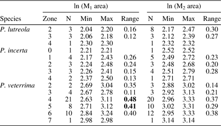
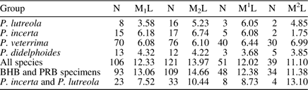
Palaeosinopa veterrima is the most abundant pantolestid in the Willwood Formation with a long stratigraphic range, and its teeth not only range widely in morphology, but also in size (Figs. 1–6). However, when viewed stratigraphically, most of the variation in size within a specific biochron is encompassed within the range of one single species, i.e., within 0.4 ln units (Fig. 7, Table 1), which includes 95% of the variation in a single species and is the standard used to recognize fossil species (e.g. Gunnell and Gingerich, Reference Gunnell and Gingerich1991; Bloch et al., Reference Bloch, Secord and Gingerich2004). This suggests that much of the variation in size occurs through time. In fact, M1 of P. veterrima appears to increase slightly in size through time (Spearman’s rho=0.32, p<0.05), but M2 does not. Despite this increase in size, the coefficient of variation (CV) for M1 length in the entire sample of P. veterrima is only 6.08 (Table 2), which is comparable to values calculated from single species (Gingerich, Reference Gingerich1974a; Gingerich and Schoeninger, Reference Gingerich and Schoeninger1979). A more striking pattern is the increase in relative width of the molars (=molar width / molar length) through the stratigraphic range of the species (Figs. 7 and 8). Although there is always a range of relative widths, the narrowest specimens of P. veterrima are the lowest and the widest occur higher in the section. Both M1 and M2 exhibit significant correlations of relative molar width with stratigraphic age (M1: Spearman’s rho=0.45, p<0.01; M2: Spearman’s rho=0.48, p<0.01).
Boxplots of molar areas (ln [length x trigonid width]) and relative widths (trigonid width / length). Center line=median, box=interquartile range, whiskers=points within 1.5× interquartile range, *=outliers. Pl=Palaeosinopa lutreola, Pi=P. incerta, Pv=P. veterrima, Pd=P. didelphoides.
Natural log of molar area (length x trigonid width*) for M1 and M2 (top). Ratio of trigonid width* / length for M1 and M2 (bottom). Meter level is plotted on the left vertical axis and age on the right. Biochrons are indicated by the gray box on the right side and dotted horizontal lines. Biohorizons A and B are plotted as gray bars. Diamonds=Palaeosinopa lutreola, triangles=P. incerta, and circles=P. veterrima. Black symbols indicate specimens that can be tied to a locality of known meter level. Gray transparent symbols indicate specimens from a locality for which the biochron is known, but a precise meter level is lacking. These specimens were plotted in the middle of their respective biochrons. White symbols indicate measurements from Schankler (unpublished data). Small gray symbols with whiskers indicate the range and approximate stratigraphic position of specimens from the Powder River Basin. Ambiguous or problematic specimens are indicated with letters or numbers: Pi: holotype of Palaeosinopa incerta; 1: USGS 3964; 2: USNM 521506; 3: USNM 538357; 4: UM 65413; 5: UM 65177; 6: UM 68718; 7: USGS 9653; 8: UM 76749; 9: USNM 533488; 10: UM 78989; 11: UM 93659; 12: USNM 493930; 13: UM 78972; 14: UM 94975; 15: UM 86788; 16: YPM 32585; 17: UM 76793; 18: UM 96132; 19: USGS 25138; 20: YPM 25648; 21: YPM 25892; 22: USNM 491896. *Area for USNM 493930 is calculated as length x talonid width.
Ratio of trigonid width / length for M1 (left) and M2 (right) for Palaeosinopa veterrima only. See Figure 7 caption for explanation of axes. Triangles represent individuals with more acute, crestiform morphology, circles represent individuals with more bunodont morphology, and squares represent individuals with intermediate morphology.
Range of ln (molar area) for species by biochronological zone
Values over 0.4 ln units highlighted in bold
Coefficients of variation for teeth of Palaeosinopa
Guthrie (Reference Guthrie1971) attributed a right M1 from the Lost Cabin Member of the Wind River Formation (biochron Wa7) to Palaeosinopa, cf. P. veterrima. Upon re-examination, we believe the tooth (CM 22042) probably represents the miacid carnivoran Vulpavus canavus and, in any case, does not belong to a pantolestid.
We assign several smaller specimens (UM 76793, 86788, 94975, 96132, USGS 25138, USNM 491896, YPM 25648, 25892, 32258) to P. veterrima that may represent the slightly smaller species P. incerta (discussed below) and recognize that this distinction is largely arbitrary. The distinction between these two species is discussed in more detail in the following sections.
Previous researchers have distinguished P. didelphoides from P. veterrima on the basis of the former having a narrower M1–2 (Matthew and Granger, Reference Matthew and Granger1918; Bown and Schankler, Reference Bown and Schankler1982). In our sample, only the M1 was significantly narrower than that of P. veterrima with an alpha-level corrected for multiple comparisons (Fig. 6). This is likely due to the small number of P. didelphoides in our sample, as the mean ratios of the M2 do appear to differ and the standard errors of the mean for P. didelphoides and P. veterrima do not overlap (Fig. 6, Table 3). The size distribution of P. didelphoides overlaps with the lower range of P. veterrima, as noted by previous authors (Matthew and Granger, Reference Matthew and Granger1918; Guthrie, Reference Guthrie1971; Bown and Schankler, Reference Bown and Schankler1982).
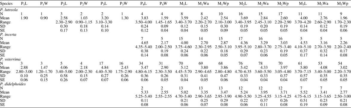
Summary of lower tooth dimensions of Palaeosinopa
SD=standard deviation; SE=standard error
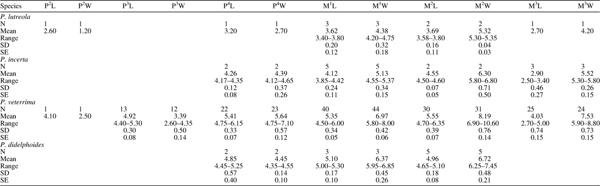
Summary of upper tooth dimensions of Palaeosinopa
SD=standard deviation; SE=standard error
We agree with Bown and Schankler (Reference Bown and Schankler1982) that Palaeosinopa didelphoides is not found in the Bighorn Basin and is restricted to the Wind River and Bridger Basins. P. veterrima can be distinguished from P. didelphoides in having a better-developed stylar shelf and generally larger conules in the upper teeth. Stratigraphically low specimens of P. veterrima are similar to P. didelphoides in their greater development of crests (Fig. 9). The variability in development of the hypocone is similar in both taxa. The lower molars of P. veterrima are generally more inflated than those of P. didelphoides, especially buccally. The most obvious difference between P. veterrima and P. didelphoides is the size and shape of the paraconid and metaconid. As noted by Guthrie (Reference Guthrie1971), the paraconid of P. didelphoides is directed anteriorly, making it appear smaller compared to the metaconid, and that of P. veterrima is more vertical. In addition, whereas P. veterrima tends to have transverse, lingually positioned paraconids, especially on M2–3, those of P. didelphoides are more cuspate and more centrally positioned throughout the molar row. Some specimens of P. didelphoides also show more developed cresting between the paraconid and metaconid on M2 (e.g., CM 22028, 22035). The metaconids of P. didelphoides are taller relative to the protoconid than in P. veterrima. The talonid of P. didelphoides is more deeply basined and tends to have a shallower talonid notch than in P. veterrima. Most specimens of P. didelphoides examined (holotype; CM 22028, 22035) have a small but distinct entoconulid anterior to the entoconid, as well as an incipient mesoconid on the cristid obliqua. Further, because we include CM 22039–22041 in P. didelphoides (rather than P. lutreola as Guthrie (Reference Guthrie1971) and Bown and Schankler (Reference Bown and Schankler1982) did), P. didelphoides is the only Palaeosinopa currently reported from the Wind River Basin.
Teeth of selected specimens of Palaeosinopa didelphoides. (1–4) lower molars in occlusal and lingual views, (5–6) occlusal view of upper molars. (1) AMNH 4804 holotype (cast) left dentary with M1–3; (2) CM 22028 left M1; (3) CM 22035 left M1; (4) left M2; (5) CM 22031 left M1; (6) right M2.
Palaeosinopa lutreola Matthew, 1918
Holotype
AMNH 15100, right dentary with M2–3.
Diagnosis, emended
Small species of Palaeosinopa, similar in size to P. aestuarium, P. russelli, and P. osborni, approximately 13%–37% smaller than P. veterrima based on ln M1 area. Generally similar to P. veterrima in morphology but differs in having upper molars with more acute cusps, smaller pre- and postcingula, less distinct hypocone, straighter buccal margin of M1 with almost no ectoflexus and slightly wider anterior stylar shelf; lower molars with less transverse paraconids.
Occurrence
Biochrons Wa2 – Wa4, Willwood Formation, Bighorn Basin, Wyoming; Early Wasatchian of the Powder River Basin, Wyoming.
Description
Palaeosinopa lutreola is significantly smaller than P. veterrima and much rarer in the Bighorn Basin. It generally occurs in the lower half of the Willwood section in the central Bighorn Basin (between about 12 m and 350 m in the FMC section). The holotype of P. lutreola (AMNH 15100) is a dentary with M2–3, in which the M2 trigonid is heavily worn and M3 is damaged such that few morphological details can be discerned (Fig. 10). The paratype (AMNH 16170) is more nearly complete, consisting of two partial dentaries with P2–M3, but P4–M3 are so heavily worn that no crown details remain. Thus, the only useful comparisons that can be made with the type specimens are largely limited to size and general proportions. Although Matthew (in Matthew and Granger Reference Matthew and Granger1918) distinguished this species from P. veterrima and P. didelphoides solely on its smaller size, these original specimens appear to have relatively shorter and wider molars than in stratigraphically low P. veterrima, with the trigonids of M2–3 slightly wider than the rather short talonids, and a relatively lingual paraconid on M3. No entoconulid is evident. In other respects, the morphology (to the extent that it can be discerned) is similar to that of P. veterrima.
Lower teeth of Palaeosinopa lutreola in occlusal and lingual views. (1) Holotype AMNH 15100 (cast), right dentary with M2–3; (2) USGS 10512, right dentary with P4–M2 (note that M2 has been glued into the M3 alveolus); (3) USNM 533549, right dentary with M1–3; (4) USGS 5971, right dentary with P1–M2; (5) USGS 9120, right dentary with M1–3; (6) USGS 5970, right dentary with P2–4, M2–3. ecld: entoconulid. Top scale bar applies to images (1–5) bottom scale bar for image (6).
A small sample of mostly very fragmentary specimens from the central Bighorn Basin can be attributed to P. lutreola based on their size (Fig. 10). Two of them (USGS 3964, USNM 521413) are virtually identical in size and proportions to the holotype. The other Willwood specimens we attribute to P. lutreola have either slightly larger molars (USGS 14729, USNM, 541892) or molars of similar length but relatively narrower, with more elongate talonids (USNM 533489). USNM 533489 is further distinguished by having small entoconulids. We consider these minor differences to reflect intraspecific variation.
Upper teeth of Palaeosinopa lutreola are preserved in USNM 511164 (left dP4–M3, right M1–2; Fig. 11). The dP4 is broken buccally, but enough remains to show that the tooth was transversely narrow and semimolariform, with an expanded posterolingual shelf bearing a tiny hypocone. The molars are similar to those of P. veterrima, but the cusps are somewhat more acute, the pre- and postcingula smaller, the hypocone less distinct, and the buccal margin of M1 straighter, with almost no ectoflexus. Despite being relatively smaller, the cingula are almost continuous lingually on M2.
Upper teeth of Palaeosinopa lutreola. (1–3) USGS 5971, skull; (2) right P2, P4–M3; (3) left P2, P4–M3; (4–5) USNM511164; (4) left dP4–M3; 5: right M1–2. Top scale applies to image (1) only.
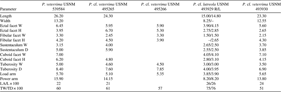
Several specimens of a small species of Palaeosinopa from the Wasatch Formation of the Powder River Basin, Wyoming (USGS 5970, 5971, 9120, 10512; USNM 493929), are here assigned to P. lutreola due to their small size and similar morphology to the Bighorn Basin P. lutreola, insofar as they can be compared (Figs. 10 and 11). These specimens are likely to be no younger than Wa3 in age, and more likely Wa2, conforming with the early Wasatchian distribution of the Bighorn Basin fossils (P. Robinson, pers. comm., 8/19/2013). They include a flattened skull with partial dentition, several dentaries, and a few postcranial bones. Tarsal elements from this Powder River Basin sample were described by Rose and Koenigswald (Reference Rose and von Koenigswald2005). This sample provides the following additions to the dental anatomy of P. lutreola.
The skull (USGS 5971) is roughly 53 mm long, about the size of that of Tupaia glis, but is severely crushed, obscuring any meaningful understanding of its anatomy. Two incisor alveoli are present on the left side, but little else can be discerned because of the damage to the front of the skull. The maxillae contain P2 and P4–M3 on both sides. The P2 is a simple, two-rooted, laterally-compressed tooth dominated by a high, acute cusp, which is joined to a small distal cusp by a sharp postprotocrista. There are no cingula. The P4 is triangular and three-rooted, with a large paracone and smaller protocone. The parastyle and metastyle are better developed than in P. veterrima, but both taxa have well developed pre- and postparacristae. In contrast to P. veterrima, P4 is slightly longer than wide, and there is a distinct ectocingulum on the metastylar lobe. M1 has a relatively wider stylar shelf than in the larger species, especially buccal to the paracone. The buccal margin of the tooth is almost straight, whereas M2 has a marked ectoflexus. The M1–2 hypocone is small but well defined and well separated from the protocone on the postcingulum, similar to the condition in most P. veterrima. It is posterolingual to the protocone. The precingulum on M1 and M3 is weaker than that of M2; the postcingulum of M1 and M2 is broad and shelf-like. Upper molar conules are well developed. Compared to USNM 511164, the posterolingual cingulum is more expanded, the hypocone more distinct, and the stylar shelf wider anteriorly, illustrating the variation in this species. In nearly all respects the upper molars of USGS 5971 are essentially the same as in P. veterrima, but smaller.
The lower premolars of the Powder River Basin specimens are on average relatively narrower than in Palaeosinopa veterrima (P1–3 are 31–50% as wide as long, P4 is 41–42% as wide as long, compared to P1–3: 39–68%, P4: 47–62% in P. veterrima). The P1 (USGS 5971) is single rooted with a tall main cusp, a small but distinct talonid cusp, and a crest joining them. The P2 (USGS 5970, 5971) is two rooted and is also dominated by a tall primary cusp (protoconid?). It exhibits a small or weakly developed anterior cusp (paraconid?) and a well developed talonid cusp (hypoconid?). There is a distinct crest running down the back of the main cusp to the tip of the hypoconid, with a shallow fossa lingual to it. The P3 (USGS 5970) preserves a better developed paraconid and a more complex distal morphology than the P2, with an incipient metaconid (?) situated half way down the crest running posteriorly from the protoconid and slightly buccal to the midline, and a distinct hypoconid joined to the back of the trigonid by a short cristid obliqua. Several Powder River Basin specimens preserve a P4 with a well developed paraconid and hypoconid, a short cristid obliqua, and a small, shallow talonid basin. A variable metaconid is present on the crest on the posterior slope of P4; it is weakly developed in USGS 10512, slightly stronger in USGS 5970, and relatively prominent on USGS 5971. Short anterolingual cingulids are present on P2–4, and posterolingual cingulids (alternatively interpreted as lingual talonid crests) are present on all four premolars. Buccal cingulids are absent.
The lower molars resemble those of P. veterrima except for being smaller and having a slightly less transverse paraconid than in the larger species. The small sample shows considerable variation in molar breadth, USGS 5971 having noticeably narrower molars than the others. Tiny entoconulids are present on M1–2 of the two better preserved specimens (USGS 5971, 9120). USGS 9120 is particularly similar to USNM 533489 from the Bighorn Basin.
Material
AMNH 15102, 16170 (paratype), 56223, 56309; UM 63295, 83068, 86747, 96061, 96594; USGS 3964, 5970, 5971, 9120; USNM 511164, 521413, 533489, 541892; YPM 23082, 25843, 26175, 26604.
Remarks
Delson (Reference Delson1971) previously reported two upper molars (AMNH 56223, 56309) attributed to Palaeosinopa, cf. P. lutreola, from the Wasatch Formation of the Powder River Basin. They are comparable in size and morphology to the specimens we describe above. There are no discernable morphological or size differences between the small Palaeosinopa specimens from the Bighorn and Powder River Basins, nor do there appear to be any stratigraphic changes in morphology or size in this lineage. Given the overall similarity of the Powder River and Bighorn Basin specimens, the close proximity, and temporal overlap of the fossil assemblages, we consider it reasonable to refer these specimens to the same species.
Guthrie (Reference Guthrie1971) assigned three teeth (CM 22039–22041) from the Lostcabinian (Wa7) of the Wind River Basin to Palaeosinopa lutreola, which would be a significant range extension if confirmed. These teeth, however, appear to represent P. didelphoides, as discussed below. CM 22042 was also listed under P. lutreola (Guthrie, Reference Guthrie1971: 60) but this was a lapsus calami, as Guthrie subsequently attributed the same specimen to Palaeosinopa, cf. P. veterrima.
Palaeosinopa lutreola intergrades in size with the slightly larger P. incerta, and thus the distinction between these species is largely arbitrary (discussed below). One specimen (USGS 3964) has a relatively large M1, close to the range seen in P. incerta (Fig. 7, no. 1) but the M2 fits better within the size range of P. lutreola. We assign this specimen to P. lutreola but acknowledge that it could represent a small individual of P. incerta.
Two small species of Palaeosinopa that occur elsewhere merit discussion and comparison with P. lutreola: P. russelli and P. aestuarium. Palaeosinopa russelli Smith, Reference Smith1997, occurs in basal Eocene strata of Dormaal, Belgium, and is comparable in size to P. lutreola. P. russelli is said to differ from P. lutreola principally in having narrower molars. Based upon the measurements given by Smith (Reference Smith1997), the relative widths of M1 (62–63% as wide as long) and M2 (67%–72%) overlap with the low end of the range of P. lutreola only slightly (M1=63%–73%; M2=69%–88%), indicating that they are proportionately narrower than those of most P. lutreola, but are not extremely so. The ln M1 area of P. russelli (1.912–1.951) suggests an animal just outside the lower tail of the size range of P. lutreola (2.012–2.302), whereas the ln area of M2 (2.128–2.291) is within the range of P. lutreola (2.120–2.505). Like P. lutreola, P. russelli has small entoconulids. The two species seem likely to be closely related.
Palaeosinopa aestuarium Beard and Dawson, Reference Beard and Dawson2009, from the early Wasatchian Red Hot Local Fauna of Mississippi is comparable in size to both P. russelli and P. lutreola, and differs from both species in having a lower-crowned M2 with more acute protoconid and hypoconid. The authors report that it differs from P. lutreola and resembles P. russelli in having an entoconulid and a large, posteriorly projecting hypoconulid. As noted above, the extreme state of wear exhibited by the holotype and paratype of P. lutreola, and indeed by a large proportion of pantolestid specimens, makes it difficult to compare features such as crown height. However, the presence of an entoconulid in P. aestuarium does not necessarily ally this species particularly with P. russelli, as entoconulids are variably present in P. veterrima, P. lutreola, and P. incerta (e.g., P. lutreola: USGS 5971, 9120, USNM 533489; P. incerta: UM 69722; P. veterrima: AMNH 2851, 94456; UM 86985, 91614, 94621, 73269). When unworn specimens are available, a projecting hypoconulid is seen to be present in most species of Palaeosinopa (e.g., P. lutreola: USGS 5971, 9120, 10512; P. incerta: UM 69722; USNM 538357; P. veterrima: UM 91648, USGS 302, 9860, 13719, USNM 491897, 533488, 537835, 539584). The relative width of the M2 of P. aestuarium (69% as wide as long) overlaps slightly with the range seen in P. lutreola and is comparable to P. russelli. Beard and Dawson (Reference Beard and Dawson2009) distinguished P. aestuarium from P. didelphoides based upon the weak expression of the preparacrista in the P4 of P. aestuarium. This crest is also weakly developed in P. lutreola and P. incerta and is variably expressed in P. veterrima. The straight buccal margin of the M1 in P. aestuarium is also approximated by some specimens of P. lutreola and P. veterrima. In summary, P. aestuarium is very similar in size and morphology to P. lutreola and P. russelli and the three species are probably very closely related.
Palaeosinopa incerta Bown and Schankler, Reference Bown and Schankler1982
Holotype
UM 69722, partial skull and dentaries with right P2–M3 and right P4–M3.
Diagnosis, emended
Intermediate in size between P. veterrima and P. lutreola, and overlapping in linear dimensions of M1–2 with both taxa. Similar to P. veterrima in morphology, but approximately 5%–33% smaller based on ln M1 area, and M1 relatively narrower on average. Up to 24% larger than P. lutreola, but overlaps in ln M1 area (Fig. 6).
Occurrence
Biochrons Wa0–Wa4, Willwood Formation, Bighorn Basin, Wyoming.
Description
The P4 of the holotype preserves a recurved protoconid that dominates the crown; a small, low paraconid; and a distinct metaconid just posterior and very slightly lingual to the apex of the protoconid (Fig. 12). The talonid is prominent and bears a centrally located cristid obliqua descending from a weakly developed cusp, and a very shallow basin lingual to the cristid obliqua. The molar teeth of P. incerta are morphologically very similar to stratigraphically low specimens of P. veterrima in being relatively narrow with high, cusps and well developed crests.
Teeth of selected specimens of Palaeosinopa incerta. (1–2) Holotype, UM 69722; (1) rostrum and mandible; (2) right dentary with P4–M1–3, in occlusal and lingual views; (3) UM 80300, left maxilla with P4–M2, right maxilla with P4–M3 in occlusal view; (4) UM 93659, left dentary with P4–M1 and right dentary with M3 in occlusal and lingual views; (5) USNM 541901, right dentary with P4, M2 in occlusal view; (6) UM 87070, right dentary with P4–M3, in occlusal and lingual view; (7) USGS 6981, right dentary with M2–3 in occlusal and lingual views; (8) USNM 510993, right dentary with M1–3 in occlusal and lingual views; (9) USNM 533488, left dentary with M2 in occlusal and lingual views. ecd: entoconulid; mcd: metaconid.
USNM 541901, attributed to P. incerta, has a relatively shorter, wider M1 than the holotype, but has an associated P4 that is reminiscent of that of the holotype with a somewhat inflated trigonid bearing a tiny paraconid anteriorly and a larger metaconid posteromedial to the tall protoconid. The talonid has a low, arcuate cristid obliqua that ascends to the posterolateral margin, though there is no distinct hypoconid, and continues lingually to meet a distinct, tall entoconid. The entocristid descends sharply to a deep talonid notch. This P4 is more robust, and has a more medially shifted metaconid than in the holotype, and differs in having an entoconid but no hypoconid.
Material
AMNH 56226; UM 64875, 65177, 65413, 68718, 69722, 76151, 76749, 77069, 77226, 78972, 78989, 80037, 80388, 83485, 85316, 85881, 87070, 87084, 93659, 99237; USGS 1807, 6981, 9653, 17642; USNM 493930, 510993, 521506, 533488, 538357, 541901; YPM 30613, 37246.
Remarks
Bown and Schankler (Reference Bown and Schankler1982) diagnosed P. incerta as being larger than P. lutreola and smaller than P. veterrima or P. didelphoides and further distinguished it from P. veterrima and P. didelphoides by its relatively narrower M1. The name reflected that this species incorporated several specimens that previous researchers had noted as being taxonomically ambiguous. Other than the narrower M1, no morphological characters were given to distinguish it from other taxa.
The lower molars of the holotype (UM 69722) are morphologically similar to the more sectorial specimens of P. veterrima, with a larger, more crestiform paraconid than P. didelphoides. The M1 of the holotype is unusually small compared to the size of the M2 and P4, being close to the mean area of M1 in P. lutreola. This is not evident in the plots of Bown and Schankler (Bown and Schankler, Reference Bown and Schankler1982) because the specimen indicated in their plots as the “type of Palaeosinopa incerta” is actually AMNH 39565, which was originally designated to be the holotype before the discovery of UM 69722 (D. Schankler, pers. comm. 7/30/2013). No other specimen in the sample has an M1 of the same size relative to M2, so we assume that this specimen is aberrant in this feature.
Based on our sample, the M1s from the original hypodigm of P. incerta are on average smaller than those of P. veterrima and larger than those of P. lutreola; however, the size range of P. veterrima in our sample overlaps substantially with that of the original P. incerta hypodigm. We find that the M1 of P. incerta is relatively narrower than that of P. veterrima in our sample, in agreement with the results of Bown and Schankler (Reference Bown and Schankler1982). However, contrary to their findings, our sample indicates that M1 of P. incerta is comparable in width to that of P. didelphoides, not narrower. Further, P. veterrima has on average a relatively wider M1 than all other species of Palaeosinopa, suggesting that P. veterrima is the unique species (Fig. 6).
When considered as a part of our expanded sample, nine of the 18 specimens comprising the original hypodigm of P. incerta appear to be more appropriately referred to P. veterrima (AMNH 15701, 16823, 56226, 56227, 94457; YPM 25506, 25648, 25892, 32258), and three to P. incerta (UM 69722 [the holotype], YPM 37246, 30613). The three specimens from the Wind River Basin (CM 22039, 22040, 22041) assigned by Bown and Schankler (Reference Bown and Schankler1982) to P. incerta we consider to be small individuals of P. didelphoides (see discussion below). The remaining three specimens (AMNH 16239, 39565, UCMP 44047) are more problematic and are discussed in more depth below.
Bown and Schankler (Reference Bown and Schankler1982) attributed several late-occurring specimens from various North American localities to P. incerta in addition to those from the Bighorn Basin, which occur exclusively below Biohorizon B (at the Wa4–5 boundary). Three of these specimens (CM 22039, 22040, 22041) are from the late Wasatchian (Wa7) of the Wind River Basin and were attributed by Guthrie (Reference Guthrie1971) to Palaeosinopa, cf. P. lutreola, due to their small size. These specimens are similar to P. didelphoides in having a small, low paraconid and a tall metaconid and, although they are slightly smaller than the other specimens of P. didelphoides from the Wind River Basin, they are not so small that they could not represent small individuals of the same species. The entire range of M1 area for the Wind River sample (including P. didelphoides and the specimens Guthrie assigned to P. lutreola and excluding CM 22042) is 0.28 ln units (Fig. 6) and the CV of M1 length is 4.32 (Table 2); both values are well within the range of a single species. Therefore, we consider CM 22039–22041 to be small individuals of P. didelphoides.
Bown and Schankler (Reference Bown and Schankler1982) allocated three other specimens (AMNH 16239 and 39565, UCMP 44047) to P. incerta, none of which come from the Bighorn Basin. AMNH 16239 is from the San Juan Basin, New Mexico, whereas the only locality data for AMNH 39565 is “near Dubois, WY”. AMNH 39565 is morphologically more similar to P. didelphoides than to P. veterrima or P. incerta and is identified as P. didelphoides in the AMNH catalog. In addition, the outcrops surrounding Dubois are likely to be late Wasatchian in age based on occurrence there of the perissodactyl Lambdotherium and the omomyid primate Absarokius abbotti (G. Winterfeld, pers. comm., 8/15/2013). Given the early Wasatchian distribution of all other P. incerta specimens, the likely late Wasatchian age of AMNH 39565, and the similarity between this specimen and P. didelphoides, we think it more likely pertains to the latter species.
AMNH 16239 is morphologically similar to P. veterrima and P. incerta in having a robust, transverse paraconid, but is smaller than typical P. veterrima specimens and is within the size range of P. incerta. This specimen was collected from the Almagre Beds of the San Jose Formation in the San Juan Basin, New Mexico, which are late Wasatchian in age (biochron Wa6; Robinson et al., Reference Robinson, Gunnell, Walsh, Clyde, Storer, Stucky, Froehlich, Ferrusquia-Villafranca and McKenna2004; Janis et al., Reference Janis, Gunnell and Uhen2008). It is the only pantolestid specimen reported from the San Jose Formation, and it may represent a distinct species known only from this formation, a late-surviving population of P. incerta, or small P. veterrima. These ambiguities can only be resolved in the context of larger samples from the San Jose Formation. Regardless, because this specimen is not from the Bighorn Basin, it is not clear how it fits into the context of our sample, and is not considered further.
UCMP 44047, from the early Wasatchian Four Mile Local Fauna in northwest Colorado, is a right dentary with M1 and is intermediate in size between P. incerta and P. veterrima, and overlaps in size with P. didelphoides in our sample (McKenna, Reference McKenna1960). UCMP 44279, an isolated M1, is not listed in the hypodigm of P. incerta, but was listed as part of UCMP 44047 by Schankler (unpubl. notes). This M1 falls within the size range of P. incerta or P. didelphoides. It is equally possible that these teeth represent P. incerta or P. didelphoides based on size, and more likely that they represent P. incerta based on stratigraphy. We have not included these specimens in our sample because it is unclear how they relate to the evolution of Palaeosinopa from the Bighorn Basin.
Several specimens that we attribute to P. incerta are ambiguous due to overlap in size with individuals of different species. These ambiguous specimens are denoted by numbers in Figures 7, 8, and 13. UM 93659 (P4–M1, M3) is cataloged as P. lutreola and either represents a small individual of P. incerta or a large individual of P. lutreola. The P4 and M1 are virtually identical in size and morphology to the holotype of P. incerta except that the paraconid of P4 is slightly smaller. The M3 is almost identical in size as far as can be compared to the holotype of P. lutreola. USNM 521506 (M2) and 538357 (M1) from Wa0 of Sand Creek Divide were assigned to P. lutreola by Rose et al. (Reference Rose, Chew, Dunn, Kraus, Fricke and Zack2012). However, these specimens are larger than most of the P. lutreola specimens in our sample and fit better within the size range of P. incerta. Until we have a better sample from Wa0, we consider both of these specimens to be P. incerta. In addition to these specimens, several others (UM 65177, 65413, 68718, 76749, 78972, 78989, USGS 9653) are assigned to P. incerta but may represent large specimens of P. lutreola.
Scatterplots of ln M1 and M2 area*. Diamonds=Palaeosinopa lutreola, triangles=P. incerta, circles=P. veterrima. Gray symbols indicate specimens from the Powder River Basin, white symbols indicate measurements from Schankler (unpublished data). Gray shaded regions indicate convex hulls for original P. incerta hypodigm from Bown and Schankler (Reference Bown and Schankler1982). See Figure 7 caption for explanation of annotations. *Area for USNM 493930 is calculated as length x talonid width.
USNM 533488 (L dentary with M2) is a juvenile specimen with partial P4 in crypt and an extremely shallow jaw. This specimen is located stratigraphically relatively low (140 m; below Biohorizon A) and like other older specimens has smaller and relatively narrow molars. The dentary of USNM 533488 is much shallower than those of P. veterrima in our sample, more similar to the holotype of P. incerta and is also very close to that species in the size of the M2. However, the dentary of Palaeosinopa increases in depth ontogenetically (Rose et al., Reference Rose, Dunn and Grande2014), so the jaw would have been much larger and deeper when fully adult. We assign this specimen to P. incerta, although it is possible that it represents a small individual of P. veterrima. USNM 493930 is intermediate in size between the holotype of P. incerta and typical specimens of P. veterrima and could reasonably be assigned to either species. Here we assign it to P. incerta based on differences in postcranial size and robusticity compared to P. veterrima, but recognize that these differences may simply be due to differences in stratigraphic position. This specimen is among the largest we attribute to P. incerta (Fig. 7, no. 12).
Additions to the postcranial anatomy of Palaeosinopa
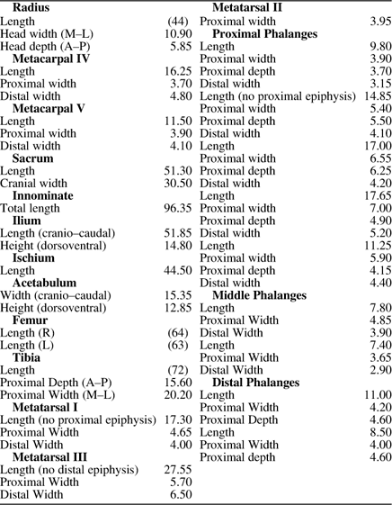
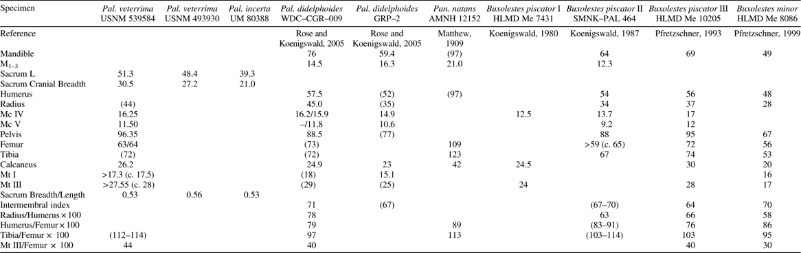
A partial skeleton associated with dentaries (USNM 539584), here referred to P. veterrima, is the most complete pantolestid skeleton known from the Willwood Formation (Figs. 2, 14, 15, Table 5). Several other specimens that include fragmentary postcranial remains or consist of isolated postcranial elements contribute to our knowledge of the anatomy of Palaeosinopa. Most epiphyses of the long bones and vertebrae in USNM 539584 remain unfused to the diaphyses, although the dentition is fully erupted and the M1 shows substantial wear. Delayed epiphyseal fusion has been described in other pantolestids and may be related to a semi-aquatic lifestyle (Kükenthal, Reference Kükenthal1891; Rose and Koenigswald, Reference Rose and von Koenigswald2005). The postcrania of several pantolestid skeletons have been described elsewhere (Matthew, Reference Matthew1909; Koenigswald, Reference Koenigswald, von1980; 1987a; Pfretzschner, Reference Pfretzschner1993; Reference Pfretzschner1999; Rose and Koenigswald, Reference Rose and von Koenigswald2005; Rose et al., Reference Rose, Dunn and Grande2014). Here, we supplement previous descriptions with those of new Willwood specimens, primarily focusing on previously unknown aspects of morphology, and noting morphological variation in Palaeosinopa.
Vertebral column (1–9), and upper limb elements (10–13) of Palaeosinopa. (1–7) USNM 539584, P. veterrima. (1) T4 lateral (top), ventral (bottom); (2) T6 anterior; (3) T11 dorsal; (4) L4 ventral (left), posterior (right); (5) proximal caudal vertebra anterior (left), ventral (right); (6) middle caudal vertebra anterior (left), dorsal (right); (7) sacrum in proximal (top), ventral (left), lateral (right); (8) USNM 493930, P. incerta, sacrum in ventral (left), lateral (right); (9) UM 80388, P. incerta, sacrum in ventral (left), lateral (right); (10) USNM 527721, P. veterrima, left proximal humerus posterior (left), medial (center), proximal (right); (11–13) USNM 539584, P. veterrima; (11) left humerus medial (left), lateral (center), anterior (right); (12) right radius, proximal (top), anterior (left), posterior (middle), lateral (right); (13) left ulna, medial (left), anterior (right). bt: bicipital tuberosity; dc: deltoid crest; dt: deltoid tubercle; gt: greater tubercle; is: infraspinatus; lt: lesser tubercle; op: olectanon process; pc: pectoral crest; sc: supinator crest; ss: supraspinatus; tf: trochlear facet; uf: ulnar facet. Scale bars in boxes=5 mm.
Hindlimb elements of Palaeosinopa. (1) USNM 539584, P. veterrima, right innominate dorsal (top) lateral (bottom); (2) UM 80388, P. incerta, right innominate lateral view (top) L innominate lateral view (bottom); (3) USNM 539584, P. veterrima, right femur anterior (left), medial (center), posterior (right); (4) USNM 539584, P. veterrima, tibia, left proximal epiphysis (left, top), left distal epiphysis (left, bottom), right tibial shaft with distal fibula (right); (5) USGS 25172, c.f. P. veterrima, right distal tibiofibula posterior (top), anterior (bottom); (6) UM 80388, P. incerta, left femur, anterior (left), posterior (right); (7) UM 80388, P. incerta, left distal epiphysis; (8) USNM 527721, c.f., P. veterrima, right distal femur; (9) USNM 539584, P. veterrima, left calcaneus, distal (left), dorsal (center), medial (right). af: astragalar facet; afo: astragalar foramen; al, attachment for the adductor longus muscle; at: adductor tubercle; ca: calcaneal facet; cu: cuboid facet fib: fibula; ff: fibular facet; gf: fossa for attachment of gastrocnemius muscle; gm: attachment for the gracilis muscle; gs: gluteal surface; gtr: greater trochanter; il: ilium; is: ischium; it: ischial tuberosity; lc: lateral condyle; lm: lateral malleolus; lr: crests for lateral rotators of the hip; ltr: lesser trochanter; mc: medial condyle; mm: medial malleolus; nf: navicular facet; pm: attachment site of pectineus muscle; pp: posterior process; pt: peroneal tubercle; rf: attachment site for rectus femoris muscle; sf: sustentacular facet; tt: tibial tuberosity; ttr: third trochanter.
Postcranial measurements of USNM 539584 (mm)
Measurements in parentheses are approximate.
Axial skeleton
Fragments of 29 vertebrae and the sacrum are preserved in USNM 539584, which is missing only one cervical and four thoracic vertebrae of the presacral series (Fig. 14). Most of the vertebrae are covered with matrix, obscuring details and making identification of exact position difficult. There are six cervical vertebrae including the axis, in which the odontoid process is broken. The cervicals are distorted, encased in matrix, and preserve only the bases of the processes. Nevertheless, the ventrally projecting transverse processes, expanded at the caudal end, indicate that these are cervical vertebrae. The transverse foramina typical of C1–6 are obscured by matrix in all but one cervical vertebra.
Nine vertebrae most likely belong to the thoracic series, as they have small centra, and several preserve transversely oriented pre- and post zygapophyses (designating them as pre-diaphragmatic vertebrae), and demi-facets for ribs. The diaphragmatic vertebra (T11) can be identified by its transverse, dorsally facing prezygapophyses and sagittally oriented, strongly convex, ventrolaterally facing postzygapophyses (Davis, Reference Davis1964). Many of the prediaphragmatic thoracic vertebrae preserve a strong ventral keel, whereas T11 and T13 (the last thoracic) do not. In those prediaphragmatic vertebrae that retain a partial spinous process, it is craniocaudally short and strongly inclined posteriorly. T1 can be identified by the widely spaced, flat, postzygapophyses. Broken posterior bases of anapophyses are evident inferolateral to them.
Six vertebrae are identified as part of the lumbar column due to their short, wide centra, strongly concave, dorso-medially directed prezygapophyses with strong metapophyses (mammillary processes), strongly convex, sagittally oriented postzygapophyses, cranio-ventrally oriented transverse processes, and lack of rib facets. Post-zygapophyses become farther apart and the transverse processes become more cranio-caudally elongate progressively from the cranial to caudal lumbar vertebrae. The anterior centrum of L1 is inclined ventrally, whereas the posterior centrum is more vertical, and only the anterior epiphysis is present. A small transverse process is present on the anterior centrum. Ventrally, a broad, rounded ventral keel is present with shallow depressions on either side. Although broken on L1, L2 preserves one cranio-caudally short anapophysis on the left side of the body, suggesting the presence of well developed anapophyses in the lumbar vertebrae.
The sacrum consists of three fused vertebral elements (Fig. 14). The auricular surface for the innominate extends cranio-caudally across the transverse process of the first sacral element and onto the proximal portion of the second. The pre- and postzygapophyses of all sacral vertebrae are completely fused in all specimens. The anatomy is very similar to the sacrum of Palaeanodon, but the latter differs in having four sacrals. Two additional Willwood specimens, attributed to P. incerta (UM 80388 and USNM 493930), also include sacra, which are essentially the same morphologically and differ only in size and robusticity. USNM 539584 is considerably more robust than the other two, although it is only slightly longer than USNM 493930. UM 80388 is significantly shorter than the other two.
Eight caudal vertebrae have been identified in USNM 539584. All are robust with centra similar in size to those of the larger lumbar vertebrae. In lateral profile the centra are strongly concave ventrally, with a variably developed median keel. The ends of the centra in several of the vertebrae are rough, suggesting unfused annular epiphyses, as seen in the subadult skeletons from the Fossil Butte Member of the Green River Formation (Rose and Koenigswald, Reference Rose and von Koenigswald2005). One caudal preserves a complete transverse process that is cranio-caudally almost as long, and approximately as wide as the centrum; two more preserve the proximal portion of a similar process and prezygapophyses with prominent metapophyses, and three more also appear to have had cranio-caudally extensive transverse processes. Based upon these features, these vertebrae can most likely be allocated to the proximal caudal region (Ca 1–8; Rose and Koenigswald, Reference Rose and von Koenigswald2005). A single caudal vertebra displays a partial “double” transverse process, with laterally expanded cranial and caudal regions, and is similar to Ca 9–12 in other skeletons of Palaeosinopa (Rose and Koenigswald, Reference Rose and von Koenigswald2005).
Several rib fragments are associated with USNM 539584. They are wider on the outer curvature (craniolaterally) than on the inner curvature, and a distinct crest extends along the cranial aspect of several of them beginning at the tubercle. The caudal surface is marked by a longitudinal groove, more marked at the vertebral end. Similar features are present in middle ribs of Castor.
Humerus
Portions of the right and left humeri are present in USNM 539584 (Fig. 14.11). The proximal epiphyses of both are unfused, but a detached partial proximal epiphysis is present for the right humerus. Both humeri in USNM 539584 are broken just distal to midshaft. The poorly preserved distal end of the left humerus shows that the distal epiphysis was closed, and displays a prominent medial entepicondyle. USNM 542054 is an isolated right humeral shaft preserving the distal deltopectoral crest, proximal supinator crest and proximal portion of the entepicondylar foramen; it agrees well with USNM 539584 in size. The deltopectoral crest in USNM 542054 is more shelf-like than in other Palaeosinopa humeri compared, with the pectoral crest projecting more medially than anteriorly—resemblances to Palaeanodon; but it compares more closely to Palaeosinopa in the less abrupt distal end of the deltopectoral crest and in the smaller (though still well developed) supinator crest. USNM 527721 includes proximal ends of both humeri, the more complete left one preserving the articular surface and greater and lesser tubercles (Fig. 14.10). Epiphyseal lines are evident in both, indicating recent attachment to the diaphysis. This specimen is slightly larger than USNM 539584, but probably within the size range of P. veterrima. The greater tubercle is significantly larger than the lesser tubercle and is raised proximally above the head. Pits for the supra- and infrasipinatus muscles are well defined. The lesser tubercle is a low protuberance, located well distal to the articular surface of head. The articular surface of the head is broad and asymmetrical (slightly ellipsoid, with an oblique long axis), and slightly more elevated medially, similar to the condition in Pantolestes. The head is less elongate than in Palaeanodon but otherwise quite similar in configuration of the tubercles.
The humerus of Willwood pantolestids is robust with flaring muscle attachments on the shaft, similar to the condition in previously described pantolestids from North America and Europe (Matthew, Reference Matthew1909; Koenigswald, Reference Koenigswald, von1980; Reference Koenigswald, von1987; Pfretzschner, Reference Pfretzschner1993; Reference Pfretzschner1999; Rose and Koenigswald, Reference Rose and von Koenigswald2005). The deltopectoral crest inclines medially on the anterior portion of the shaft. It begins at the distal end of the epiphyseal surface and terminates in a rough tubercle located close to the midshaft of the humerus. The medial surface of the pectoral crest is concave. The deltoid crest is continuous with the greater tubercle as in Pantolestes. It extends distally about two-thirds the length of the pectoral crest and projects straight laterally. The supinator crest originates on the dorsal surface of the humerus, opposite and just proximal to the termination of the pectoral crest. The supinator crest is not completely preserved on any single Willwood specimen, but it was large and laterally flaring, as in P. didelphoides from Fossil Butte (Rose and Koenigswald, Reference Rose and von Koenigswald2005).
Radius and ulna
The only pantolestid radius known from the Willwood Formation is the right radius of USNM 539584, which is complete except for the missing distal epiphysis (Fig. 14.12). It is bowed anteriorly and increases in anteroposterior depth distally. Proximally, the articular surface for the humerus is fully co-ossified with the shaft, with no evidence of an epiphyseal line. It is transversely almost twice as wide as it is anteroposteriorly deep and is irregularly rectangular in outline. The articular surface for the humeral capitulum is gently concave and that for the trochlea is offset and inclines medially. There is a large capitular eminence on the anteromedial edge of the radial head. The articular facet for the ulna is located posteriorly and is elongate and nearly flat. The bicipital tuberosity is a moderate eminence situated on the posterior surface of the proximal shaft just distal to the ulnar articular facet. The interosseous crest is a rounded ridge on the lateral surface of the radial shaft extending from just proximal to midshaft to about 3/4 of the way down the shaft. There is a low tubercle on the distal portion of the anterior border of the radial shaft probably delineating the insertion of the pronator teres medially and the supinator laterally. The distal extent of this tuberosity cannot be determined due to damage.
Only the olecranon process and a portion of ulnar shaft are preserved in USNM 539584, and no other ulnae are known from Willwood Palaeosinopa (Fig. 14.13). The proximal epiphysis was unfused and is missing. The olecranon process is robust and medially inflected, with a deep fossa on the medial surface, as is the condition in previously described pantolestid skeletons (Matthew, Reference Matthew1909; Rose and Koenigswald, Reference Rose and von Koenigswald2005). The ulnar shaft was distinctly wider craniocaudally than mediolaterally.
Metacarpals
USNM 539584 includes two complete metacarpals: a left Mc IV and a right Mc V. Both metacarpals are stout; and distal epiphyseal lines are visible, signifying that they fused recently. Mc IV is narrower at the proximal end and widens distally so that the head is wider than the base. The base is slightly offset from the long axis of the shaft, resulting in the articular surface for the unciform facing at an oblique angle to the primary digital axis. The facet for the unciform is wider dorsally than ventrally and dorsoventrally than mediolaterally. It is convex dorsoventrally and almost flat mediolaterally. The facet for the fifth metacarpal is crescentic and weakly spiraled, flat to slightly concave, and tapering ventrally. Medially, there are two facets for the third metacarpal. The dorsal facet is continuous with the unciform facet and is gently convex, whereas the ventral one is flat and separated from the facet for the unciform by a sharp ridge. Both facets are separated by a wide nonarticular area. Distally, there is a distinct ventral keel on the head of the metacarpal, but it does not project strongly ventrally and does not extend onto the distal surface of the head. There is a shallow extensor fossa on the dorsal aspect of the metacarpal head.
Metacarpal V is shorter than Mc IV and slightly wider at the base. The facet for the unciform is strongly convex dorsoventrally and relatively uniform in width. A lateral crest extends from the proximal end of the shaft to midshaft, perhaps for insertion of the extensor carpi ulnaris (Davis, Reference Davis1964). The ventral keel of the head is smaller than on Mc IV and there is no extensor sulcus present.
Innominate
The right innominate of USNM 539584 is complete except for the pubic symphysis, a portion of the ischio-pubic ramus, and possibly lacking epiphyses on the cranial ilium and ischial tuberosity (Fig. 15.1). It is the most complete innominate of Palaeosinopa for which both the medial and lateral surfaces are visible (other specimens such as those described by Rose and Koenigswald (Reference Rose and von Koenigswald2005), Koenigswald (Reference Koenigswald, von1980; Reference Koenigswald, von1987), and Pfretzschner (Reference Pfretzschner1993; Reference Pfretzschner1999) are partially obscured by matrix). The left innominate is also present but less complete. UM 80388 (P. incerta) includes partial left and right innominates consisting of the acetabulum and portions of the ischium and ilium (Fig. 15.2), and USNM 493930 (small P. veterrima) includes a crushed left acetabulum. Except for minor details noted below, these more fragmentary specimens are essentially the same as USNM 539584, and the following description is based mainly on the latter. In lateral view, the long axes of the ilium and ischium are aligned, resulting in a straight dorsal border of the innominate. The ilium is slightly more than half the length of the innominate (54% measured from the ilium-ischium suture to the cranial tip of the iliac blade). In dorsal view, the iliac blades flare laterally at the cranial end, but not quite as much as in Palaeanodon. The auricular surface for articulation with the sacrum is extensive, covering approximately two-thirds of the medial surface of the iliac blade. The lateral surface of the ilium is divided into two distinct surfaces by a raised midline ridge, the (wider) gluteal surface dorsally and the iliac surface ventrally. The ridge dividing the two planes is stronger in USNM 539584 than in the smaller UM 80388. Where the ridge meets the anterior acetabular rim a slight eminence is present that together with a shallow triangular fossa anterior to it marks attachment of the rectus femoris muscle.
The acetabulum of both specimens is oval, slightly wider cranio-caudally than dorsoventrally deep, and faces dorso-laterally. The articular surface of the acetabulum is widest cranially and ventrally and narrow dorsally and caudally. There are three distinct tubercles or projections on the outer margin of the acetabulum: one cranially for the attachment of the rectus femoris (noted above), one caudally, and one ventrally where the semilunar surface terminates.
The ischium is just under half the length of the innominate in USNM 539584. It lacks a discrete ischial spine, but the dorsal border of the cranial half of the ischium is sharp and forms a blade-like crest in both USNM 539584 and UM 80388. In USNM 539584 there is a second, shorter crest caudal and lateral to the first one, extending cranially from the ischial tuberosity. These crests may mark the attachment sites for the gemelli and quadratus femoris muscles (Davis, Reference Davis1964; Argot, Reference Argot2002), which extend and laterally rotate the hip. The ischial tuberosity, for attachment of the hamstrings, is rough and rounded and projects slightly dorso-caudally.
The pubis is the least robust component of the innominate and is nearly uniform in width throughout its length. At the ilio-pubic juncture, there is a low, rough tubercle that probably marked the attachment for the pectineus (Davis, Reference Davis1964), although Argot (Reference Argot2002) describes the psoas minor inserting at this location in marsupials. The lateral surface of the pubis preserves a low ridge extending obliquely from the cranial margin of the obturator foramen to the ventral border of the pubic ramus approximately at its midpoint. This may divide the attachment surfaces for the gracilis (cranially and ventrally) and the adductor longus muscles (dorsally and caudally).
Femur
USNM 539584 includes right and left femora that are complete except for the proximal and distal epiphyses (Fig. 15.3). USNM 527721 (P. cf. veterrima) includes a distal right femur (Fig. 15.8), USNM 493930 (P. veterrima) includes a right proximal and distorted distal femur, and UM 80388 (P. incerta) includes partial right and left femora (Fig. 15.6, 15.7). The epiphyses remain unfused in USNM 539584 and UM80388 (distal only). In USNM 493930 and USNM 527721 the epiphyses are fused, but epiphyseal lines remain evident. The shafts of both femora of USNM 539584 are craniocaudally compressed and transversely wide throughout their length, whereas the femoral shafts of the two smaller specimens (USNM 493930 and UM 80388) are rounder in cross section and appear to be more gracile. Although the flatter condition of the larger femora may be due in part to postmortem distortion, we believe the preserved shape to a large extent reflects their original anatomy, as it is similar to that seen in other pantolestid skeletons (Matthew, Reference Matthew1909; Rose and Koenigswald, Reference Rose and von Koenigswald2005). The femoral neck (observable in USNM 539580 and USNM 493930) is short and oriented cranially, with an obtuse angle from the long axis of the femoral shaft. The articular surface of the head, preserved and visible only in USNM 493930, extends onto the posterior aspect of the neck. A deep trochanteric fossa is present on the posterior surface of the greater trochanter of USNM 493930. The height of the greater trochanter relative to the head cannot be accurately determined in USNM 539584 due to trochanteric damage and the absence of the epiphyses, but in USNM 493930 the greater trochanter extends proximal to the level of the femoral head. In USNM 539584 the third trochanter is well developed and part of a laterally projecting crest that extends from the greater trochanter to just proximal to midshaft. In the smaller specimens the third trochanter is smaller and less projecting, and relatively slightly longer with a more gradual profile and appears to have been situated slightly more proximally, although the exact position cannot be determined in the incomplete elements. The third trochanter in USNM 539584 is a wide flange that grades gradually into the distal shaft and the greater trochanter rather than a distinct tubercle creating a broad, plate-like surface for attachment of the gluteal muscles on the posterior aspect of the femur similar in morphology to Castor. The lesser trochanter (preserved only in USNM 539584) lies just distal to the femoral head and projects posteromedially.
At the distal end, the breadth across the epicondyles in USNM 527721 is slightly greater than that in USNM 539584 (measured in the absence of the epiphysis), which suggests that the two animals were similar in size. The medial and lateral patellar rims are similar in height and the trochlea well-grooved, as is the case in USNM 493930 and UM 80388. As in other pantolestids, the medial condyle of all Willwood specimens extends conspicuously farther posteriorly than the lateral one. The articular surface of the lateral condyle is flatter than that of the medial one, which is uniformly convex. Two shallow depressions are present on the posterior aspect of the epicondyles (one on each side, most evident in USNM 539584), possibly for attachment of the medial and lateral heads of the gastrocnemius muscle (Rose and Koenigswald, Reference Rose and von Koenigswald2005). In some modern mammals, the caudofemoralis also attaches proximal to the lateral condyle, so it is possible that the pit served as an attachment site for both muscles (Argot, Reference Argot2002). A small supratrochlear fossa is present in all specimens just proximal to the patellar groove. USNM 527721 also displays a distinct adductor tubercle on the medial epicondyle.
Tibia and fibula
The right tibial shaft of USNM 539584 is complete, but lacks epiphyses. The left tibia consists of proximal and distal epiphysis, and two shaft fragments (Fig. 15.4). UM 80388 (P. incerta) includes a tibial shaft lacking proximal and distal ends. USGS 25172 (cf. P. veterrima) consists of a fused distal tibiofibula with complete epiphyseal fusion, but broken medially (Fig. 15.5). This specimen is approximately the same size as USNM 539584. The Willwood specimens do not differ appreciably in morphology from other known pantolestids, and are similar to previously described Wasatchian and Bridgerian pantolestid tibiae. Pantolestid tibiae show the following distinctive features: The proximal end is wider than deep, the medial condyle concave, and the lateral condyle convex. The shaft is concave caudally in lateral view and displays torsion such that the distal articulation is rotated medially approximately 45° relative to the proximal end. As in other pantolestids, the tibial tuberosity is relatively distally placed, and tubercle-like, forming the termination of a long, poorly developed cnemial crest. A low, anteromedial crest near the proximal end probably marks the insertion of the gracilis, sartorius, and semitendinosus tendons, as has been noted for other pantolestids (Rose and Koenigswald, Reference Rose and von Koenigswald2005).
Both USGS 25172 and USNM 539584 have a fully fused distal tibiofibula. UM 80388 lacks proximal and distal ends, so this feature cannot be assessed. It is interesting to note that USNM 539584 exhibits complete co-ossification of these elements despite the lack of fusion seen in the majority of the epiphyses. Rose and Koenigswald (Reference Rose and von Koenigswald2005) speculated that distal fusion was ontogenetically related, and thus more likely in older individuals, but fusion in this otherwise subadult specimen suggests the explanation may not be so simple. Full tibiofibular fusion is variable in pantolestids (Rose and Koenigswald, Reference Rose and von Koenigswald2005), but these two elements always show an expanded distal articulation. The distal articular surface (seen in USGS 25172 and USNM 539584) is moderately keeled for articulation with the astragalus, as in other pantolestids. The medial malleolus is small and shifted anteriorly. A deep groove posterior to the medial malleolus probably transmitted the tendons of the tibialis posterior and flexor digitorum longus muscles. A distinct posterior process is present and projects more distally than the anterior margin or the medial malleolus. There is a wide groove on the postero-lateral aspect of the posterior process, probably for the tendon of the flexor fibularis muscle. The lateral malleolus of the fibula (USGS 25172) is robust, with a vertical facet on the medial surface for articulation with the lateral surface of the astragalar body, and a concave, postero-distally oriented facet for articulation with the ectal process of the calcaneus. A strong process is present laterally with a groove posterior to it, which probably marks the course of the peroneus longus and brevis tendons.
Calcaneus
The calcaneus is best preserved in USNM 539584 (Fig. 15.9, Table 6). The epiphysis on the calcaneal tuberosity is unfused and missing. The tuberosity is deep dorsoventrally and narrow mediolaterally and approximately half the length of the calcaneus. The plantar border of the calcaneus is concave, mostly due to the presence of a large plantar tubercle, located beneath the cuboid facet. The ectal process consists of a fibular facet laterally and an astragalar facet medially. The fibular facet is approximately half the width of the astragalar facet, convex, and directed disto-laterally. The astragalar facet is almost transversely oriented and faces disto-medially; it is predominantly flat, but the dorso-lateral edge is convex. There is a shallow fossa behind the fibular facet. The sustentaculum is small and trapezoidal in shape, and does not extend to the distal border of the calcaneus. The proximal end of this facet overlaps slightly with the ectal process. The cuboid facet is triangular with the apex pointing plantarly. It is slightly concave in a medio-lateral direction, mostly due to the fact that the dorso-lateral corner extends onto the base of the peroneal process, which is robust and extends beyond the distal end of the calcaneus.
Calcaneal dimensions of Wasatchian Palaeosinopa
D=depth; H=height; W=width; L=length.
Variation in the calcaneal and astragalar morphology in Wasatchian Palaeosinopa was described in detail by Rose and Koenigswald (Reference Rose and von Koenigswald2005).
Metatarsals and phalanges
USNM 539584 includes four incomplete metatarsals (a left Mt I, lacking a proximal epiphysis, the base of a left Mt II, a right Mt III lacking the distal epiphysis, and an isolated distal epiphysis), six proximal phalanges (four complete, one lacking the proximal epiphysis, and one distal end broken at midshaft), three middle phalanges (two complete, one broken distally), and two distal phalanges (one complete, the other nearly so). The base of Mt I is dorsoplantarly deeper than wide, and triangular with the apex oriented dorsally. In lateral and dorsal views, the shaft is narrow overall, tapering slightly distally. The articular surface for the proximal phalanx covers the plantar and distal surfaces of the head, but is limited in its dorsal extent. There is a low but distinct keel on the plantar surface.
The base of Mt II is wedge-shaped, wider dorsally than plantarly, concave mediolaterally, and slightly convex dorsoplantarly. The plantar apex of the base is curved laterally. The articular surface for the mesocuneiform covers the dorsal half of the base. Laterally, there are two facets for articulation with the ectocuneiform, and perhaps for the third metatarsal. The dorsal facet is flat and is directed laterally and slightly plantarly; the more plantar facet is directed dorso-laterally and is slightly concave. In dorsal view, the shaft is sharply constricted just distal to the base but immediately begins to widen.
The third metatarsal lacks a distal epiphysis and is significantly different in morphology and robustness than Mt I, being approximately 60% longer. The articular surface for the ectocuneiform is “T” shaped and slightly convex dorsoplantarly. On the medial surface of the base is a small, dorsal facet for articulation with the Mt II that is convex and continuous with the facet for the ectocuneiform. Laterally, there are dorsal and plantar facets for articulation with Mt IV. The dorsal facet faces planto-laterally and the plantar facet faces laterally and slightly dorsally. The shaft is uniform in width and thickness for the proximal quarter of the bone and widens distally, in contrast to that of Mt I, which tapers distally. The shaft is roughly circular in cross section at the distal end. The differences in morphology between Mt I and Mt III approximate those seen in the pes of Castor.
Five relatively complete proximal phalanges and one fragmentary proximal phalanx are present. While it is possible that they all come from the pes, this cannot be determined for certain given the fragmentary state of most pantolestid material. The specimens from the Green River and Messel are more complete, but many of the individual bones of the manus and pedes are obscured. The proximal phalanges from USNM 539584 show a mixture of morphologies, some being relatively narrow and deep, especially at the base, and others being wider and flat. This might seem to suggest that they are from different limbs (manus vs. pes), but the differences in width and robustness seen in the phalanges are similar to the differences between the first and third metatarsals. Comparable differences in proportions are seen in the pes of Castor, where the first and second metatarsals and corresponding phalanges are less robust than metatarsals III–V, with the phalanges of the lateral three digits being wider and flatter than those of the medial two. The distal articular surface for the middle phalanx is restricted to the plantar portion of the phalangeal head and is slightly grooved, suggesting limited mobility of the proximal interphalangeal joint.
Two complete middle phalanges and one partial middle phalanx either from the manus or pes are present with USNM 539584. These phalanges are relatively much shorter than the proximal phalanges. All are slightly deeper and wider proximally than distally. The proximal articular surface is faces proximo-dorsally and preserves an indistinct keel. The distal facet for articulation with the terminal phalanx extends from the plantar surface of the phalangeal head onto the dorsal surface, allowing some degree of extension at this joint.
Two ungual phalanges are present with the specimen. They are relatively uncompressed laterally and widest at the base. The dorsal margin is moderately convex and the plantar margin flat and slightly less curved (concave) in profile. They are of different lengths but are similar in proximal depth and width. There is a distinct groove on each side of each phalanx extending from the plantar nutrient foramina along the lateral surface toward the tip, which preserves a small dorsal fissure. There is a prominent flexor tubercle and small extensor tubercle. Overall these phalanges are similar in morphology to those of other pantolestid specimens and fossorial mammals (MacLeod and Rose, Reference MacLeod and Rose1993; Rose and Koenigswald, Reference Rose and von Koenigswald2005).
Functional interpretations
At first glance the most obvious feature of this skeleton of Palaeosinopa veterrima is that many of the epiphyses remain unfused despite a fully-erupted adult dentition, including permanent premolars and third molars and substantial dentine exposure on the first two molars. This pattern of early eruption of permanent premolars and late epiphyseal closure has been observed in other species of Palaeosinopa and other pantolestids in general and may reflect the semi-aquatic habits of the group (Rose and Koenigswald, Reference Rose and von Koenigswald2005; Rose et al., Reference Rose, Dunn and Grande2014).
As in other pantolestids, the large, robust caudal vertebrae with bifid spinous processes suggest a very robust tail that is consistent with use of the tail in propulsion, as would be expected in a swimming animal. The robust nature of the humerus, radius, and ulna in all the Willwood specimens suggest proficient scratch-digging ability, and is also seen in other pantolestids. The sinuous profile of the humerus and asymmetry of the humeral tubercles are similar to other aquatic and semi-aquatic mammals such as Castor and Lutra. The radial head associated with USNM 539584 is the first to be described in three dimensions for pantolestids. The flat ulnar facet and large capitular eminence, together with the high radial head index (maximum diameter / minimum diameter x 100=186), suggest restricted supinatory abilities, and are most similar to terrestrial mammals including fossorial forms (Rose, Reference Rose1990; MacLeod and Rose, Reference MacLeod and Rose1993; Dunn and Rasmussen, Reference Dunn and Rasmussen2007). The morphology of the fourth and fifth metacarpals is also consistent with scratch-digging in having pronounced ventral keels and an extensor fossa on MC IV to prevent hyperextension of the phalanges, and in the pronounced attachment for the extensor carpi ulnaris on MC V. The uncompressed and distally fissured ungual phalanges further support this interpretation: the plantar surface is relatively wide and flat, and the unguals are relatively elongate and dorsoplantarly shallow, features typical of extant digging mammals (MacLeod and Rose, Reference MacLeod and Rose1993; Rose and Koenigswald, Reference Rose and von Koenigswald2005). Indeed, the forelimb elements of P. veterrima show numerous derived resemblances to palaeanodonts such as Palaeanodon and Metacheiromys, suggesting both functional similarity and phylogenetic relationship.
As noted above, the innominate of USNM 539584 is the first complete pantolestid innominate to be described. The shape of the lunate surface of the acetabulum (being widest cranially and ventrally) suggests habitual use of extended postures of the thigh, such as used by some semi-aquatic mammals when swimming. The strong muscle attachment sites interpreted to be for the gemelli and quadratus femoris muscles, as well as the dorsally and caudally projecting ischial tuberosity, suggest that muscles for extending and laterally rotating the hip were well developed. The femora of the large Willwood Palaeosinopa specimens are similar in morphology to previously described pantolestid femora, being anteroposteriorly flattened with wide, asymmetrical epicondyles. The distal femur of USNM 527721 is very similar in morphology and proportions to that of Pantolestes natans (Rose and Koenigswald, Reference Rose and von Koenigswald2005: fig. 8G). The two smaller specimens (USNM 493930 and UM 80388) are more gracile, and are round in cross-section rather than flat. Like those of other pantolestids, the tibiae of the Bighorn Basin Palaeosinopa specimens are sinuous in lateral profile, with a medially rotated distal end. These elements support previous observations of morphological similarity between pantolestids and aquatic mammals such as Castor and Lutra (Matthew, Reference Matthew1909; Koenigswald, Reference Koenigswald, von1980; Rose and Koenigswald, Reference Rose and von Koenigswald2005).
While the morphology of the hind limb in USNM 539584 agrees very closely with that of other pantolestid specimens, the proportions of the hind limb are slightly divergent (Table 7). Rose and Koenigswald (Reference Rose and von Koenigswald2005) reported that the ilium of P. didelphoides from the Green River Formation is 40% longer than the ischium, which reflects the primitive mammalian condition. Swimmers tend to have shorter ilia than non-swimmers. In Castor and Lontra, for example, the ilia are approximately 10% longer than the ischia (Gingerich, Reference Gingerich2003; Rose and Koenigswald, Reference Rose and von Koenigswald2005). The ilium of USNM 539584 is 16% longer than the ischium, more closely resembling proportions in aquatic mammals than generalized mammals or P. didelphoides. Further, the tibia of USNM 539584 is approximately 11–14% longer than the femur, whereas the two elements are more or less equal in length in P. didelphoides (Rose and Koenigswald, Reference Rose and von Koenigswald2005). In this feature, P. veterrima is more similar to the other pantolestids Pantolestes natans and Buxolestes piscator (tibia 13% and 3–14% longer respectively) and to aquatic mammals such as otters (with a tibia 5–17% longer than the femur) than to P. didelphoides. It is tempting based upon these differences in limb proportions to suggest that P. veterrima was more aquatically adapted than P. didelphoides, and this may be the case; however, the differences in proportion may reflect ontogenetic differences in the specimens rather than behavioral differences. Buxolestes minor (known from one specimen) has a crural index similar to that of P. didelphoides, with a femur slightly longer than the tibia, and different from Buxolestes piscator, which (in the two specimens preserving these elements) has a longer tibia than femur (Koenigswald, Reference Koenigswald, von1980; Reference Koenigswald, von1987; Pfretzschner, Reference Pfretzschner1993; Reference Pfretzschner1999; Rose and Koenigswald, Reference Rose and von Koenigswald2005). The specimen from which B. minor is known is a very young individual that quite possibly conspecific with B. piscator (Rose et al., Reference Rose, Dunn and Grande2014), suggesting that juveniles may simply have longer femora and that the tibiae grow more than the femora during ontogeny. This might be supported further by the fact that tibial epiphyses fuse later than femoral ones in pantolestids (Rose et al., Reference Rose, Dunn and Grande2014).
Comparison of pantolestid elements and limb proportions (lengths in mm)
R/L; measurements in parentheses are approximate.
Discussion
Several factors influence the difficulty of diagnosing different species of Palaeosinopa, foremost among them being conservative yet highly variable dental morphology and rapid dental wear. However, larger sample sizes of Palaeosinopa now available allow us to make some progress on the front of pantolestid systematics. We recognize three species of Palaeosinopa from the Willwood Formation, in order of increasing size, P. lutreola, P. incerta, and P. veterrima. Because there do not appear to be any morphological features of the available dentition distinguishing P. incerta, molar size may be the only diagnostic feature, as is often the case in closely related extant and fossil sympatric species (Gingerich, Reference Gingerich1974b; Reference Gingerich1974a). The first molar (upper and lower) has been shown to be the least variable tooth in the dentition of many extant mammals and best correlated with body size, with the M2 being slightly more variable (Gingerich, Reference Gingerich1974a; Gingerich and Schoeninger, Reference Gingerich and Schoeninger1979; Gingerich, Reference Gingerich1980). The coefficient of variation (CV) of M1 length in the entire sample of specimens from the Bighorn and Powder River Basins is 12.27, and the CV of M2 length is 13.73 (Table 2). We use molar length to assess size variability within Palaeosinopa because dental length is often the least variable measurement associated with the dentition (Gingerich, Reference Gingerich1974a). A CV of greater than 10 is generally considered to indicate the presence of more than one species in the sample (Simpson et al., Reference Simpson, Roe and Lewontin1960; Gingerich, Reference Gingerich1974a; Plavcan and Cope, Reference Plavcan and Cope2001). Scatter plots of the natural logarithm of molar area (length x width) show the presence of at least two clusters of points (Fig. 13). In plots of M1 dimensions P. lutreola and P. incerta largely group together, whereas when M2 dimensions are considered, most specimens representing P. incerta occupy a more intermediate position between the smaller P. lutreola group and larger P. veterrima group. The CV of M1 length for the combined sample of P. incerta and P. lutreola is 7.52, within the range expected for a single species, and that for M2 length is 10.44, just above the single-species threshold.
When specimens associated with stratigraphic data (N=175) are considered stratigraphically, it appears that the smaller specimens are present only low in the Bighorn Basin section, below Biohorizon B, or within biochrons Wa0–4, and the majority cluster below Biohorizon A (Wa0–3). Above Biohorizon B only the larger species P. veterrima is present, but when P. veterrima is present below Biohorizon B, those specimens tend to be smaller than average for P. veterrima, intergrading with P. incerta (Fig. 7.). This suggests that P. veterrima evolved from a smaller species, either P. incerta or P. lutreola and explains why diagnostic morphological features and size cut-offs might be difficult to define. Nevertheless, separation of Palaeosinopa into distinct species is warranted due to the large range of variation in size within individual biochrons, especially below Biohorizon B. Within the Wa2 biochron, for example, the total range of the natural logarithm of M1 area is 2.04–3.04, more than twice the amount of variation expected in a single species. Indeed, we infer the presence of three species in this interval (Table 1). Ranges of M1 areas within Wa3 and 4 are similarly high (ranges of 0.72 and 0.81 ln units respectively), suggesting at least two species are represented in the Bighorn Basin below Biohorizon B. M2 shows even more variation in size in Wa3 and 4 (1.01 in both biochrons), which is expected given the greater variability of M2 in general. Within the combined sample of P. lutreola and P. incerta, size ranges for M1 in biochrons Wa2–4 are relatively low (0.44, 0.35, and 0.2). Area ranges for M2 are higher, the highest being for Wa3 (0.67). Although the lower levels of size variability for M1 in these zones is consistent with the presence of just one small species, the high variability of M2 is consistent with the presence of more than one species in Wa3. The lack of detailed stratigraphic data for many specimens in our sample means that not all specimens can be considered stratigraphically, and further suggests that the size ranges we see in each biochron are probably conservative estimates of the true variation. In consideration of the preceding discussion, we think it is likely that the group of small pantolestids contains more than one species although they are difficult to differentiate, and thus, we retain P. incerta Bown and Schankler, Reference Bown and Schankler1982 as a distinct species.
The teeth of Palaeosinopa veterrima are exceedingly variable, ranging from narrow and crestiform to wide and bunodont (Figs. 1–5). When this variation is considered in a stratigraphic context, it becomes apparent that the oldest P. veterrima have the narrowest and most crestiform molars, whereas the youngest specimens have wider, more bunodont molars (Fig. 8). At first we were tempted to place these different morphologies in distinct species. However, rather than an abrupt shift (punctuation) from one morph to the other, there appears to be a temporal gradation within the same lineage from the sectorial morph to the bunodont morph, with substantial overlap in characteristics (Fig. 8). Stratigraphically intermediate specimens exhibit a mosaic of characteristics of both morphs, making it difficult or impossible to objectively separate the two into distinct species. Furthermore, if successive samples we refer to P. veterrima are parts of an unbranched evolving continuum, as interpreted here, species separation would be unwarranted. To complicate matters further, the relatively small, narrow, and sectorial molars of stratigraphically older P. veterrima approach those of coeval P. incerta in size and morphology, which in turn are difficult to distinguish from P. lutreola of similar age. This suggests that the three lineages of Palaeosinopa converge towards a more restricted size and morphological range at the beginning of the Wasatchian, and that part of the difficulty distinguishing between species at lower stratigraphic levels is due to an adaptive radiation of Palaeosinopa during this time.
The new data presented here make possible an assessment of the evolutionary patterns within the genus Palaeosinopa in the Bighorn Basin. The first species of Palaeosinopa to appear in the Bighorn Basin is P. incerta, which is present in the earliest Wasatchian Wa0 fauna from the Sand Creek Divide (referred to P. lutreola by Rose et al., Reference Rose, Chew, Dunn, Kraus, Fricke and Zack2012). As was discussed above, these earliest specimens consist of isolated teeth that represent either small specimens of P. incerta or large P. lutreola. Specimens more easily attributed to P. incerta appear in Wa1, followed by P. veterrima in Wa2. All three of these species exist together through and beyond Biohorizon A, an episode of faunal turnover associated with the beginning of an interval of climatic cooling (Schankler, Reference Schankler1980; Bown et al., Reference Bown, Rose, Simons and Wing1994a; Reference Bown, Holroyd and Rose1994b; Chew, Reference Chew2009), until the beginning of Biohorizon B at the Wa4–5 boundary. Biohorizon B coincides with the beginning of a period of climatic warming and is also associated with increased appearance and disappearance rates (Schankler, Reference Schankler1980; Bown et al., Reference Bown, Holroyd and Rose1994b; Chew, Reference Chew2009). Although many mammalian lineages from the Willwood Formation exhibit cladogenesis in or just above Biohorizon B (Bown et al., Reference Bown, Holroyd and Rose1994b), Palaeosinopa veterrima persists as the sole Willwood pantolestid species from Wa5 into Wa7.
The patterns of size evolution are slightly different among the three species. There is no evidence of an increase or decrease in M1 or M2 area throughout the early Wasatchian in P. lutreola or P. incerta. P. veterrima is consistently larger than the others and shows a slight but statistically significant increase in M1 area, although this appears to occur primarily below Biohorizon B. In contrast, the M2 of P. veterrima does not increase in size, but appears static throughout the early Wasatchian.
Several evolutionary scenarios might explain the patterns we see in molar size. When all taxa are considered together, there is a cluster of small taxa early in the Wasatchian (Wa0–3) consisting of P. lutreola, P. incerta, and small individuals of P. veterrima. The small and large groups diverge in Wa4, and by Wa5 only P. veterrima remains. This suggests that the earliest species is ancestral to the others. If the two specimens from Sand Creek Divide are attributable to P. incerta, then this species likely was ancestral to the smaller P. lutreola and the larger P. veterrima. However if these early specimens are better attributed to P. lutreola, this species was likely the founding population. More specimens from early Wasatchian biochrons especially Wa0–2 are needed improve our understanding of this initial diversification. The few known specimens from the Clarkforkian do not resolve the situation. Rose (Reference Rose1981) referred a single specimen from Cf2 to cf. Palaeosinopa lutreola, and it appears to conform in size to that species. However, he also reported isolated teeth of a larger species comparable in size to P. didelphoides and Tiffanian P. simpsoni. It seems most likely that these larger specimens represent a relict Paleocene species. If correct, that would support P. lutreola as the progenitor or sister taxon of other Wasatchian species. Most early specimens representing P. veterrima are among the smallest for that species and approach the upper size limits of P. incerta. These species overlap stratigraphically and agree with each other in morphology, with older P. veterrima having narrower, more crestiform teeth. This suggests that P. veterrima may have evolved from P. incerta. On the other hand, the presence of a large M2 (UM 77119) and M3 (UM 85803) from approximately 55.4 and 55.6 ma, respectively, may indicate that P. veterrima was already a well established large species of Palaeosinopa that migrated into the Bighorn Basin in Wa2, or was perhaps already in the basin (though rare) since the Clarkforkian.
Like the teeth of pantolestids, the postcranial skeleton is conservative in morphology across time and space: the North American late Wasatchian P. didelphoides closely resembles Bridgerian Pantolestes, and both resemble Buxolestes from Messel, Germany. The new skeleton associated with USNM 539584 is the most complete skeleton known from the Bighorn Basin and the first associated with Palaeosinopa veterrima. It is also notable in being associated with both the upper and lower dentition. Whereas fragmentary postcranial elements of similar age and slightly older have been described (Rose and Koenigswald, Reference Rose and von Koenigswald2005), this skeleton is also the oldest associated pantolestid skeleton. The skeleton exhibits many features that have been described in the pantolestids Buxolestes from Europe and Pantolestes from North America. In comparison to Palaeosinopa didelphoides, the skeleton of P. veterrima has a relatively shorter ilium and femur relative to the ischium and tibia respectively, which may indicate a stronger commitment to a semi-aquatic lifestyle, but may also represent different stages of ontogenetic development or normal variation within the genus.
Acknowledgments
We thank D. Schankler for use of unpublished data, G. Winterfeld, P. Robinson, G. F. Gunnell, T. M. Bown, K. C. Beard, and R. K. Stucky for information about fossil localities and specimens, A. E. Chew for access to biostratigraphic data, K. E. Jones for advice on vertebral anatomy and allocation, and V. DeLeon for general assistance. Thanks also to J. Galkin and C. M. Mehling (AMNH), P. D. Gingerich and G. F. Gunnell (UM), C. Norris and D. Brinkman (YPM), and A. Tabrum and K. C. Beard (CMNH) for loan of specimens and casts, and to K. C. McKinney for access to Hanley’s field notes. W. von Koenigswald and G. Gruber provided access to and information on Buxolestes specimens. W. von Koenigswald and G. F. Gunnell provided helpful comments on the manuscript. We are grateful to the National Geographic Society and the National Science Foundation (grants BSR-8215099, BSR-8500732, DEB-8918755, IBN-9419776, EAR-0000941, and EAR-0616376, to KDR; and EAR-0739718 to A.E. Chew) for support of field work resulting in the collection of many of the specimens reported here. Fossils were collected under Bureau of Land Management permits, most recently #183-WY-PA95 to KDR.
Accessibility of supplemental data
Supplemental data deposited in Dryad data package: http://datadryad.org/handle/ doi:10.5061/dryad.b1d63.