

Introduction
Fertility is a biological trait that involves, and is determined by, several factors, not only biological but also social, cultural and economic (Bongaarts & Potter, Reference Bongaarts and Potter1983; Robinson, Reference Robinson1987; Van Landingham et al., Reference Van Landingham, Trusell and Grummer-Strawn1991; Clegg & Cross, Reference Clegg and Cross1995; Omariba, Reference Omariba2006; Mcallister et al., Reference Mcallister, Gurven, Kaplan and Stieglitz2012; Johow & Voland, Reference Johow and Voland2012; Stanford & Smith, Reference Stanford and Smith2013). In pre-transitional populations, fertility is regulated by a set of factors known as proximate determinants (Bongaarts, Reference Bongaarts1978). Duration of lactation, postpartum amenorrhoea, irregularity in menstrual cycles and coital frequency are the proximate determinants that are mainly influenced by the nutritional and health status of women (Ellison, Reference Ellison2003; Jasienska et al., Reference Jasienska, Thune and Ellison2006; Nenko & Jasienska, Reference Nenko and Jasienska2013; Jasienska, Reference Jasienska2013), but also by other factors such as the existence of kin networks (Sear et al., Reference Sear, Mace and McGregor2003; Tymicki, Reference Tymicki2004; Lahdenperä et al., Reference Lahdenperä, Lummaa, Helle, Tremblay and Russell2004). This multifactorial determination is especially evident in pre-transitional and historical populations, where there is no intentional fertility control, which is the most important determining factor in post-transitional populations’ fertility. In general, these pre-transitional populations show high fertility rates, early first conception, stable inter-birth intervals and long reproductive spans. However, there are specific characteristics in each population that depend on the relationship of the population with its environment, its adaptation to it, and the nutritional and health status of the individuals, especially, as has been previously stated, that of the women.
According to life history theory, changes in fertility imply changes in longevity, ageing and mortality. This theory suggests the existence of trade-offs between growth, maintenance and reproduction mechanisms throughout life. Depending on environmental conditions, natural selection favours the fittest distribution of resources, which are limited among growth, maintenance and reproduction (Stearns, Reference Stearns1992; Muñoz-Tudurí, Reference Muñoz-Tudurí2008; Le Cunff et al., Reference Le Cunff, Baudisch and Pakdaman2014). Therefore, an early high inversion in reproduction can have a negative effect on survival at older ages. This relationship between longevity and reproduction follows Kirkwood’s somatic theory about ageing (Kirkwood, Reference Kirkwood1977). This theory, which is the most accepted nowadays, proposes that energy resources of the body are limited and reproduction, as well as maintenance and repair mechanisms, have to compete for them. This theory complements Williams’ antagonistic pleiotropy theory (Williams, Reference Williams1957). Williams asserted that genes that have a beneficial effect on an organism’s fitness in early life (i.e. increasing reproductive success) had been selected despite being detrimental to the organism’s fitness later in life (i.e. accelerating ageing).
However, there is some controversy in the literature related to this trade-off: some studies have found an adverse effect of a high early reproductive effort on later survival (Westendorp & Kirkwood, Reference Westendorp and Kirkwood1998; Doblhammer, Reference Doblhammer2000; Kemkes-Gottenthaled, Reference Kemkes-Grottenthaler2004; Henretta, Reference Henretta2007), whereas others have not (Le Bourg et al., Reference Le Bourg, Thon, Légaré, Desjardins and Charbonneau1993; Smith et al., Reference Smith, Mineau and Bean2002; Gagnon et al., Reference Gagnon, Smith, Tremblay, Vézina, Paré and Desjardins2009). Other studies have related a late age of last maternity with a longer lifespan (Westendrop & Kirkwood, Reference Westendorp and Kirkwood1998; Doblhammer, Reference Doblhammer2000; Sear, Reference Sear2007), as a sign of a joint senescence of both somatic and reproductive organs. Late childbearing has been related to extreme longevities, as evidence of selection for pleiotropic genes positively affecting both late reproduction and longevity (Perls et al., Reference Perls, Alpert and Fretts1997; Helle et al., Reference Helle, Lummaa and Jokela2005). Nevertheless, other studies have not found any relationship between late maternity and lifespan (Muñoz-Tudurí, Reference Muñoz-Tudurí2008).
The analysis of fertility in colonizing populations is of great interest, since its individuals experience a major environmental change, and fertility rates can reflect the level of adaptation of the population to its new conditions. Unfortunately, demographic data of historical colonizing populations are often unavailable and only a few studies have analysed their fertility patterns (Leonetti, Reference Leonetti1978; Kashiwazaki, Reference Kashiwazaki1979; Le Bourg et al., Reference Le Bourg, Thon, Légaré, Desjardins and Charbonneau1993; Gagnon et al., Reference Gagnon, Smith, Tremblay, Vézina, Paré and Desjardins2009).
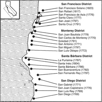
The occupation of Alta California was one of the last colonial efforts of the Spanish Empire and consequently several historical records have survived. Spanish colonization of California began in earnest in the late 1700s, after several brief encounters between the previous explorers and the Indians who used to live there (Walker & Johnson, Reference Walker and Johnson1992). The colonization involved two consecutive processes. First, between 1769 and 1823, the Spanish colonists gained control over Californian’s indigenous population by means of 21 Franciscan missions established along Alta California (Fig. 1), which were protected by the presidios – strategically located garrisoned forts. The mission system grew rapidly until the beginning of the nineteenth century.
Alta California’s missions.

Immigration from Mexico began in 1769 with the arrival of a small expedition of soldiers and priests and the establishment of the San Diego mission and presidio. By 1776, three presidios and seven missions had been settled and California’s Hispanic population consisted of 300–350 people, including nineteen missionaries and about 160 soldiers. In 1785 it had increased to 900 people and by 1820 the total non-native population of Alta California was about 3000 (Costello & Hornbeck, Reference Costello and Hornbeck1989). After that time, Spanish control over California rapidly diminished. In 1821 Mexico gained independence from Spain and California became a province of the new nation. This period represented the beginning of the second phase in California’s colonization, which coincided with the westward expansion of the United States. During the 1840s Mexican control over this territory weakened and US citizens began to move into California. In 1846, the United States declared war on Mexico and in 1847 California was officially transferred to the United States.
Spain’s colonization of California was not a natural response to population growth in Mexico, but a reaction to the directives of political leaders who wanted to establish a military presence in Alta California that would stop the expansion of other colonial powers, especially to prevent Russians from settling in the region (Farris, Reference Farris1989). Because of this, the fundamental economic role of the early Spanish colonists was to provide support to military garrisons. The missions were also important in that they offered a way to gain control over the indigenous population and the labour force these Native Americans represented.
For the above reasons, the colonization of Alta California was closely monitored by both government and church officials. As a result, there are detailed records of the births, baptisms, marriages and deaths of the settlers as well as the natives. This documentation has been used extensively to analyse the demographic collapse of California’s indigenous population (Walker & Johnson, Reference Walker and Johnson1992, Reference Walker and Johnson1994, Reference Walker and Johnson2002). Comparatively less research has been done regarding the demographic characteristics of the Spanish colonists (García-Moro et al., Reference García-Moro, Toja and Walker1997, Reference García-Moro, Pascual, Toja and Walker2000).
The aims of the present study were to analyse the fertility pattern of women of the Spanish-Mexican families who colonized Alta California (now the state of California) between 1742 and 1876, analyse the fitness of these women measured as their lifetime reproductive success, observe the relationships among the fertility variables and between them and longevity, and analyse their temporal variation.
Methods
Data
To analyse the fertility pattern of the women who initially colonized California in the 18th and the 19th centuries, data were derived from the genealogical compilations of M. E. Northrop (Reference Northrop1976, Reference Northrop1984). These books gather information from Alta California mission and parish records corresponding to the period 1742–1876 (the Spanish-Mexican period), and are presented sorted by families. For each family, data included in the book are: name, dates of birth and death both of the husband and the wife, names of their parents, date of marriage (and of additional marriages if there are more than one), and dates of birth, death and marriage of their children. Although Northrop’s compilations are not exhaustive, they do provide considerable information about the settlers who arrived in California during this period. The problem of missing data is more significant in some missions than in others. Thus, for five of the 21 missions (La Purísima, Santa Cruz, San Miguel, San Rafael and San Francisco Solano) no marriages and only a few baptisms were recorded. In the cases of San Francisco Solano and San Rafael this may be because these missions were founded late in the colonial period, when Spanish administrative control over California had started to weaken, registration was not so exhaustive and record-keeping deteriorated. Missions do not show differences among them in any of the analysed variables (Del Hoyo, unpublished observations).
The genealogies contain a few mistakes and inconsistencies, especially regarding dates, which can sometimes be resolved using substitutions. For example, in the absence of birth or death date, the baptismal and burial dates have been used respectively. In order to include only women who had completed their reproductive histories, the sample included 197 of the 599 women from the 657 families compiled by Northrop. These women had at least one child and died beyond 45 years in age.
The variables taken into account were: Fertility, calculated as the number of children produced in a lifetime; Lifetime reproductive success, measured as the total number of children produced in a lifetime that survived to 15 years of age; Age at marriage, age in years when a woman got married for the first time; Age at first child, defined as the age in years when a woman gave birth to her first child; Age at last child, calculated as the age in years when a woman gave birth to her last child; Longevity, age at death in years of individuals and obtained as the difference between the date of death and the date of birth; Protogenesic interval, measured as the difference in years between the date of the first child’s birth and the date of first marriage; Mean inter-birth interval, measured as the average time in months between successive births. For women who had married more than once, mean inter-birth intervals were obtained for each relationship separately and the final value is the mean of the total. Reproductive span was calculated as the difference between the dates of last child’s and first child’s births.
To analyse the temporal variation, five sub-periods were considered: 1742–75, 1776–1800, 1801–25, 1826–50 and 1851–76. In order to distribute the women in the sample equally, the first and last sub-periods were slightly longer than the rest.
Analysis
Data analysis was performed by means of the SPSS 18.0 statistical package. Mean values of each variable were obtained to define the fertility pattern. Before analysing the relationships between variables, the Kolmogorov–Smirnov test of normality was performed. All variables showed a non-normal distribution (z-values between 1.313 and 12,064; p-values <0.007 in all cases), therefore a non-parametric correlation test, the Kendall test, was used to analyse their relationships. To study how the different variables related to the fertility pattern and the influence of longevity and fertility, multiple regression analyses were performed using longevity and fertility as dependent variables. Comparisons among sub-periods were performed by means of the Kruskal–Wallis test.
Results
Fertility pattern
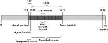
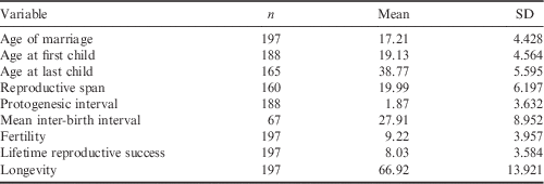
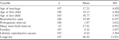
Table 1 shows the descriptive statistics of the variables analysed, and Fig. 2 summarizes the mean values for the reproductive pattern. The women analysed show a traditional reproductive pattern with an early marriage (at age 17) and first delivery (at age 19). They had their last child at the age of almost 39, and thus their reproductive span was 20 years. These women had on average nine children, with a mean inter-birth interval of 28 months. With regard to longevity, Spanish-Mexican women in the settler population lived roughly 67 years. Regarding fertility and fitness, 3720 out of the 4309 births compiled by Northrop reached the age of 15 (3690 out of 4271 if only children from women who survived to 45 years of age were taken into account), i.e. 86% of the children born between 1742 and 1876 reached adulthood. Considering women who survived to 45, 8.03 out of the 9.22 children every woman gave birth to lived up to reproductive age.
Fertility patterns of the women of Spanish-Mexican families that settled in Alta California.

Descriptive statistics for post-reproductive women, Spanish-Mexican colonists, Alta California
Relationship among variables
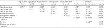
Table 2 shows the correlations matrix among variables. The results show a high, positive and significant correlation between age at marriage and age at first child, demonstrating that marriage acted as the beginning of the reproductive period in this population. The reproductive span shows a significant correlation with the age at first child, but less strong correlation than with the age at last child, which appears to be the variable that determines the width of the reproductive span, despite only women who had finished their reproductive period being taken into account.
Correlation matrix among the analysed variables, post-reproductive women, Spanish-Mexican colonists, Alta California

*p<0.05; **p<0.01.
Logically, there is a high, positive and significant correlation between fertility and reproductive span, so the longer the reproductive period, the higher the probability of having children. Finally, the highest correlation is that between fertility and lifetime reproductive success. Therefore, as stated above, the number of children who reach age 15 mainly depends on the fertility, and the effect of low infant mortality.
With a multiple regression analysis using longevity as dependent variable, the results show that none of the variables had a significant influence over longevity. In contrast, the number of children is positively associated with the reproductive span, i.e. by the age at first and last child. To a lesser extent, mean inter-birth interval also has a positive influence on fertility. The regression model obtained is:
 $${\rm Fertility}=0.{\rm 449}\,{\rm ALC}{\minus}0.{\rm 461}\,{\rm AFC}{\minus}0.{\rm 21}0\,{\rm MIBI}{\plus}{\rm 5}.{\rm 957}$$
Temporal trend
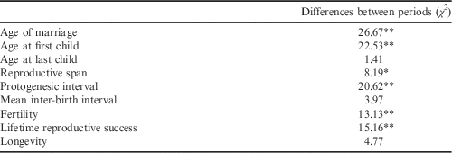
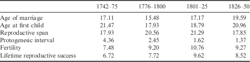
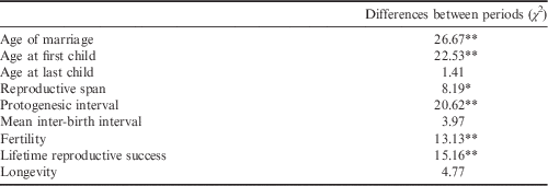
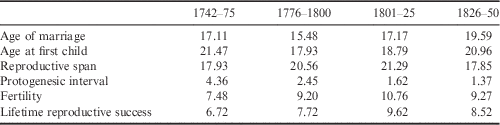
The colonizing period was divided into five sub-periods (1742–75, 1776–1800, 1801–25, 1826–50 and 1851–76) in order to analyse the temporal trends of the variables. Only two women who married between 1851 and 1876 survived to the age of 45, so the comparison excluded this last period. By means of a Kruskal–Wallis test, significant differences among periods were found for the variables age at marriage, age at first child, reproductive span, protogenesic interval, fertility and lifetime reproductive success (Table 3). Both ages at first marriage and at first child present a decrement in the second period, followed by an increase since the beginning of the 19th century (Table 4). Protogenesic interval decreases with time. Regarding fertility and lifetime reproductive success, these parameters show a clear increase until 1825 and a decrement in the last stage, according to the time trend of reproductive span.
Kruskal–Wallis test results analysing differences between time periods, post-reproductive women, Spanish-Mexican colonists, Alta California
*p<0.05; **p<0.01.
Time trend of age at marriage, age at first child, protogenesic interval, fertility and lifetime reproductive success, post-reproductive women, Spanish-Mexican colonists, Alta California
Discussion
The fertility pattern of California’s Spanish-Mexican settlers corresponds to that of a population with a natural fertility pattern (Bongaarts & Potter, Reference Bongaarts and Potter1983) – a pattern of fertility in the absence of deliberate birth control (Henry, Reference Henry1961). This kind of pattern is generally characterized by a number of around seven children, spread along a reproductive span of about 20 years, with an early first delivery. Natural fertility patterns show slight variations in accordance with the environmental conditions, with variations mainly in the age at first child and the mean inter-birth interval. For example, Hadza and !Kung are both hunter–gatherer populations that present differences in their inter-birth interval, being larger in the !Kung, who have fewer available resources. Therefore, there can be differences in the number of children per woman depending on the availability of resources (Blurton Jones et al., Reference Blurton Jones, Smith, O’Connell, Hawkes and Kamuzora1992). Pre-transitional European populations show a late age at marriage, but the reduction in the mean inter-birth interval allows families to reach a number of children similar to that of hunter–gatherers (Hajnal, Reference Hajnal1965; Knodel, Reference Knodel1978; Alfonso-Sánchez et al., Reference Alfonso-Sánchez, Peña and Calderón2003; Gögele et al., Reference Gögele, Pattaro, Fuchsberger and Pramstaller2009). For the Spanish-Mexican colonizers of Alta California, these differences are clearly seen in the length of the reproductive span, which are comparatively longer in this sample than in other contemporary south-western European populations (Alfonso-Sánchez et al., Reference Alfonso-Sánchez, Peña and Calderón2003; Muñoz-Tudurí, Reference Muñoz-Tudurí2008).
The inter-birth intervals obtained in this study are relatively short compared with those found in other populations (Helle et al., Reference Helle, Lummaa and Jokela2005; Muñoz-Tudurí, Reference Muñoz-Tudurí2008). As postpartum amenorrhoea is one of the most influential factors on inter-birth intervals, and is mainly determined by length and intensity of lactation (Wrigley, Reference Wrigley1998), Spanish-Mexican settlers in California probably had a short lactation period.
Regarding longevity, the results are similar to those obtained for contemporary European populations (Hollingsworth, Reference Hollingsworth1957; Korpelainen, Reference Korpelainen2000), and even longer in certain cases (Rüttimann & Loesch, Reference Rüttimann and Loesch2012). In their analysis of the Chumash Indians, former inhabitants of California, Walker and Johnson (Reference Walker and Johnson1994) reported that the average life expectancy may have been only about 35 years, or even less, clearly lower than the values obtained in the present study for the settlers, and slightly higher than those obtained by Jackson (Reference Jackson2004) for contemporary Paraguayan Indians. Thus, California’s settlers show a comparatively long lifespan, indicating good adaptation to their new environmental situation.
Fertility values are similar to those found for other populations in the same period; however, fitness values are clearly higher (Livi-Bacci, Reference Livi-Bacci2001; Lahdenpera et al., Reference Lahdenperä, Lummaa, Helle, Tremblay and Russell2004; Jackson, Reference Jackson2004; Pettay et al., Reference Pettay, Kruuk, Jokel and Lummaa2005). Finns and Canadians in the 18th and 19th centuries (Lahdenpera et al., Reference Lahdenperä, Lummaa, Helle, Tremblay and Russell2004), for instance, also had high numbers of children (6.8 and 9.1, respectively, on average), but only 55% of the newborns reached adulthood (3.8 and 5.1 on average). This 45% infant mortality was due to poor nutrition and to the socioeconomic and health conditions of the time (Knapp, Reference Knapp1998; Edvinsson & Janssens, Reference Edvinsson and Janssens2012). These data support the idea that the life conditions of the colonizers were favourable for population growth, since infant mortality is the main sign of a population’s healthiness. Thus, it seems that there was not a negative effect of the new environmental conditions on the survival of the individuals. García-Moro and Hernández (Reference García-Moro and Hernández1997) found high child mortality rates in Magellanes’ settlers. The high mortality in Magellanes was related to epidemics, especially respiratory disease epidemics, and so differences in climatic conditions could be the main reason explaining the differences with Alta California’s lower child mortality. However, it is not only environmental factors that determine child mortality. Walker and Johnson (Reference Walker and Johnson1992, Reference Walker and Johnson2002) found high child mortality levels among the Chumash, the former Indian inhabitants of Alta California. During the colonization process, the Chumash were converted to Catholicism and entered the California mission system, where they suffered new diseases that drastically reduced the number of surviving children. The same effect was observed in Paraguayan missions (Jackson, Reference Jackson2004) with the native population. In summary, the living conditions, both environmental and social, of the settlers of California’s missions were sufficient to reduce child mortality to low levels in comparison with other contemporary populations.
As stated in the Introduction, Williams’ antagonistic pleiotropy theory (Williams, Reference Williams1957) and Kirkwood’s disposable soma theory of ageing (Kirkwood, Reference Kirkwood1977) suggest a relationship between reproduction and lifespan. Their idea was that organisms only have a limited amount of energy that has to be divided between reproductive activities and the maintenance of non-reproductive aspects of the organism. Thus, those organisms that invest high energy levels in reproduction will suffer a reduction in their longevity.
Nevertheless, in the literature, there is some controversy about the influence of reproductive variables on longevity. Some authors demonstrate a trade-off between early childbearing and longevity, in agreement with classical theories (Westendorp & Kirkwood, Reference Westendorp and Kirkwood1998; Doblhammer, Reference Doblhammer2000; Kemkes-Gottenthaled, Reference Kemkes-Grottenthaler2004; Mirowsky, Reference Mirowsky2005; Henretta, Reference Henretta2007), whereas others report no influence of the age at first child on lifespan (Le Bourg et al., Reference Le Bourg, Thon, Légaré, Desjardins and Charbonneau1993; Smith et al., Reference Smith, Mineau and Bean2002; Muller, Reference Muller2004; Gagnon et al., Reference Gagnon, Smith, Tremblay, Vézina, Paré and Desjardins2009). The results obtained in the present study agree with those of the latter group, with no influence of the age at first child over longevity. The analysed database is slightly reduced and this could be the reason for the lack of long-term effects of reproduction on survival. In pre-transitional populations, female mortality related to pregnancy and childbearing is significantly higher (Kiple, Reference Kiple1993). Thus, the number of women who lived beyond 45 is limited, and it could mask the long-term effects that should be observed in a larger sample of post-reproductive women.
Furthermore, fertility is clearly influenced by the reproductive span: this is, by the ages at first and at last child, and to a lesser extent by the inter-birth interval and the lifespan. These results show a clear differentiation of the fertility pattern of California’s settlers from the European one, which shows a clear influence of the inter-birth interval over the number of children (Henry, Reference Henry1961; Nenko & Jasienska, Reference Nenko and Jasienska2013). As stated above, the women in this study show long reproductive spans, which are also very variable, so the influence of reproductive span on fertility could be greater than in other contemporary populations.
Significant differences among time periods have been observed for age at marriage, age at first child, reproductive span, protogenesic interval, fertility and lifetime reproductive success. Changes during the intermediate periods result in a temporary increase in the reproductive span and in fertility. This trend changes at the end of the studied period, resulting in a clear reduction in the reproductive span. These final changes result in a fertility pattern similar to the contemporary European one (Korpelainen, Reference Korpelainen2000), with a later age of childbearing and a reduction of the reproductive span.
The main reason for these fluctuations in the fertility variables may be the living conditions of the colonizing population, since both the start and the end of the colonization process were unstable periods: the beginning due to the conquest of the new territory, and the end because of conflicts between the Spanish crown and Mexico for the control of the land. In contrast, the stability of the intermediate period is manifested in an increase in the number of children, with a larger reproductive span. This last period of war must suppose, not only a period of instability, but also a period of disruption in the sex ratio of the population and consequently a bad time for reproduction.
In conclusion, the Spanish-Mexican settlers of Alta California show a natural fertility pattern, with high levels of infant survival, which result in high fitness levels, clearly different to the contemporary European pattern.
Acknowledgments
The authors thank V. Jalencas and J. Degon for the helpful comments they provided on earlier drafts of the manuscript. They also thank the two anonymous reviewers of this article for their helpful comments. Part of this work is included in the dissertation thesis of I. del Hoyo for the Degree in Biology from the University of Barcelona. The paper is dedicated to Dr Phillip L. Walker in memoriam.