Introduction
Right ventricular mechanics in preterm neonates begin to undergo maturational changes during the early post-natal period that have prognostic implications for clinical status and long-term outcomes.Reference Levy, El-Khuffash and Patel1–Reference Lewandowski, Bradlow and Augustine3 The determinants of right ventricle performances involve interactions between its contractility, afterload, preload, and morphology.Reference El-Khuffash and Mcnamara4 Preterm infants have maladaptive changes in right ventricle performance when compared to healthy term infants,Reference Breatnach, Levy, James, Franklin and El-Khuffash2, Reference Aye, Lewandowski and Lamata5, Reference Schubert, Muller, Abdul-Khaliq and Norman6 where important morphological differences that explain the relative dysfunction are exacerbated as prematurity exposes the right ventricle to the stress of post-natal cardiovascular performance before development is concluded.Reference Bensley, Moore, De Matteo, Harding and Black7 Recent work has validated emerging quantitative methods to characterise neonatal right ventricle performance,Reference Levy, El-Khuffash and Patel1, Reference Breatnach, Levy, James, Franklin and El-Khuffash2 including myocardial tissue deformation by two-dimensional speckle-tracking echocardiography,Reference Levy, El-Khuffash and Patel1, Reference Breatnach, Levy, James, Franklin and El-Khuffash2 fractional area of change,Reference Levy, Dioneda and Holland8 tricuspid annular plane systolic excursion,Reference Aye, Lewandowski and Lamata5 pulmonary artery acceleration time (estimate of right ventricle afterload),Reference Levy, Patel, Choudhry, Hamvas and Singh9 right ventricle areas,Reference Levy, Dioneda and Holland8, Reference James, Corcoran, Franklin and EL-Khuffash10 and linear dimensions.Reference Levy, Patel, Choudhry, Hamvas and Singh9 Our group has also recently demonstrated that common cardiorespiratory complications of prematurity, including bronchopulmonary dysplasia and pulmonary hypertension, negatively impact right ventricle performance over the first year of age.Reference Levy, El-Khuffash and Patel1, Reference Levy, Dioneda and Holland8, Reference Levy, Patel, Choudhry, Hamvas and Singh9 However, longitudinal comparisons of right ventricle performance in robust healthy birth cohorts and birth cohorts affected by prematurity from the neonatal period through 1 year of age are lacking, but are necessary prerequisites for clinical adoption in evaluating pathologic changes and progression.
We hypothesise that developmental and maturational disruption to the right ventricle from preterm birth results in dysfunction and remodelling as a distinct pathophysiology from bronchopulmonary dysplasia and pulmonary hypertension that is evident at 32 weeks post-menstrual age and persists through 1 year corrected age. Accordingly, the objective of this study was to identify and longitudinally track right ventricle performance using emerging measures of right ventricle mechanics in a cohort of infants born preterm (asymptomatic and those with bronchopulmonary dysplasia and/or pulmonary hypertension) and compare them with healthy infants born at term at 1 month and 1 year of age.
Materials and methods
Study design and population
To define the cardiac phenotype, we retrospectively analysed prospectively acquired data from two cohorts: (1) a cohort of preterm-born infants and (2) a cohort of full term-born infants. Each cohort was recruited at birth and longitudinally followed to 1 year of age. The cohort of 80 preterm infants were enrolled through the Prematurity and Respiratory Outcomes Program at Washington University School of Medicine/Saint Louis Children’s Hospital neonatal intensive care unit between August 2011 and November 2013. The detailed design of the Prematurity and Respiratory Outcomes Program Study (ClinicalTrials.gov: NCT01435187) has been described.Reference Pryhuber, Maitre and Ballard11, Reference Poindexter, Feng and Schmidt12 This was a retrospective secondary analysis of prospectively acquired data. Preterm subjects were eligible for inclusion in this study if they were born between 23 0/7 weeks and 28 6/7 weeks gestational age and had an echocardiogram at 32 weeks post-menstrual age and 1 year corrected age (patients were excluded if they had any suspected congenital anomalies of the airways, congenital heart disease (except age-appropriate atrial septal communications), intrauterine growth restriction (< 3rd percentile), or small for gestational age (birth weight < 10th centile for gestation)). We have provided a list of terms (Supplementary Information, Appendix 1) that describes the definitions for length of gestation and age in the perinatal period based on the policy statement from the American Academy of Pediatrics.Reference Engle13
A cohort of 50 healthy, age-, weight-, and sex-matched infants born at term (>37 weeks gestation at birth) were included as the control group from the University of Nebraska Medical Center from February 2013 to September 2016. Echocardiograms were obtained at 1 month and 1 year of age. The data were also prospectively acquired and retrospectively matched to the preterm cohort. Specific exclusion criteria for the control cohort consisted of multiple gestation pregnancy, maternal self-reported use of illegal drugs, maternal smoking, maternal diabetes, antepartum haemorrhage, pre-eclampsia, and/or proven chorioamnionitis. Infants with intrauterine growth restriction, perinatal acidosis (umbilical cord pH < 7.10), suspected congenital or chromosomal anomalies, neuro-muscular disorders, abnormal cardiac rhythms, heart failure, and infants readmitted to the hospital within the first year of age were also excluded. The Institutional Review Board for human studies at Washington University and the University of Nebraska Medical Center approved the study. Written informed consents were obtained from the parents/guardians of the study subjects.
Clinical demographics
For both cohorts, perinatal clinical information and demographic characteristic were obtained. In addition for the preterm infants, clinical cardio-respiratory characteristics during the two echocardiographic assessments were collected and included: systolic and diastolic blood pressure, heart rate, oxygen requirements, and oxygen saturation. The following clinical outcomes were also obtained in the preterm cohort: intraventricular haemorrhage classified according to Papile Classification,Reference Papile, Burstein, Burstein and Koffler14 necrotising enterocolitis with radiological evidence of pneumatosis, retinopathy of prematurity (stage 2 or higher), pulmonary hypertension,Reference Mourani, Sontag and Younoszai15 and bronchopulmonary dysplasia (using a modified definition of the 2001 National Institutes of Health bronchopulmonary dysplasia workshop, Supplementary Information, Appendix 2).Reference Poindexter, Feng and Schmidt12
Echocardiography
Echocardiograms were performed with commercially available ultrasound imaging systems (Vivid 7 and E9; General Electric Medical Systems, Milwaukee, Wisconsin) at each centre. Images were obtained at 32 weeks post-menstrual age and 1 year corrected age in infants born preterm and at 1 month and 1 year of age in infants born at term. One designated experienced paediatric cardiac sonographer at each centre obtained all the echocardiographic images using a phased array transducer (7.5–12 MHz).Reference Levy, Holland, Sekarski, Hamvas and Singh16 The echocardiographic images were acquired using a standardised image acquisition protocol based on the guidelines of the American Society of Echocardiography.Reference Lang, Badano and Mor-Avi17, Reference Mitchell, Rahko and Blauwet18 The image data were acquired digitally and stored in raw Digital Imaging and Communications in Medicine cine-loop format for offline analysis for the following functional and morphometric parameters.
Deformation analysis
Two-dimensional speckle tracking imaging
Myocardial mechanics were analyzed by the quantification of right ventricle free wall longitudinal strain (%) and systolic longitudinal strain rate (1/second) using previously published image acquisition and data analysis protocols from our laboratories.Reference Levy, Holland, Sekarski, Hamvas and Singh16, Reference Sanchez, Levy, Sekarski, Hamvas, Holland and Singh19 Right ventricle segmental longitudinal strain was obtained from the segments at the apical, mid-ventricular, and basal levels of the right ventricle free wall from a focused right ventricle apical 4-chamber view.Reference Levy, El-Khuffash and Patel1, Reference Cade, Tinius, Reeds, Patterson and Cahil20 Peak strain for each index was measured as end-systolic strain at the closure of the pulmonic valve,Reference Voigt, Pedrizzetti and Lysyansky21 (Supplementary Information, Figure S1). Two observers (PL and CE), who were blinded to the maternal and infant clinical and cardio-respiratory conditions, analysed deformation using vendor customised commercially available software (EchoPAC; General Electric Medical Systems, Waukesha, WI, USA, version 112).
Other measurements of right ventricle performance
We assessed and compared additional indices of 1) right ventricle function: tricuspid annular plane systolic excursion,Reference Aye, Lewandowski and Lamata5 fractional area of changeReference Levy, Dioneda and Holland8; 2) right ventricle morphology: right ventricle areas from the apical 4-chamber view in systole and diastole,Reference Levy, Dioneda and Holland8, Reference James, Corcoran, Franklin and EL-Khuffash10 right ventricle linear dimensions at the basal and mid ventricular levels and in the longitudinal direction from the apical 4-chamber view at end-diastoleReference Levy, Patel, Choudhry, Hamvas and Singh9, Reference Lang, Badano and Mor-Avi17, Reference Mitchell, Rahko and Blauwet18; and 3) right ventricle afterloadReference Levy, Patel, Choudhry, Hamvas and Singh9: pulmonary artery acceleration time and ratio of pulmonary acceleration time to right ventricle ejection time between the term and preterm infants at each time point.Reference Levy, Patel, Choudhry, Hamvas and Singh9 All measures were generated according to guidelines of the American Society of EchocardiographyReference Lang, Badano and Mor-Avi17, Reference Mitchell, Rahko and Blauwet18 and recent validated protocols in neonates.Reference Breatnach, Levy, James, Franklin and El-Khuffash2
Confounders
We evaluated the effects of common cardiorespiratory complications of prematurity, including bronchopulmonary dysplasia and pulmonary hypertension, on right ventricle structure and function.Reference Levy, El-Khuffash and Patel1, Reference Levy, Dioneda and Holland8, Reference Levy, Patel, Choudhry, Hamvas and Singh9 We utilised a modified definition of the 2001 National Institutes of Health bronchopulmonary dysplasia workshopReference Poindexter, Feng and Schmidt12 (Supplementary Information, Appendix 2) and a broad echocardiogram-based definition of pulmonary hypertension,Reference Levy, El-Khuffash and Patel1, Reference Mourani, Sontag and Younoszai15 in the assessment of their contributions on right ventricle performance indices at 32 weeks post-menstrual age, and at 1 year corrected age. Preterm infants were classified with pulmonary hypertension at 32 weeks post-menstrual age if they had one or more of the following conventional echocardiographic measures of pulmonary hemodynamics: (1) an estimated right ventricle systolic pressure > 40 mmHg, using the Doppler velocity of tricuspid regurgitation; (2) a ratio of right ventricle systolic pressure to systemic blood pressure > ½; (3) bidirectional or right to left shunt at the atrial level or patent ductus arteriosus; (4) septal wall flattening as defined as decreased septal curvature into the right ventricle at end systole; or (5) dilated/hypertrophy of the right ventricle and right atrial morphology. The independent effect of the risk factors for bronchopulmonary dysplasia and pulmonary hypertension, including gestational age, sex, total oxygen days, length of hospital stay, perinatal factors, neonatal morbidities (necrotising enterocolitis, intraventricular hemorrhage, and retinopathy of prematurity), and presence of a patent ductus arteriosus at 32 weeks post-menstrual ageReference Schena, Francescato and Cappelleri22 was also accounted for in the analysis.
Statistical analyses
Continuous data were summarised as mean ± standard deviation or as medians (interquartile range) as appropriate. Categorical data were summarised as count and per cent. Continuous variables of right ventricle longitudinal strain, longitudinal systolic strain rate, and segmental longitudinal strain at the base, mid-ventricular, and apical levels were tested for normality using the Shapiro-Wilk test and histogram of the data. Analysis of variance tests were used to compare the changes in measures of right ventricle function and structure amongst preterm infants with and without bronchopulmonary dysplasia and/or pulmonary hypertension at 32 weeks post-menstrual age with term born control infants. Student t-test and Bonferroni adjustment where appropriate were separately utilised to compare the patterns between uncomplicated preterm infants and those with bronchopulmonary dysplasia and/or pulmonary hypertension. All outcome variables with non-normal distributions were analysed in simple comparisons using Wilcoxon rank sum tests or Kruskal-Wallis one-way analysis of variance for tests with more than two independent groups. Chi-square tests (or Fisher Exact test as appropriate) were used to assess the association between categorical variable. Univariate analysis was used to determine the best predictors to enter in the model and then backward step-wise regression was performed to assess the independent effect of gestational age, sex, total oxygen days, length of stay, and common neonatal morbidities, while adjusting for weight at examination. We performed stepwise regression to analyse the influence of pulmonary hypertension and bronchopulmonary dysplasia on right ventricle performance patterns at 32 weeks post-menstrual age and 1 year corrected age.Reference Mourani, Sontag and Younoszai15, Reference Mourani and Abman23 All results were considered statistically significant for values of p ≤ 0.05. The statistical analysis was performed using SAS v9.4 (SAS Institute, Cary, NC).
Results
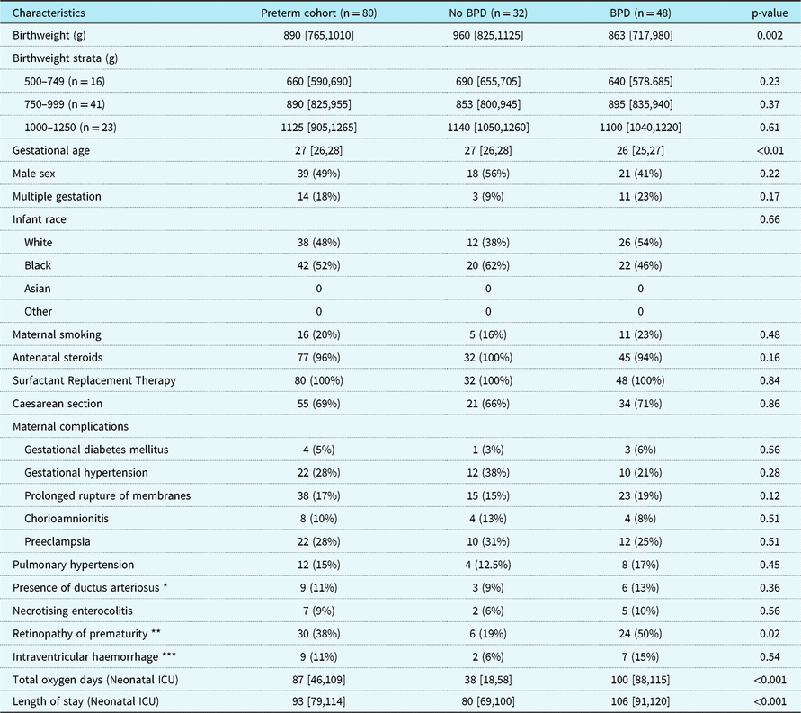
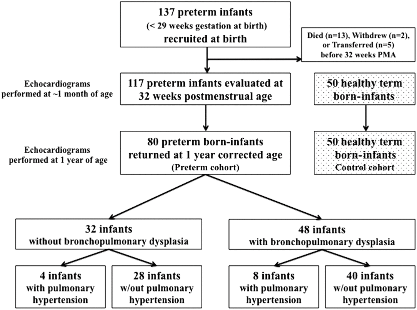
A total of 130 infants (80 preterm and 50 term) were included in this study (Fig 1). We compared the maternal and infant demographic characteristics and clinical data between preterm infants with and without bronchopulmonary dysplasia in Table 1, and with and without pulmonary hypertension in Supplementary Information, Table S1. Clinical data not described in Table 1 or Table S1 are detailed in the Supplementary Information.
Infant characteristics and clinical outcomes.

Data are presented as median (interquartile range) or number (Percentage). Data are presented as column percentages
* Presence of ductus arteriosus at 32 weeks post-menstrual age
** Retinopathy of prematurity threshold (stage 2 of higher)
*** Intraventricular haemorrhage (Grade 3 or 4)
Study subject flow diagram.

Maturational patterns of RV performance in term and preterm infants
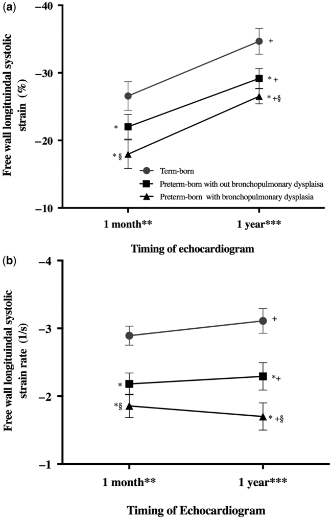
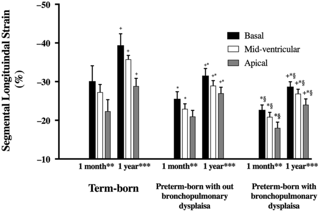
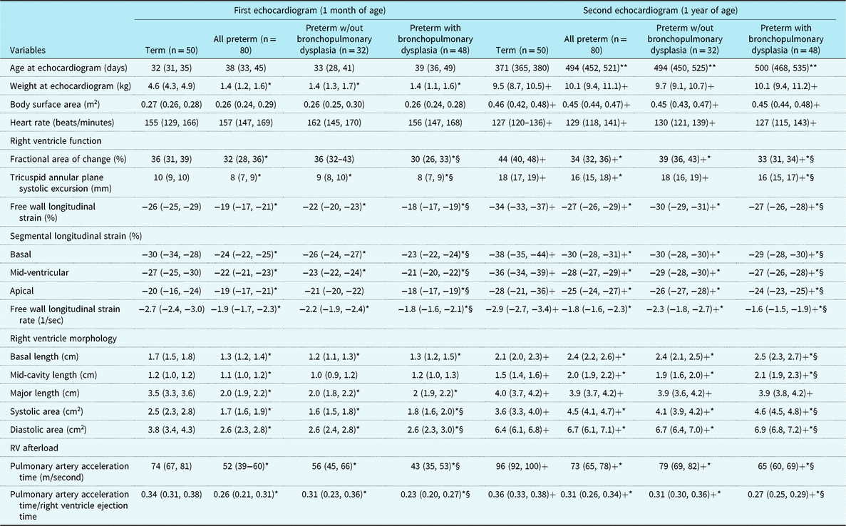
The magnitudes of right ventricle free wall longitudinal strain, fractional area of change, tricuspid annular plane systolic excursion, pulmonary artery acceleration time, and the ratio of pulmonary artery acceleration time to right ventricle ejection time increased from 32 weeks post-menstrual age to 1 year corrected age in the preterm infants and from 1 month of age to 1 year of age in the term infants (p < 0.01 for all measures, Fig 2a, Table 2). The magnitude of RV free wall systolic longitudinal strain rate in infants born at term increased from 1 month to 1 year of age (p < 0.01) but remained unchanged in infants born preterm over the same time period (p = 0.5) (Fig 2b). A significant base-to-apex (the magnitude was highest-to-lowest) segmental longitudinal strain gradient (p < 0.001) was observed in the right ventricle free wall in both the preterm and term infants (Fig 3). Right ventricle morphology measures of systolic and diastolic areas, basal, mid-cavity, and apex-to-base linear dimensions also increased in preterm infants from 32 weeks post-menstrual age to 1 year corrected age (p < 0.01 for all measures) (Table 2).
Comparison of right ventricle free wall longitudinal systolic strain (a) and strain rate (b) in term and preterm infants at 1 month of age and 1 year of age. *p < 0.01 vs. Term; § p < 0.01 vs. Preterm without BPD; +p < 0.001 vs. first echocardiogram. **In the preterm-born cohort, echocardiograms were performed at 32 weeks post-menstrual age with a median of 38 days (interquartile range: 33–45). In the term-born cohort, echocardiograms were performed at a median of 32 days (interquartile range: 31–35). ***In the preterm-born cohort, echocardiograms were performed at 1 year corrected of 494 days (interquartile range: 452–521). These numbers reflect actual days of age, but corrected age in days for all preterm infants was a median of 381 days (interquartile range: 355–424). In the term-born cohort, echocardiograms were performed at a median of 371 days (interquartile range: 365–380).

Comparison of right ventricle segmental longitudinal systolic strain in term and preterm infants at 1 month and 1 year of age. * p < 0.01 vs. Term; § p<0.01 vs. Preterm without BPD; +p < 0.001 vs. first echocardiogram. **In the preterm-born cohort, echocardiograms were performed at 32 weeks post-menstrual age with a median of 38 days (interquartile range: 33–45). In the term-born cohort, echocardiograms were performed at a median of 32 days (interquartile range: 31–35). *** In the preterm-born cohort, echocardiograms were performed at 1 year corrected of 494 days (interquartile range: 452–521). These numbers reflect actual days of age, but corrected age in days for all preterm infants was a median of 381 days (interquartile range: 355–424). In the term-born cohort, echocardiograms were performed at a median of 371 days (interquartile range: 365–380).

Comparison of RV performance between term and preterm infants
All preterm infants had significantly lower values of right ventricle free wall longitudinal strain, longitudinal systolic strain rate, tricuspid annular plane systolic excursion, pulmonary artery acceleration time, pulmonary artery acceleration time/right ventricular ejection time (p < 0.01 for all measures) than those of term infants at 1 month and 1 year of age (Table 2, Fig 2). Segmental longitudinal strain at the basal, mid-ventricular, and apical levels of the right ventricle free wall were also decreased at 1 month and 1 year corrected age in preterm infants compared to term infants (p < 0.01 for all measures). As expected, the systolic and diastolic areas, the basal, mid-cavity, and the longitudinal diameter from the apex to the base were all smaller in the preterm infants at 1 month of age (p < 0.01 for all measures). At 1 year of age, all morphometric measures, except the longitudinal diameter from the apex to the base were larger in the preterm infants (p < 0.01 for all measures). All values for comparisons can be found in Table 2.
Bronchopulmonary dysplasia
Since measures of right ventricle function performed better in infants born at term, pairwise comparison was conducted using the Bonferroni method to detect any difference in right ventricle function in a) preterm infants with and without bronchopulmonary dysplasia and b) preterm infants with and without pulmonary hypertension at 32 weeks post-menstrual age. Adjustments for gestational age at birth, sex, and heart rate revealed significantly lower values in the magnitudes of right ventricle free wall longitudinal strain, longitudinal systolic strain rate, fractional area of change, tricuspid annular plane systolic excursion, pulmonary artery acceleration time, pulmonary artery acceleration time/right ventricle ejection time, and segmental longitudinal strain at the basal, mid-ventricular, and apical levels of the right ventricle free wall at 32 weeks post-menstrual age in preterm infants with bronchopulmonary dysplasia (p < 0.0001 for all). The same patterns persisted at 1 year corrected age (p < 0.0001 for all). The systolic and diastolic areas were larger in the preterm infants with bronchopulmonary dysplasia at both time points. The basal, mid-cavity, and longitudinal diameter from the apex to the base were similar between the preterm groups at 32 weeks post-menstrual, but the basal and mid-cavity diameter were significantly larger in the preterm infants with bronchopulmonary dysplasia by 1 year corrected age (p < 0.01 for both measures).
Pulmonary hypertension
In preterm infants with and without pulmonary hypertension, there was also a decrease in right ventricle free wall longitudinal strain, free wall longitudinal systolic strain rate, fractional area of change, tricuspid annular plane systolic excursion, pulmonary artery acceleration time, pulmonary artery acceleration time/right ventricle ejection time, and segmental longitudinal strain at the basal, mid-ventricular, and apical levels of the right ventricle free wall at 1 year corrected age in those with pulmonary hypertension (p < 0.0001 for all). The base-to-apex segmental longitudinal strain gradients were preserved in all preterm infants at both time points, irrespective of the presence of pulmonary hypertension. These trends persisted even after adjusting for the presence of bronchopulmonary dysplasia (for all measure, β > 2.4, p < 0.01) and presence of patent ductus arteriosus (for all measures β > 2.6, p < 0.01). The dimensions and areas were similar between preterm infants with and without pulmonary hypertension at 32 weeks post-menstrual age. Right ventricle basal and mid-ventricular diameter and both right ventricle areas were increased in preterm infants with pulmonary hypertension at 1 year corrected age (p< 0.01 for both). There was no difference in the longitudinal right ventricle dimension from the apex to the base at any time point between the groups (Supplementary Information, Table S1).
Term vs. uncomplicated preterm
We compared the measures between term infants and preterm infants without bronchopulmonary dysplasia and/or pulmonary hypertension at each time point. Adjustments for weight at exam, sex, and heart rate revealed significant lower values of the magnitudes of right ventricle free wall longitudinal strain, free wall longitudinal systolic strain rate at both time points, and decreases in segmental longitudinal strain at the basal and mid-ventricular levels of the right ventricle free wall at 1 month, and all segmental longitudinal strain levels by 1 year of age. At 1 month, fractional area of change was similar between the term and preterm without bronchopulmonary dysplasia and/or pulmonary hypertension, but tricuspid annular plane systolic excursion was decreased (p < 0.01). At 1 year of age, tricuspid annular plane systolic excursion was similar, and fractional area of change was decreased in the preterm infants without bronchopulmonary dysplasia and/or pulmonary hypertension. All morphometric measures were decreased, as expected, in preterm infants at 1 month of age, but by 1 year of age, the systolic and diastolic areas and the basal and mid-cavity diameters were significantly larger in the preterm infants without bronchopulmonary dysplasia and/or pulmonary hypertension (p < 0.01 for both measures). The longitudinal diameter from the apex to the base was similar between the groups.
Discussion
In this study, we observed right ventricle dysfunction in asymptomatic infants born preterm at 32 weeks post-menstrual age (∼1 month of age) that persisted to 1 year corrected age when compared to healthy infants born at term over the same time period. In addition, right ventricle morphologic differences existed between preterm and term infants at 1 year of age, irrespective of the degree of lung disease during the neonatal period. Subclinical right ventricle dysfunction assessed using deformation, fractional area of change, and tricuspid annular plane systolic excursion, as well as alteration in right ventricle morphology, appears to be a distinct pathology of prematurity that is present even after resolution of bronchopulmonary dysplasia and pulmonary hypertension.
Right ventricle mechanics undergo post-natal physiologic adaptations over the first year of age that have long-term influence on myocardial performance.Reference Aye, Lewandowski and Lamata5 Preterm birth is associated with alterations in myocardial performance that appear in the neonatal period,Reference Aye, Lewandowski and Lamata5 persist through 1 year corrected age,Reference Levy, El-Khuffash and Patel1 early childhood,Reference Mohlkert, Hallberg and Broberg24 and are present at an early adult age with a disproportionate increase in ventricular mass and decrease in right ventricle function.Reference Lewandowski, Bradlow and Augustine3 In neonates, strain analysis has been shown to be more sensitive in detecting early myocardial changes than conventional echocardiography in health and disease states.Reference Breatnach, Levy, James, Franklin and El-Khuffash2 Kwon et al. found a lower magnitude of right ventricle strain in children born preterm with a history of pulmonary hypertension during the neonatal period compared to non-pulmonary hypertension children born preterm.Reference Kwon, Kim and An25 Lewandowski showed reduced right ventricle strain in adults born preterm compared to adults born at term, which was gestational age dependent, but unrelated to the premature lung disease.Reference Lewandowski, Bradlow and Augustine3 In the present study, we observed decreased right ventricle free wall longitudinal and longitudinal systolic strain rate in preterm infants compared to term infants at 32 weeks post-menstrual age and 1 year corrected age. Taken all together, these studies from the early neonatal period through adulthood suggest that the factors that contribute to the right ventricle maladaptation following premature birth are complex and may differ substantially between infants with different antenatal and post-natal experiences.
The exact mechanism that explains why preterm infants have subclinical right ventricle dysfunction and alterations in right ventricle morphology that persists to 1 year of age is not yet known. We propose two theories that may explain this observation: (1) changes in post-natal loading conditionsReference Kang, Kim, Hwang and Kim26 and (2) loss of the third trimester myocardial structural programming.Reference Bensley, Moore, De Matteo, Harding and Black7 The exposure of an immature preterm right ventricle to a sustained increase in hemodynamic load of post-natal circulation (increased right ventricle afterload), at a time in the development when the right ventricle primarily supports a low-resistance circulation, may induce myoarchitectural adaptation that can lead to changes in geometry, structure, and function, “a process known as ventricular remodeling.”Reference Haddad, Hunt, Rosenthal and Murphy27 In this study, measures of right ventricle function were decreased and indices of right ventricle morphology (right ventricle areas and right ventricle linear dimensions/length at the base and mid-cavity regions) were increased in preterm infants by 1 year corrected age. Functional measures were further decreased and morphological measures increased at 32 weeks post-menstrual in preterm infants with bronchopulmonary dysplasia, suggesting early dysfunction and remodelling.Reference Aye, Lewandowski and Lamata5 However, these observations do not explain the increased incidence of subclinical right ventricle dysfunction and remodelling in a subset of premature infants without apparent clinical signs of lung disease of prematurity.Reference Levy, El-Khuffash and Patel1, Reference Mourani, Sontag and Younoszai15, Reference Weismann, Asnes, Bazzy-Asaad, Tolomeo, Ehrenkranz and Bizzarro28 Since premature birth results in the loss of the normal maturational process that occurs in utero during the third trimester, the evidence of disproportionate increase in ventricular size and decrease in right ventricle function in the post-natal period,Reference Aye, Lewandowski and Lamata5 over and above what would be expected, indicates that loss of a primary myocardial structural programming event may also lead to post-natal ventricular remodelling.Reference Bensley, Moore, De Matteo, Harding and Black7 Bensely et al. demonstrated that preterm birth may lead to an abrupt reduction in cardiomyocyte cell division, adversely impact upon the final number of cardiomyocytes, reduce right ventricle functional reserve, and impair the reparative capacity of the myocardium.Reference Bensley, Moore, De Matteo, Harding and Black7
Strain is influenced by preload and afterload.Reference Levy, El-Khuffash and Patel1, Reference Breatnach, Levy, James, Franklin and El-Khuffash2, Reference Geyer, Caracciolo and Abe29 Strain rate correlates well with load-independent measures of contractility and is a more accurate reflection of intrinsic myocardial contractile function.Reference Breatnach, El-Khuffash, James, McCallion and Franklin30–Reference Ferferieva, Van den Bergh and Claus32 The force–frequency relationship reflects alteration of the contractile force of cardiac myocytes through the neurohumoral effects, which are influenced by changes in the frequency of stimulation.Reference Alvarez, Fortin-Pellerin and Alhabdan31 Both preclinicalReference Alvarez, Fortin-Pellerin and Alhabdan31, Reference Ferferieva, Van den Bergh and Claus32 and human studiesReference Breatnach, El-Khuffash, James, McCallion and Franklin30, Reference Alvarez, Fortin-Pellerin and Alhabdan31 have demonstrated that strain rate has a force–frequency relationship behaviour that is consistent with a measure of contractility that is mostly load independent within physiological ranges. We speculate that the decreased strain in preterm infants reflects the myocardium’s response to post-natal-loading conditions, according to the Frank Starling phenomenon, while the decreases in strain rate possibly reflect a less mature myocardial programming mechanism stemming from preterm birth that alters contractility, according to the force–frequency relationship. While all measures of right ventricle function increased from 32 weeks post-menstrual to 1 year corrected age, strain rate was unchanged in preterm infants, and decreased in preterm infants with bronchopulmonary dysplasia and/or pulmonary hypertension, indicating a less developed intrinsic myocardial function response in preterm infants.
In the right ventricle, free wall a base-to-apex segmental longitudinal strain gradient (highest-to-lowest magnitude of segmental longitudinal strain values) has been observed in extreme preterm population,Reference Levy, El-Khuffash and Patel1 late preterm infants,Reference Aye, Lewandowski and Lamata5 healthy term neonates,Reference Schubert, Muller, Norman and Abdul-Khaliq33 infants,Reference Schubert, Muller, Abdul-Khaliq and Norman6 children,Reference Levy, Sanchez, Machefsky, Fowler, Holland and Singh34 and adults.Reference Fine, Chen and Bastiansen35 This physiologic pattern reflects the base-to-apex alignment of the dominant deep longitudinal layers of the right ventricle that allows for greater longitudinal shortening during contraction.Reference Levy, Sanchez, Machefsky, Fowler, Holland and Singh34 All preterm infants had a decreased magnitude of segmental longitudinal strain at the basal and mid-ventricular and apical levels of the right ventricle free wall at 32 weeks post-menstrual age and 1 year corrected age. Focusing on the right ventricle free wall provides an assessment of the predominant wall contributing to right ventricle contractile function.Reference Fine, Chen and Bastiansen35 We have previously demonstrated that the presence of bronchopulmonary dysplasia and/or pulmonary hypertension during the neonatal period leaves a negative impact of global and regional strain values.Reference Levy, El-Khuffash and Patel1 However, even those preterm infants without significant morbidity have decreased regional strain values, further suggesting that the post-natal period appears to be a critical period of cardiac development and may offer a window for interventions to prevent the long-term cardiovascular consequences of preterm birth.Reference Aye, Lewandowski and Lamata5 We observed a difference of segmental longitudinal strain at the basal and mid-ventricular levels of the right ventricle free wall between preterm infants without pulmonary hypertension and/or bronchopulmonary dysplasia compared to healthy term infants. Interestingly, the apical segmental longitudinal strain values were similar between these two groups. We have previously demonstrated that only the basal and mid-ventricular segments of the right ventricle free wall increase throughout maturation, while the apical segmental longitudinal strain was unchanged from birth to 1 year of age in healthy term infants.Reference Erickson, Levy, Craft, Li, Danford and Kutty36 Since the right ventricle is composed of superficial oblique fibres that are arranged in parallel with the AV groove and turn obliquely towards the cardiac apex and continue into the superficial myofibers of the left ventricle,Reference Sadeghpour and Alizadehasl37 the expected physiologic increase of segmental longitudinal strain at the right ventricle side of the apical free wall is likely masked by the contribution of the left ventricle to the contraction patterns of the right ventricle free wall.Reference Levy, Holland, Sekarski, Hamvas and Singh16 Therefore, we would expect not to see a difference between the segmental longitudinal strain at the apical level, unless a significant disease process like pulmonary hypertension or bronchopulmonary dysplasia alters these “physiological” gradients.
Study limitations
We acquired images of the right ventricle free wall from a right ventricle-focused apical four-chamber view. Recently, several authors have outlined an approach for assessment of right ventricle function that utilises three-chamber right ventricle-focused views to capture the inlet, apical, and outlet in one image view.Reference Jain, Mohamed and El-Khuffash38, Reference Forsha, Risum and Kropf39 Currently there is no consensus on the best approach, but more studies have utilised the right ventricle-focused apical four chamber method.Reference Levy, Sanchez, Machefsky, Fowler, Holland and Singh34 Since the right ventricle lacks a middle layer of circumferential fibres, and the major contribution to ejection fraction and stroke volume during systole is provided by the dominant deep longitudinal fibre shortening, right ventricle circumferential and radial strain patterns were not assessed.Reference Atsumi, Seo and Ishizu40 Future work is needed to investigate right ventricle circumferential, radial, and circumferential-longitudinal shear strain (rotational mechanics) in neonates with the use of three-dimensional deformational approaches.Reference Atsumi, Seo and Ishizu40 Finally, we chose to exclude intrauterine growth-restricted neonates from the analysis, since it has been documented that foetal growth restriction alone may induce cardiac remodelling that persists beyond the neonatal period.Reference Sarvari, Rodriguez-Lopez and Nunez-Garcia41
Conclusion
This study tracks the maturational patterns of global and regional right ventricle performance by two-dimensional speckle-tracking echocardiography in extremely preterm infants and term infants over the first year of age. Infants born preterm exhibit decreased right ventricle performance at 1 month of age that persists to 1 year of age when compared to infants born at term. Right ventricle remodelling is evident by 1 year corrected age in preterm infants, irrespective of the degree of neonatal lung disease, suggesting a less developed intrinsic myocardial function response following preterm birth.
Echocardiographic data of term and preterm infants at 1 month of age and 1 year of age.

All data presented as median (interquartile range)
Right ventricle strain rate significantly decreased from the first echocardiogram to the second echocardiogram for preterm infants with bronchopulmonary dysplasia (p < 0.001)
* p < 0.01 vs. Term; § p<0.01 vs. Preterm without bronchopulmonary dysplasia; + p < 0.001 vs. first echocardiogram
** These numbers reflect actual days of age, but corrected age (days) for all preterm infants was 381 (355,424), and for preterm without bronchopulmonary dysplasia was 381(357, 428) and preterm infants with bronchopulmonary dysplasia was 398 (377, 428)
Supplementary Material
To view supplementary material for this article, please visit https://doi.org/10.1017/S1047951119001161.
Author ORCIDs
Philip T. Levy, 0000-0001-5103-5834
Acknowledgements
None.
Financial Support
This study was supported by a grant from Premature and Respiratory Outcomes Program (NIH 1U01 HL1014650, U01 HL101794).
Conflicts of Interest
None.
Ethical Standards
The Institutional Review Board for human studies at Washington University and the University of Nebraska Medical Center approved the study. Written informed consents were obtained from the parents/guardians of the study subjects.