Introduction
Lemierre's syndrome in its classical clinical form involves a young patient with a recent history of oropharyngitis, high fever, rigors, lymphadenopathy and tenderness along the angle of the jaw and the lateral aspect of the sternocleidomastoid muscle. When infection finds its way through the parapharyngeal space into the internal jugular vein, the consequent suppurative thrombophlebitis results in persistent bacteraemia and various life threatening complications, as metastatic septic emboli are spread towards multiple organs. Lungs, liver, joints and kidneys are the most frequently affected organs. Osteomyelitis, endocarditis, thyroiditis, mediastinal infection with pericardial tamponade and meningitis are less common complications.Reference Hagelskjaer, Prag, Malczynski and Kristensen1–Reference Goyal, Sharma, Jain, Gupta and Berry6
Fusobacterium necrophorum, a gram-negative anaerobic rod, has been identified as the causative pathogen. Although it is a common saprophyte of the human alimentary tract, it is also a potential primary pathogen capable of invasive infection. Unlike other gram-negative bacteria which tend to cause septicaemias in elderly people or people who are chronically ill, after the host has been compromised by other bacteria, Fusobacterium necrophorum is capable of causing life threatening infections in previously healthy adolescents.Reference Sinave, Hardy and Fardy3, Reference Eykyn7–Reference Leugers and Clover9
Case report
A 22-year-old man presented to the emergency room after five days of high fever (up to 39°C), odynophagia and malaise. For the past four days he had been treated with a macrolide per os, without any improvement. On admission he appeared very ill. His temperature was 39°C and his blood pressure 105/50 mmHg. Oral examination revealed enlarged exudative tonsils. No uvula deviation was noted and cervical motion was of full range. The lymph nodes were palpable and tender in both submandibular regions. Lung and heart auscultation revealed no abnormal findings. No abdominal tenderness was observed.
Laboratory work up on admission included white blood cells count 6200/mm3, with 87 per cent granulocytes, platelets 150 000/mm3 and haematocrit 42 per cent. Erythrocyte sedimentation rate was 58 mm/h. Coagulation rates were elevated with prothrombin time at 14 seconds and partial thromboplastin time at 63 seconds. Serum D-dimers were measured 970 mg/l. Liver function was estimated as normal, since oxaloacetate aminotransferase, pyruvate aminotransferase and bilirubin were within normal limits. On the contrary, serum urea (200 mg/dl) and creatinine (2.8 mg/dl) values revealed kidney dysfunction. Moreover, urine was amber with 15–20 red blood cells, 2–3 white blood cells, protein 30 mg/dl and cylinders. Blood cultures and serology for infectious mononucleosis were obtained and lincomycin (600 mg twice daily intravenously) was initiated.
His clinical profile deteriorated in the afternoon and from his laboratory work up a decrease of platelets to 73 000/mm3 and an increase of white blood cells to 14 000/mm3 was noted. The patient remained febrile (39°C) and his medication was changed to ciprofloxacin (400 mg twice daily) and clindamycin (600 mg thrice daily) intravenously.
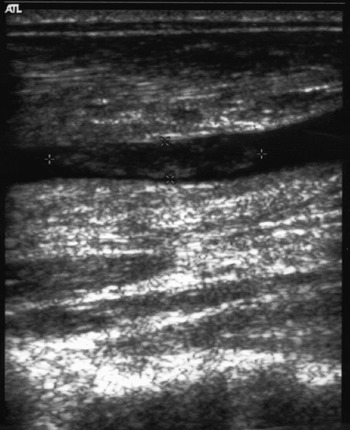
During the next morning he complained of precordium pain. Auscultation revealed tachypnoea, pleural and pericardial rub. Rhonchi were heard at both lung bases. The subsequent ultrasonography of the heart demonstrated no abnormal findings. Ultrasound study of the neck and triplex ultrasonography of the vessels of the neck detected lymphadenopathy and thrombophlebitis of the left internal jugular vein (Figure 1). The patient was desaturated requiring transfer to the intensive care unit, where he responded well to oxygen supplementation via a Venturi mask. His white blood cell counts continued to increase (19 300/mm3 with 91 per cent neutrophils). A further decrease of his haematocrit (34 per cent) and his platelets' count (40 000/mm3) in combination with haematuria and elevated serum urea (140 mg/dl) and creatinine (1.8 mg/dl) were noted. D-dimers remained within normal limits, but coagulation rates were still elevated.
Ultrasound demonstrating thrombosis of the left internal jugular vein.

His blood cultures were positive for a gram-negative anaerobic bacterium, which was eventually identified as Fusobacterium necrophorum. Following these blood culture indications, high dose intravenous penicillin (24 million IU) was added to his treatment and clindamycin was substituted by metronidazole (500 mg thrice daily). Serology for infectious mononucleosis was negative.
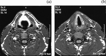
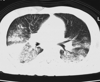
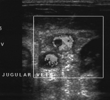
The contrast-enhanced computerised tomography (CT) scan of the neck, which was performed the following morning, confirmed thrombosis of the left internal jugular vein (Figure 2). No anticoagulation therapy was administered. A CT scan of the chest revealed multiple confluent densities of the lower as well as the upper lobes, bilaterally. The right lower lobe demonstrated more pulmonary infiltrates and pleural effusion than the left one, with an additional triangular atelectasis, but fortunately no intrapulmonary abscess (Figure 3). Few microcavitary lesions and bronchiectasis were identified in bilateral lung fields. CT scan of the brain revealed no abnormal findings. After five days of hospitalisation in the intensive care unit and eight days in the internal pathology department he was discharged home in good condition. He continued per os penicillin and metronidazole and recovered without sequelae after four more weeks of antibiotic treatment. Two months later magnetic resonance imaging of the neck revealed that thrombosis of the left internal jugular vein had not resolved. Five months after his discharge home triplex ultrasonography of the neck revealed that the internal jugular vein had recanalised (Figure 4).
(a) and (b) CT scan of the neck showing thrombosis of the left internal jugular vein.

A CT scan of the chest revealed multiple pulmonary infiltrates bilaterally and an atelectasis in the right lower lobe.

Five months after hospitalisation triplex ultrasonography of the neck revealed that the internal jugular vein had recanalised.

Discussion
Lemierre's syndrome is a rare clinical entity,Reference Hagelskjaer, Prag, Malczynski and Kristensen1, Reference Brazier, Hall, Yusuf and Duerden10 characterised by a high degree of variability due to different patterns of organ involvement in the septic embolic process. Pulmonary impairment, as seen in our patient, is a common manifestation of Lemierre's syndrome. Pulmonary septic emboli manifested as pleuritic chest pain, dyspnoea, haemoptysis or respiratory distress are seen in 95 per cent of Lemierre's syndrome cases reported. Within only a few hours after the initial symptoms, multiple bilateral necrotic pulmonary infiltrates frequently associated with pleural effusion, empyema, and/or pulmonary abscesses often develop. The consequent respiratory distress can be severe enough to necessitate intensive care observation and ventilatory support.Reference Sinave, Hardy and Fardy3, Reference Koay, Heyworth and Burden4, Reference Barker, Winer-Muram and Grey11, Reference DeSena, Rosenfeld, Santos and Keller12 Mild renal lesions manifested as albuminuria accompanied by increase of blood urea are not uncommon in Lemierre's syndrome. Some elevations in serum creatinine or blood urea nitrogen as well as transient haematuria have also occasionally been reported.Reference Sinave, Hardy and Fardy3, Reference Williams, Nagy, Wingate, Bailey and Wax13
The special clinical interest of this patient refers to his haematological and renal complications and namely the acute onset of both microangiopathic coagulation abnormalities and severe renal injury. Significant acute renal failure additionally complicated with thrombocytopenia, coagulopathy and haemolytic anaemia have not been associated with Lemierre's syndrome before. Differential diagnosis probably includes mild disseminated intravascular coagulopathy and haemolytic uraemic syndrome. These consumption coagulopathy situations are multiform and differential diagnosis between them is often difficult. Haemolytic uraemic syndrome is a heterogeneous group of devastating, multisystem disorders that affect predominantly young children with a history of gastrointestinal infection. In the absence of gastrointestinal infection or Shiga toxin-producing pathogens, acute onset of both microangiopathic haemolytic anaemia and renal injury evidenced by either haematuria, proteinuria, or elevated serum creatinine and urea levels and leukocytosis, as described in our patient, may compose the clinical profile of an atypical haemolytic urasemic syndrome. Thrombocytopenia can usually be detected early in haemolytic uremic syndrome. However, coagulation studies (prothrombin, partial thromboplastin time) are usually normal (in contrast to the abnormal values of our patient) and so are D-dimers. The syndrome has not so far included Fusobacterium necrophorum among the causative pathogens.Reference Elliott and Robins-Browne14 Disseminated intravascular coagulopathy, on the other hand, is more often correlated with pathological prothrombin, partial thromboplastin time values. D-dimers are usually elevated in disseminated intravascular coagulopathy (but normal in our patient) and are considered a sensible index for its diagnosis.Reference Wieding, Eisinger and Kostering15
Despite the serious renal and pulmonary complications, our patient responded well to the combination of high dose penicillin and metronidazole administered as recommended by most authors. Monotherapy with clindamycin for two to three weeks in patients without metastatic infection and for at least four to six weeks in patients with metastatic infection has also been proposed.Reference Gilbert, Moellering, Sande, Gilbert, Moellering and Sande16, Reference Appelbaum, Spangler and Jacobs17 Since the bacterium shows varying sensitivity to macrolides, these antibiotics may not be considered as the treatment of choice in Lemierre's syndrome.Reference Finegold18 In our case also, the initial treatment of the patient with macrolides was ineffective and led to deterioration of his general condition. Finally, although the role of early and prolonged intravenous antibiotic therapy in eradication of the multiple septic emboli is well recognised, at present ligation and resection of the internal jugular vein are no longer advocated and anticoagulation therapy is controversial.Reference Barker, Winer-Muram and Grey11
• Acute tonsillitis or pharyngitis may lead to suppurative thrombophlebitis of the internal jugular vein known as Lemierre's syndrome
• Respiratory deficiency and renal impairment are often reported in patients suffering from Lemierre's syndrome
• Fusobacterium necrophorum, a gram-negative anaerobic rod, has been identified as the causative pathogen
• Early identification of the syndrome may be crucial for patients who present in emergency rooms severely ill after insufficient treatment of a head and neck infection
The presentation of this case aims at correlating intravascular consumption of coagulation factors secondary to circulating septic emboli or secondary to vascular injury and acute renal failure to Lemierre's syndrome. Recognition of these haematological complications is critical, particularly in view of the fact that this life threatening syndrome usually involves previously healthy young patients.