Key findings
• This study documented the in vitro anti-trichinellosis effect of Annona muricata (Graviola) leaves, a drug of plant origin.
• Graviola leaves have potential activity against both intestinal and muscular phases of T. spiralis and could serve as an adjuvant to anti-trichinellosis drug therapy, especially in combination with albendazole.
• Besides, A. muricata has a prophylactic effect, thus adding to its value.
• Further studies are needed to elucidate the actual mode of action of A. muricata and explore its effect in combination with other anti-trichinellosis drugs.
Introduction
Trichinellosis is a zoonotic parasitic disease caused by nematodes of the genus Trichinella, which possesses great clinical importance and continues to be a public health concern worldwide (Wang et al., Reference Wang, Li, Wei, Wang, Zhang, Shi, Liu, Yang and Fang2018). It has a cosmopolitan distribution, especially in developing countries, with about 10,000 cases infected yearly (Gottstein et al., Reference Gottstein, Pozio and Nöckler2009; Wang et al., Reference Wang, Li, Wei, Wang, Zhang, Shi, Liu, Yang and Fang2018). Trichinella spiralis, the first discovered species, is the most virulent and pathogenic species to humans and has two lifecycle phases: an intestinal phase and a muscular phase. Humans can get infected by ingesting infective T. spiralis larvae in undercooked pork meat (Gottstein et al., Reference Gottstein, Pozio and Nöckler2009; Pozio, Reference Pozio2019).
Anthelmintic drugs, especially benzimidazole derivatives (such as albendazole and mebendazole), were used to treat human trichinellosis, but with no efficacy in killing the encysted larvae (Attia et al., Reference Attia, Mahmoud, Farrag, Makboul, Mohamed and Ibraheim2015). Furthermore, some of these drugs showed limited bioavailability and a high degree of resistance. Certain drugs are contraindicated in children below three years old and in pregnancy, while others are thought to be carcinogenic (Yadav, Reference Yadav2012). Therefore, establishing new safe and efficient anti-trichinellosis drugs is a pressing need, and a drug of plant origin may be a promising option.
Annona muricata (or, as the fruit is known, Graviola) is a well-known plant worldwide and contributes significantly to alternative medicine (Vila-Nova et al., Reference Vila-Nova, de Morais, Falcão, Alcantara, Ferreira, Cavalcanti, Vieira, Campello and Wilson2013). The key active orchestrating biomolecules responsible for the bioactive properties of Graviola are the alkaloids and acetogenins. They showed many anti-parasitic effects against various external and internal parasites (Rady et al., Reference Rady, Bloch and Chamcheu2018).
Many studies highlighted Graviola's anthelmintic activity against different nematodes (Souza et al., Reference Souza, Bevilaqua, Morais, Costa, Silva and Braz-Filho2008; Kamaraj & Rahuman, Reference Kamaraj and Abdul Rahuman2011).
In respect to the evidence regarding the medicinal aspects of Graviola, in our work, we have attempted validation of Graviola extract activity against T. spiralis both in vivo and in vitro.
Materials and methods
Animals, parasites and materials
Swiss albino male mice (n = 90) aged 5–6 weeks and weighing 20–25 g at the beginning of the experiment were obtained from the biological unit of Theodor Bilharz Research Institute (TBRI) (Giza, Egypt). The mice were kept on a standard commercial pelleted diet with free accessible water and ensuring good sanitary conditions throughout the time of the study.
The T. spiralis strain was obtained from the Parasitology department, TBRI. Mice were orally infected with 200 T. spiralis larvae (Wassom et al., Reference Wassom, Debra and Dick1988; Abou Rayia et al., Reference Abou Rayia, Saad, Ashour and Oreiby2017).
Albendazole was purchased as Bendax 200 mg tablets from Sigma Pharmaceutical Industries, Egypt, whereas A. muricata was purchased as Graviola 500 mg capsules from NOW® foods, USA. Annona muricata was used as standardized commercial 500 mg vegetarian capsules. Each capsule contained 1 g Graviola leaf, as the manufacturer recommends its supportive effects on healthy cell growth and function, and its free radical scavenging properties.
Isolation of T. spiralis adult worms and muscle larvae
Trichinella spiralis adults and muscle larvae were obtained from the infected mice according to Ozkoc et al. (Reference Ozkoc, Tuncay, Delibas and Akisu2009).
Swiss albino mice infected with T. spiralis for 30 days were sacrificed, muscles were separated and minced, and muscle larvae were digested by immersion in the acid pepsin solution (Dennis et al., Reference Dennis, Despommier and Davis1970). The mixture was incubated at 37°C for 2 h, continuously mixing with an electric stirrer (Dunn & Wright, Reference Dunn and Wright1985). Filtration of the digest was done according to Kapel et al. (Reference Kapel, Webster and Gamble2005). The collected larvae were washed two to three times with tap water and suspended in a conical flask for half an hour to allow sedimentation.
Trichinella spiralis adult worms were isolated from the small intestines of infected untreated mice six days post-infection (p.i.). The intestine was washed, opened longitudinally along its entire length, cut into small pieces 2 cm each and placed in normal saline at 37°C for three to four hours to allow the worms to migrate out of the tissue (Wakelin & Margaret, Reference Wakelin and Margaret1980).
In vitro and in vivo experimental design
Trichinella spiralis adult worms (25 parasites per well) were cultured in a 24-well tissue culture plate prepared with an incubation medium consisting of RPMI-1640 Medium (containing 20% fetal bovine serum, 200 U/ml penicillin and 200 μg/ml streptomycin). Three groups were established in this study: group I – adult worms cultured in the incubation medium only; group II – adult worms cultured in the incubation medium containing Graviola that was dissolved in dimethyl sulfoxide (DMSO) at a concentration of 100 μg/ml (Vila-Nova et al., Reference Vila-Nova, de Morais, Falcão, Alcantara, Ferreira, Cavalcanti, Vieira, Campello and Wilson2013); and group III – adult worms cultured in the incubation medium containing albendazole that was dissolved in DMSO at a concentration of 100 μg/ml (Tritten et al., Reference Tritten, Nwosu, Vargas and Keiser2012). Three wells were used for each group, and the plate was placed in the incubator at 37°C and 5% carbon dioxide for 24 h. Then, all adult worms were collected for scanning electron microscopic study.
In the in vivo study, mice were divided into five groups, with 18 mice in each group:
• Group I: infected non-treated (control positive).
• Group II: receiving Graviola for seven days before infection (as prophylaxis).
• Group III: infected and treated with Graviola (given orally at a dose of 1 g/kg) and dissolved in distilled water (Somsak et al., Reference Somsak, Polwiang and Chachiyo2016).
• Group IV: infected and treated with albendazole (given orally at a 50 mg/kg dose and suspended in 10% Tween 80 and 90% deionized water just before oral administration) (Attia et al., Reference Attia, Mahmoud, Farrag, Makboul, Mohamed and Ibraheim2015).
• Group V: infected and treated with a combination of Graviola plus albendazole in half doses.
Groups III–V were divided into three subgroups (a, b and c); each comprised six animals to assess the effect of the drugs given during the intestinal phase only (a) (3–5 days p.i.), muscular phase only (b) (30–32 days p.i.) and intestinal then muscular phases (c) (3–5 days p.i. and 30–32 days p.i.) separately.
Detection of T. spiralis adult worms and muscle larvae burden
In subgroups (a), to evaluate the effects of the treatment against the intestinal phase, the mice were sacrificed on day 6 p.i., and the small intestine was processed as previously described (Wakelin & Margaret, Reference Wakelin and Margaret1980). Trichinella spiralis adults were obtained, and the worm reduction rate was calculated.
In subgroups (b), to evaluate the effects of the treatment against the muscular phase, the mice were sacrificed on day 35 p.i., and the muscle larvae were obtained by the pepsin digestion method. The larvae were counted microscopically using the McMaster counting chamber, Faust-Germany. Parasite burdens were expressed as the number of larvae per gram of carcass digested (ML/g) (Nuñez et al., Reference Nuñez, Gentile, Costantino, Sarchi and Venturiello2005).
Scanning electron microscopy (SEM)
After 24 h, adult worms were processed as described by Abou Rayia et al. (Reference Abou Rayia, Saad, Ashour and Oreiby2017). Worms from each group were directly pipetted and immediately fixed in a fresh fixation solution of 2.5% glutaraldehyde solution buffered with 0.1 M sodium cacodylate at pH 7.2 and left overnight at 4°C. The fixed specimens were then washed in 0.1 M sodium cacodylate buffer at pH 7.2 for 5 min, post-fixed in 2% osmium tetroxide for 1 h and washed in distilled water. The specimens were dehydrated in ascending grades of ethyl alcohol and then mounted on carbon-coated adhesive material and examined using a FEI-Philips scanning electron microscope, Thermo Fisher Scientific, USA (Kim & Myron, Reference Kim and Myron1980; Bughdadi, Reference Bughdadi2010).
Histopathological studies
Parts of the small intestine and skeletal muscles from the studied groups were fixed in 10% formalin for 24 h, washed in water for 12 h, dehydrated in ascending grades of alcohols and cleared in xylene. Impregnation was done in pure soft paraffin for 2 h at 55°C. Then, hard paraffin sections of 5 μm thickness were cut by microtome. Sections were stained with haematoxylin and eosin stain (Drury & Wallington, Reference Drury and Wallington1980).
Statistical analysis
The data were analysed using Microsoft Excel 2016 and statistical package for social science IBM SPSS Statistics for Windows, version 26 (IBM Corp., Armonk, New York, USA). Continuous normally distributed variables were represented as mean ± standard deviation. With a 95% confidence interval, a P-value < 0.05 will be considered statistically significant. Student's t-test was performed to compare the means of normally distributed variables between groups, and analysis of variance (ANOVA) followed by Tukey honestly significant difference (HSD) as a post-hoc test in multiple groups were performed.
Results
SEM findings
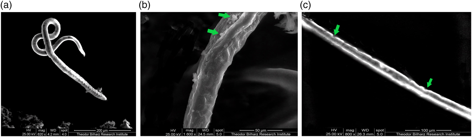
As regards the infected control group, when cultured in the incubation medium only, the cuticle of the adult worm retained the normal structure in the form of ridges, transverse creases and annulations, with the appearance of openings of the hypodermal gland (fig. 1a). In Graviola-treated groups, there was severe destruction of the adult worm, marked cuticle swelling, areas with vesicles, blebs and loss of annulations (fig. 1b). In the albendazole treated group, there was severe destruction of the adult worm and multiple fissures in the cuticle, with loss of annulations (fig. 1c).
SEM findings of the cultured T. spiralis adult: (a) normal adult worm; (b) Graviola-treated group, showing severe destruction of the adult worm, with loss of annulations and areas with blebs and vesicles (green arrows); (c) albendazole-treated group, showing severe destruction of the adult worm and multiple fissures in the cuticle (green arrows).

In vivo studies
Adult worm count in the small intestine
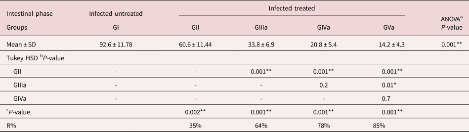
Prophylactic treatment of the infected mice by Graviola (GII) significantly reduced (P < 0.001) the mean adult worm count (60.6 ± 11.44) with an efficacy of 35% compared to the control infected untreated group (GI) (92.6 ± 11.78). A significant decrease in the mean number of adult worms was obtained in all treated groups (P < 0.001) in comparison to the control infected untreated group. The least mean adult count was found in GVa, which received combination therapy (14.2 ± 4.32) and showed the most effective eradication of T. spiralis adult worms, with a drug efficacy of 85%, followed by GIVa, which received albendazole (20.8 ± 5.4), with an efficacy of 78%. In comparison, the mean adult worm count was found to be 33.8 ± 6.87 in GIIIa, which received Graviola with a satisfactory percentage of reduction of 64% (table 1).
Trichinella spiralis adult worm count in the small intestine at the day 6 post-infection.

a P-value is significantly different comparing between groups, depending on the one-way ANOVA test.
b P-value is significantly different comparing between groups, depending on a post-hoc test (Tukey HSD).
c P-value is significantly different comparing with control, depending on Student t-test.
* Initial P-value < 0.05 is significant.
** Initial P-value < 0.01 is highly significant.
GI, infected untreated, GII, prophylactically treated with Graviola for seven days before infection; GIII, infected and treated with Graviola; GIV, infected and treated with albendazole; GV, infected and treated with the combination of Graviola plus albendazole in half doses; R%, percentage of the reduction.
Encysted larvae count in muscles
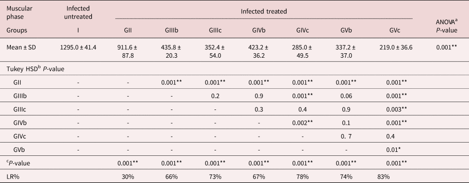
Concerning the drug effects on the muscle phase, prophylactic treatment of the infected mice by Graviola significantly reduced (P < 0.001) the mean larval count per gram muscle (911.6 ± 87.82) with an efficacy of 30% compared to the control infected untreated group (1295 ± 41.38). A significant decrease in the mean larval count per gram muscle was detected in all treated groups (P < 0.001) compared to the control infected untreated group. In comparing the drug effects on the muscular phase while giving the drugs in two regimens, either giving the drugs for a single dose in the muscular phase only or giving the drugs for two doses, one dose in the intestinal phase followed by a second dose of the same drugs during the muscular phase, a better larvae eradication was found in groups that received the drugs in two doses than groups that received the drugs for a single dose. As regards the two doses regimen, the best reduction of the mean larval count was found in group GVc, which received combination therapy (219 ± 36.63) with an efficacy of (83%), followed by the mice group that received albendazole (GIVc) (285 ± 49.48) with an efficacy of 78%. In the mice group that received Graviola (GIIIc), the mean larval count was 352.4 ± 53.96 with a 73% reduction percentage. Concerning the single-dose regimen, the best reduction of the larval count was found in group GVb, which received combination therapy (337.2 ± 37.02) with an efficacy of 74%. Nearly similar effectiveness was detected in mice groups that received Graviola (GIIIb) and albendazole (GIVb), with mean larval counts of 435.8 ± 20.34 and 423.2 ± 36.17, and percentages of reduction of 66% and 67%, respectively (table 2).
Trichinella spiralis encysted larvae count per gram muscle at the day 35 post-infection.

a P-value is significantly different comparing between groups, depending on the one-way ANOVA test.
b P-value is significantly different comparing between groups, depending on a post-hoc test (Tukey HSD).
c P-value is significantly different comparing with control, depending on Student t-test.
* Initial P-value < 0.05 is significant.
** Initial P-value < 0.01 is highly significant.
GI, infected untreated; GII, prophylactically treated with Graviola for seven days before infection; GIII, infected and treated with Graviola; GIV, infected and treated with albendazole; GV, infected and treated with the combination of Graviola plus albendazole in half doses; R%, percentage of the reduction. Groups III–V were divided into three subgroups (a, b, c), each composed of six animals to assess the effect of the drugs given during the (a) intestinal phase only (3–5 days p.i.); (b) muscular phase only (30–32 days p.i.); and (c) intestinal then muscular phases (3–5 days p.i. and 30–32 days p.i.) separately.
No remarkable behavioural changes were observed in mice treated with Graviola leaf extract, and clinical signs were absent.
Histopathological results
Small intestine changes
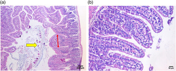
Histopathological examination of sections from the small intestine of the infected control group (GI) showed dense intravillous inflammatory cellular infiltration mainly consisting of mononuclear cellular infiltrate in the form of lymphocytes and plasma cells. There was broadening and atrophy of the intestinal villi with crypt hyperplasia. Moreover, fragments of the adult worms were detected within the intestinal lumen (fig. 2a).
Histopathological findings of sections from the small intestine: (a) GI, showing fragments of the worm within the intestinal lumen (yellow arrow), broadening (red double-headed arrow) and atrophy of the intestinal villi, as well as dense intravillous plasma-lymphocytic cellular infiltration (H&E stain, X100). (b) GVa, showing return of the normal villous pattern (H&E stain, X400).

Concerning the results of the sections examined from the treated groups, an evident decrease in the intensity of the inflammatory cellular infiltration was observed, together with remarkable improvement of the other histopathological changes of the intestine, with a returning of the normal villous pattern in GVa (fig. 2b).
Skeletal muscle changes
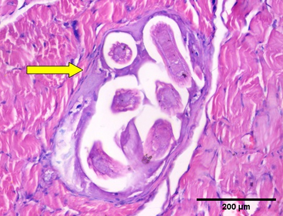
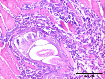
Histopathological examination of muscular sections from the infected control group (GI) revealed the presence of a massive number of encysted T. spiralis larvae present diffusely in the sarcoplasm of the muscles and a number of chronic inflammatory cells in the form of lymphocytes, plasma cells and histiocytes infiltrating muscle bundles and surrounding the encysted larvae (fig. 3). The muscles from the Graviola prophylaxis group (GII) showed a large number of cysts with focally degenerated capsule and pericapsular histio-lymphocytic inflammatory cellular infiltration (fig. 4).
Histopathological examination of sections from skeletal muscle of GI, showing intact capsule (yellow arrow) and pericapsular plasma-lymphocytic inflammatory cellular infiltration (H&E stain X200).

Histopathological examination of sections from skeletal muscle of GII, showing focally degenerated capsule and pericapsular histio-lymphocytic inflammatory cellular infiltration (H&E stain X200).

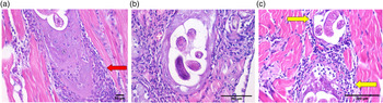
Concerning the histopathological examination of muscular sections from mice groups that received the drugs for a single dose in the muscular phase only (GIIIb, GVIb, GVb), there was a decreased number of cysts with focally degenerated capsule and dense pericapsular plasma-lymphocytic inflammatory cellular infiltration (fig. 5a–c).
Histopathological examination of sections from skeletal muscle of (a) GIIIb, showing mostly intact capsule and pericapsular histio-lymphocytic inflammatory cellular infiltration (red arrow)(H&E stain X200); (b) GIVb, showing focally degenerated capsule and dense pericapsular plasma-lymphocytic inflammatory cellular infiltration (H&E stain X200); (c) GVb, showing degenerated capsule (yellow arrows) and pericapsular histio-lymphocytic inflammatory cellular infiltration (H&E stain X200).

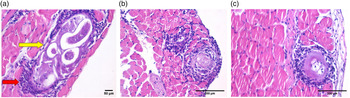
Examination of muscular sections from mice groups that received the drugs for two doses showed that one dose in the intestinal phase followed by a second dose of the same drugs during the muscular phase revealed marked improvement of the histopathological finding compared to the infected control group (GI). GIIIc and GIVc showed a decreased number of cysts with degenerated capsule and focal pericapsular plasma-lymphocytic inflammatory cellular infiltration (fig. 6a, b), while GVc showed the best improvement in the presence of the least number of cysts with degenerated larvae capsules. There was also a decrease in larvae size and destruction of the larvae's internal structure (fig. 6c).
Histopathological examination of sections from skeletal muscle of (a) GIIIc, showing degenerated capsule (yellow arrow) and focal pericapsular plasma-lymphocytic inflammatory cellular infiltration (red arrow) (H&E stain X200); (b) GIVc, showing degenerated capsule and pericapsular plasma-lymphocytic inflammatory cellular infiltration (H&E stain X200); (c) GVc, showing degenerated capsule and focal pericapsular plasma-lymphocytic inflammatory cellular infiltration (H&E stain X200).

Discussion
The medical treatment of trichinellosis is a matter of much debate. Albendazole, one of the benzimidazoles, is still the available drug of choice in trichinellosis treatment. However, it is reported that albendazole can cause multiple systemic serious adverse drug reactions, as severe drug eruptions, encephalitis, epilepsy and even death (Shalaby et al., Reference Shalaby, Moghazy, Shalaby and Nasr2010; Yadav, Reference Yadav2012). Besides, it shows poor susceptibility to migrating and encapsulated muscle larvae (Djurkovc et al., Reference Djurkovc, Bobic, Nikolic, Klun and Dupouy-Camet2013). These data elucidate the urgent need for a new, safe and effective treatment capable of eradicating the Trichinella spp. infection.
Annona muricata (Graviola) is a widely known fruit due to its sour and sweet taste. It has been a matter of dedicated experimental studies, being a safe, natural agent. The anti-parasitic activity was tested (Othman & Shoheib, Reference Othman and Shoheib2016; Abdul Wahab et al., Reference Abdul Wahab, Jantan, Haque and Arshad2018). Moreover, Graviola has a wide range of safety. Research showed that the oral median LD50 was estimated to be ≥2500 mg/kg, and no mortality occurred within the observation period of seven days in mice given 4000 mg/kg (Somsak et al., Reference Somsak, Polwiang and Chachiyo2016).
In the present study, the anthelmintic activity of Graviola against Trichinella spp. was tested both in vitro and in vivo. To the best of our knowledge, this is the first report to evaluate this plant activity against Trichinella spp.
The body wall of Trichinella spp. consists of the cuticle, the hypodermis and the somatic musculature. The shape of the parasite, nutrition and protection is dependent on cuticle integrity. The cuticle is also essential for osmoregulation (Djurkovc et al., Reference Djurkovc, Bobic, Nikolic, Klun and Dupouy-Camet2013). In our study, the electron microscopy scans showed severe destruction of the adult worm, marked cuticle swelling, areas with vesicles, blebs and loss of annulations in Graviola- and albendazole-treated groups. At the same time, it retained its normal morphology when incubated in the culture medium only. The main route of drug passage into nematodes is transcuticular passive diffusion with subsequent destruction of the worm's surface. The appearance of surface blebs on the worm's surface is attributed to the response of the worm to replace its destroyed surface membrane. So, all these findings indicate efficient anti-parasitic activity (Abdul Wahab et al., Reference Abdul Wahab, Jantan, Haque and Arshad2018). This may help to explain the mechanism of action of Graviola.
The in vitro anti-parasitic effects of Graviola have been previously documented in many studies (Nguyen-Pouplin et al., Reference Nguyen-Pouplin, Tran and Tan2007; Osorio et al., Reference Osorio, Arango, Jiménez, Alzate, Ruiz, Gutiérrez and Robledo2007; Boyom et al., Reference Boyom, Fokou and Yamthe2010; Ross, Reference Ross2010; Ferreira et al., Reference Ferreira, Castro, Chagas, Franca and Beleboni2013). Nevertheless, most of these studies tested the activity against eggs or larval stages. Few studies tested the anti-parasitic action against the adult worm as performed in our study. Hounzangbe-Adote et al. (Reference Hounzangbe-Adote, Paolini, Fouraste, Moutairou and Hoste2005) and Ferreira et al. (Reference Ferreira, Castro, Chagas, Franca and Beleboni2013) found that extracts of Graviola were active against eggs, infective larvae and adult forms of the parasite. The effect was comparable to levamisole, which is considered a promising result, as its activity during all the worm's lifecycle stages could decrease the parasite's drug resistance (Ferreira et al., Reference Ferreira, Castro, Chagas, Franca and Beleboni2013).
The potential anti-parasitic activity of an agent is usually tested in vitro first before attempting in vivo studies, as this is practical to save time and money. When an agent is concluded to be effective in vitro, this does not necessarily mean that it will have an in vivo activity. This discrepancy is secondary to variable factors as the bioavailability and the pharmacology of such agents in the host (Boyom et al., Reference Boyom, Fokou and Yamthe2010). Therefore, a successful in vitro agent must be tested in vivo as well. Because of this, we performed in vivo testing also for Graviola using experimental mice.
In this study, the administration of Graviola, albendazole and combined therapy (Graviola and albendazole) significantly reduced the total adult worm count compared to the infected control group. The reduction percentage was 35% in the Graviola prophylactic group and 64% in the Graviola-treated group – less than that found in the albendazole-treated group (78%). However, the best response was found in the combined (Graviola and albendazole) treated group (85%).
Concerning the drug effects on the muscular phase, a significant decrease in the mean larval count per gram muscle was detected in all treated groups (P < 0.01) compared to the control infected untreated group. Their levels of efficacy varied according to the drug regimen. Better larvae eradication was found in the subgroups that received the two-doses drug regimen (the drugs were given during intestinal then muscular phases). The best reduction of the mean larval count per gram muscle was found in the combination therapy group (83%), followed by the albendazole-treated group (78%) and Graviola-treated group (73%).
Prophylactic treatment against T. spiralis has been evaluated in previous studies. Abu El Ezz (Reference Abu El Ezz2005) reported a significant reduction in adult (45.3%) and larval counts (43.3%) when Nigella sativa was used as prophylaxis against T. spiralis. The same results were also documented by Nada et al. (Reference Nada, Mohammad, Moad, El-Shafey, Al-Ghandour and Ibrahim2018).
In the present study, single-drug treatment significantly decreased the count of T. spiralis, but the drug combination worked better than a single drug. In agreement with our results, a study carried out by Shoheib et al. (Reference Shoheib, Shamloula, Abdin and El-Segai2006) reported that using the anti-fibrotic agents, either αchymotrypsin or colchicine alone, results in lower worm reduction than when combined with albendazole. In the same way, Ren et al. (Reference Ren, Qin, Zhang, Zheng, Dai, Wu and Cui2018) reported better larvae eradication when using a combination of Wortmannilatone F and recombinant G31P against T. spiralis.
The effect of albendazole against T. spiralis has been documented in previous studies, with variable efficacies (Chung et al., Reference Chung, Joo, Quan, Kwon and Cho2001; Siriyasatien et al., Reference Siriyasatien, Yingyourd and Nuchprayoon2003; Shoheib et al., Reference Shoheib, Shamloula, Abdin and El-Segai2006; Nada et al., Reference Nada, Mohammad, Moad, El-Shafey, Al-Ghandour and Ibrahim2018; Huang et al., Reference Huang, Yao, Liu, Yang, Wang, Shi and Yang2020).
The variation in albendazole efficacies against intestinal and muscular stages depends on treatment dose, time and duration (Siriyasatien et al., Reference Siriyasatien, Yingyourd and Nuchprayoon2003). Its action is through the inhibition of microtubule polymerization via selective binding to beta-tubulin monomer of the parasite, with little effect on the binding of the host tubulin (Aguayo-Ortiz et al., Reference Aguayo-Ortiz, Méndez-Lucio, Medina-Franco, Castillo, Yépez-Mulia, Hernández-Luis and Hernández-Campos2013).
Few studies tested the in vivo anti-parasitic effects of Graviola. In the study by Vieira et al. (Reference Vieira, Cavalcante, Pereira, Dantas and Ximenes1999), a 40% reduction of Haemonchus contortus egg counts in the sheep was reported. Annona muricata in vivo anti-malarial activity was attributed to the effect of some bioactive compounds (diterpenoids, flavonoids, polyphenols, saponins, alkaloids, kaempferol and acetogenin). Moreover, the antioxidant effect in this plant may also contribute to the anti-malarial activity (Yamthe et al., Reference Yamthe, Fokou, Mbouna, Keumoe, Ndjakou, Djouonzo and Boyom2015). Additionally, it has been demonstrated that phenolic compounds inhibit the activity of important enzymes for fatty acid biosynthesis (Coria-Téllez et al., Reference Coria-Téllez, Montalvo-Gónzalez, Yahia and Obledo-Vázquez2018). Some of those mechanisms may apply in the case of Trichinella spp. infection, resulting in decreased adult and larval counts.
Till now, the actual mechanisms of action of the Annonaceae family's extracts as anthelmintic are still unclear. Nevertheless, studies have suggested that they cause inhibition of cell division, which hinders the formation and development of vital structures that inhibit the larval development of different parasites (Gallardo et al., Reference Gallardo, Aragón, Tormo, Blázquez, Zafra-Polo and Cortes1998; Rady et al., Reference Rady, Bloch and Chamcheu2018).
In this study, histopathological examination of sections from the small intestine of the infected control group showed dense intravillous inflammatory cellular infiltration. There was broadening and atrophy of the intestinal villi with crypt hyperplasia. Moreover, fragments of the adult worms were detected within the intestinal lumen. Muscular sections from the infected control group revealed the presence of a massive number of encysted T. spiralis larvae diffusely present in muscle sarcoplasm and a number of chronic inflammatory cells. This result agreed with Dyab et al. (Reference Dyab, Ahmed and Abdelazeem2019).
Reduction of these destructive and inflammatory changes was evident in the treated groups. The combination therapy group showed the best improvement in restoring the normal architecture, the presence of the least number of cysts with degenerated capsule and focal pericapsular plasma-lymphocytic inflammatory cellular infiltration.
In conclusion, our results conclude that Graviola could have a promising, safe, adjuvant natural prophylactic and therapeutic option both in vitro and in vivo against T. spiralis in its intestinal and muscular phases. It showed comparable results with albendazole. Furthermore, its use in combination with albendazole gives a much better response than albendazole alone.
We recommend further studies to elucidate the molecular basis of the A. muricata action in trichinellosis and extract its bioactive compounds to be more efficient in the disease treatment without undesirable side effects.
It is suggested that using natural plant extract is better than using ready-to-use capsules as this will provide us with basic information about the plant.
Financial support
This research received no specific grant from any funding agency, commercial or not-for-profit sectors.
Conflicts of interest
None.
Ethical standards
All applied experiments were carried out after the approval of the Ethical Committee at the Faculty of Medicine, Kafrelsheikh University, Egypt (IRB#1632020RA) under the ethical guidelines of animal experiments.