Introduction
The concept of ‘Catapyrenium s. lat.’ used here includes squamulose, squamulose-areolate or nearly crustose species; thalli are heteromerous with a variable degree of differentiation, and different types of tissues, both in the upper cortex, medulla and lower cortex. Attachment organs are mainly rhizohyphae and rhizines (rhizinomorphs sensu Malone Reference Malone1977; Wagner & Letrouit-Galinou Reference Wagner and Letrouit-Galinou1988). The photobiont is a green alga. Ascomata are perithecia, with involucrellum present or not; asci are cylindrical or clavate, 8-spored, with ascospores simple and colourless. Pycnidia can be present or not and both Dermatocarpon and Endocarpon types have been observed in the group. Members of Catapyrenium s. lat. occur mostly on soil, sometimes directly on rocks or in crevices and on ledges, and even living as parasites; most species occur on calcareous substrata. They are distributed mainly in temperate, semiarid or cold areas of both hemispheres (Breuss Reference Breuss1990a; Harada Reference Harada1993; Breuss Reference Breuss1996a).
Traditionally, many species included in Catapyrenium s. lat. have been placed in other genera such as Endocarpon, Dermatocarpon or Verrucaria (e.g. Endocarpon cinereum or Dermatocarpon boccanum) (Persoon Reference Persoon1794; Acharius Reference Acharius1810; Servít Reference Servít1955). Nevertheless, Breuss (Reference Breuss1990a) emended the genus Catapyrenium Flot. to accommodate only the squamulose species with simple ascospores and without hymenial algae, recognizing six groups at infrageneric level (for a historical review on the early systematics of Catapyrenium s. lat. see Breuss Reference Breuss1990a), and thus recognising the considerable anatomical and morphological variability present in the group. Some years later, a total of eight different genera was segregated by Harada (Reference Harada1993) and Breuss (Reference Breuss1996a) based on combinations of characters such as the type of pycnidium, ascus shape and arrangement of the ascospores, thallus anatomy and morphology (structure of the upper cortex and type of attachment organs) and presence or absence of an involucrellum. These authors recognized Anthracocarpon Breuss, Catapyrenium Flot. (Catapyrenium s. str.), Clavascidium Breuss, Heteroplacidium Breuss, Involucropyrenium Breuss, Neocatapyrenium H. Harada, Placidium A. Massal and Scleropyrenium H. Harada.
Recent molecular studies have been carried out in order to understand relationships between members of Verrucariales at different levels (e.g. Heiðmarsson Reference Heiðmarsson2003; Gueidan et al. Reference Gueidan, Roux and Lutzoni2007; Amtoft et al. Reference Amtoft, Lutzoni and Miądlikowska2008; Savić et al. Reference Savić, Tibell, Gueidan and Lutzoni2008; Prieto et al. Reference Prieto, Martínez, Aragón, Otálora and Lutzoni2010). The results of these molecular analyses have shown that the main genera included in the concept of Catapyrenium s. lat. (except the genus Clavascidium) are independent and unrelated entities (Gueidan et al. Reference Gueidan, Roux and Lutzoni2007, Reference Gueidan, Savić, Thüs, Roux, Keller, Tibell, Prieto, Heiðmarsson, Breuss and Orange2009; Savić et al. Reference Savić, Tibell, Gueidan and Lutzoni2008), thus confirming that the variability of the group is related to evolutionary patterns.
Catapyrenium s. lat. has been poorly studied in the Iberian Peninsula with only scattered references in some floristic studies or in publications of some newly described species (e.g. Breuss & Etayo Reference Breuss and Etayo1992; van den Boom & Giralt Reference van den Boom and Giralt1996; Navarro-Rosinés et al. Reference Navarro-Rosinés, Etayo and Breuss1996; Aragón Reference Aragón2003); although Breuss (Reference Breuss1990a) in his monograph of the genus included this region, the study was mainly focused on central Europe. As the group has remained poorly understood in the Iberian Peninsula, with little collected material, and because of recent taxonomic changes in the group, an exhaustive revision of all taxa in the group in this region was necessary. The work formed part of the ‘Flora Liquenólogica Ibérica’ project.
Material and Methods
This study is based mainly on material collected by the authors in the Iberian Peninsula and the Balearic Islands between 2005 and 2009. The collection comprises c. 1700 specimens deposited at MA.
In addition, Iberian collections from BCC, BCN, BP, LEB, LISU, MA, MACB, MAF, SANT, TFC, VAL and VIT and European and North American herbaria ABL, ARIZ, ASU, B, BM, COLO, GB, H, HAL, L, LI, NY, PRM, S and TUR were revised. Some personal collections (C. Keller, C. Scheidegger and G. Aragón) have also been revised. A total of c. 2000 specimens were studied. A list of the material studied can be seen online (http://www.escet.urjc.es/biodiversos/espa/personal/isabel/isabel_e.htm).
Observations and measurements were made using a Nikon SMZ-800 dissecting microscope and an Olympus BX 51 microscope. The sections (14–20 µm) of thalli were made using a Leica CM 1850 UV freezing microtome and were observed and measured in water and sometimes in lactophenol cotton blue.
For anatomical studies, ten samples per species were analysed (when available), and ten measurements of each specimen on different squamules were carried out. The limited material of some species and the poor condition of others, led to a lower number of measurements in some cases. Measurements are expressed as the mean ± standard deviation (SD) with the extremes in parentheses; length/breadth ratios (l/b) were calculated for both ascospores and conidia. Only data from the Iberian material were included in the descriptions. Frequently, measurements of asci and paraphyses were not enough for descriptive purposes, in these cases we included published data. Most variables were normally distributed, in cases where they were not normal, transformations were carried out.
For the statistical analysis we conducted ANOVA in order to understand the differences between closely related species using SPSS Statistics 17.0.
Distributional maps were drawn with ArcView GIS 3.1, based on UTM coordinates (WGS84 Datum).
For each taxa, we include data of the basionym, type specimens and their location, but not previously published synonyms (see Breuss Reference Breuss1990a and Harada Reference Harada1993).
Key to genera
1 Perithecia between the squamules (sometimes in the margin), not immersed in the thallus, with involucrellum ... Involucropyrenium
[Pycnidia not very common, Endocarpon-type]
Perithecia immersed in the thallus, without involucrellum ... 2
2(1) Upper cortex cinereum-type, thin (5–40 µm) and poorly delimited from the algal layer, with small, roundish-subangular cells of 4–10 µm diam. ... ... Catapyrenium
[Pycnidia absent]
Upper cortex clearly delimited from the algal layer, from 20 to c. 100 µm thick, with roundish-angular cells 4–15 µm diam. ... 3
3(2) Pycnidia Endocarpon-type, rhizohyphae present or not, with rhizines or stipe-like structures . ... 4
Pycnidia Dermatocarpon-type, rhizohyphae always present, rhizines present or not ... 5
4(3) Exciple and rhizines brown to carbonaceous, rhizohyphae dark, lower cortex absent ... Anthracocarpon virescens
[including Clavascidium liratum]
Exciple not carbonaceous, rhizohyphae absent, squamules attached to the substratum by the basal ends or by rhizines, lower cortex clearly delimited from the medulla ... Neocatapyrenium
5(3) Squamules small, 0·3–3 mm, thallus entirely paraplectenchymatous or subparaplectenchymatous, upper cortex 20–55 µm thick, with cells of 5–13 µm, laminal pycnidia, asci clavate ... Heteroplacidium
Squamules larger, (1) 2–8 (15) mm, thallus more differentiated, upper cortex 20–100 µm thick, with cells of 5–17 µm, laminal or marginal pycnidia, asci cylindrical at least when young, clavate in some rhizinate species ... Placidium
Taxonomic treatment
Anthracocarpon Breuss
Ann. Naturhist. Mus. Wien, Ser. B, Bot. Zool. 98 (Suppl.): 40 (1996); type: A. virescens (Zahlbr.) Breuss [= Clavascidium liratum (Breuss) Breuss].
The thallus is squamulose, with an anatomy very similar to the genus Placidium but characterized by the presence of Endocarpon-type pycnidia, perithecia with a black to carbonaceous exciple (at least on the top, e.g., A. caribaeum Breuss), and rhizohyphae and rhizines as attachment organs. Of the three species reported worldwide, only A. virescens occurs in Europe.
Anthracocarpon virescens (Zahlbr.) Breuss
Ann. Naturhist. Mus. Wien, Ser. B, Bot. Zool. 98 (Suppl.): 40 (1996).—Dermatocarpon virescens Zahlbr., Österr. Botan. Z. 68: 69 (1919); type: Jugoslawien, Dalmatien, West Meleda, erdige Kalkfelsspalten über Porto Soline, c. 200 m, 17/03/1910, J. Baumgartner (W!—holotype).
Clavascidium liratum (Breuss) Breuss, Ann. Naturhist. Mus. Wien, Ser. B, Bot. Zool. 98 (Suppl.): 41 (1996).—Catapyrenium liratum Breuss, Linzer biol. Beitr. 23(2): 533 (1991); type: Mallorca, Sierra del Norte (Serra de Tramuntana), Col de Hono zwischen Buñolá und Alaró, G. B. Feige nº 6856 (HAL—holotype!; LI—isotype!).
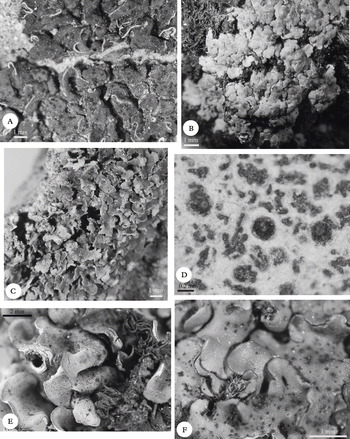
Habit of some species of Catapyrenium s. lat. A, Anthracocarpon virescens; B, Catapyrenium psoromoides; C, Heteroplacidium divisum; D, Involucropyrenium llimonae; E, Neocatapyrenium cladonioideum; F, Placidium imbecillum.

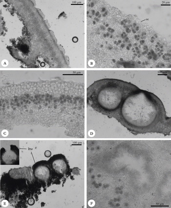
Anatomy of Catapyrenium s. lat. species, A–E, cross sections of thalli; F cross-section of pycnidia. A, Anthracocarpon virescens showing the sulcate epinecral layer; B, Catapyrenium psoromoides showing the cinereum-type upper cortex (uc); C, Heteroplacidium divisum; D, Heteroplacidium imbricatum, showing two perithecia; E, Involucropyrenium waltheri showing two perithecia with involucrellum (inv); F, Endocarpon-type pycnidia in Neocatapyrenium cladonioideum.

Thallus squamulose; squamules up to 5 mm, scattered to contiguous, sometimes overlapping, flattened to convex, rounded to slightly lobed; upper surface cream-coloured, pale, medium to dark brown, with conspicuous black perithecial ostioles (Fig. 1A); lower surface pale, brown or black with dark rhizohyphae and carbonaceous rhizines, simple or branched, one to several per squamule. Anatomy: thallus (180) 235 ± 35 (325) µm thick, with or without epinecral layer [up to 45 µm when present sulcate or not (Fig. 2A)]; upper cortex paraplectenchymatous, (37) 58 ± 15 (100) µm thick, with cells (6) 9·5 ± 2·4 (15) µm diam. Algal layer (52) 74 ± 15 (112) µm thick, with cells (4) 6·7 ± 1·5 (13) µm diam. Medulla prosoplectenchymatous, (45) 93 ± 30 (162) µm thick, with elongated cells (2) 3·8 ± 0·9 (6) µm diam.; lower cortex absent. Rhizohyphae (2·5) 3·6 ± 0·7 (5) µm, dark; rhizines up to 300 µm thick, simple or branched.
Perithecia up to 500 µm wide, pyriform to subglobose, with a brown to black and carbonaceous exciple, 20–55 µm thick. Asci clavate, 60–90 × 14–19 µm; ascospores biseriate, simple, (14) 17·5 ± 1·6 (21) × (6) 7·4 ± 0·8 (9) µm, l/b ratio (1·7) 2·4 ± 0·4 (3·5).
Pycnidia Endocarpon-type; conidia bacilliform, (5) 6·7 ± 0·9 (9) × (1) 1·1 ± 0·2 (1·5) µm, l/b ratio (4) 6·2 ± 1·3 (9).
Notes. Clavascidium liratum was described by Breuss (Reference Breuss1991) and differentiated from A. virescens mainly by the presence of a thicker and sulcate epinecral layer and a brown exciple in C. liratum (rather than carbonaceous as in A. virescens). Both shared a prosoplectenchymatous medulla, dark rhizohyphae and black rhizines. The type material of C. liratum was morphologically very similar to other specimens of A. virescens although with the upper surface paler.
Endocarpon-type pycnidia are present in A. virescens, but they were not observed in the type specimen of Clavascidium liratum; this last species was known only from the type locality in the Balearic Islands. Recently, we collected material in this locality belonging to the typical form of A. virescens. Two more specimens collected in Portugal showed a pale upper surface, with a sulcate epinecral layer, and a non carbonaceous exciple, being similar to C. liratum. These specimens presented Endocarpon-type pycnidia, until now never observed in C. liratum.
A molecular analysis of these Portuguese specimens, together with the type specimen of C. liratum and two typical samples of A. virescens showed that all formed a monophyletic clade (results not shown). This analysis supports the decision taken here to synonymize Clavascidium liratum with Anthracocarpon virescens.
Due to the presence of rhizines, the carbonaceous exciple and the colour and shape of the squamules, A. virescens resembles Endocarpon pusillum Hedw.; however, this last species is characterized by perithecia with hymenial algae and muriform ascospores.
Ecology. The species shows preferences for soil and rock ledges on calcareous and gypsiferous substrata in shrublands with Buxus sempervirens L., Lavandula latifolia Medik., Lycium sp, Rosmarinus officinalis L. and Thymus sp. in dry and open habitats; we have also collected it in Pinus halepensis Mill. forests. Anthracocarpon virescens has been frequently found together with Placidiopsis cinerascens (Nyl.) Breuss, P. custnani (A. Massal) Körb. or Placidium pilosellum. In the area studied, A. virescens was found between 170 and 1200 m altitude.
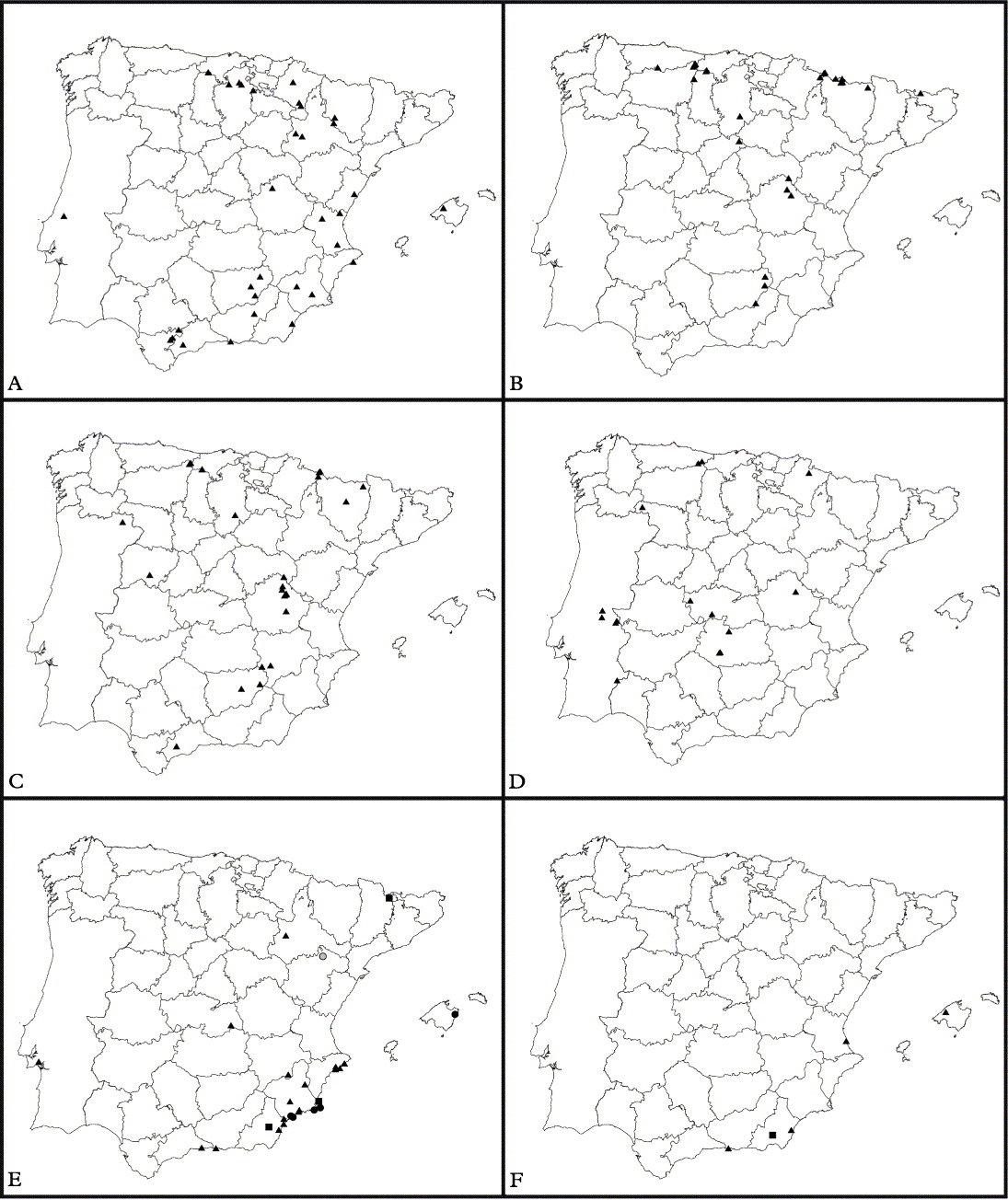
Distribution. The species is relatively frequent in xeric areas of the eastern Iberian Peninsula, being also present in the Balearic Islands (Fig. 3A). New data extend the known distribution of the species in the Iberian Peninsula, that was previously known mainly from the east coast of Spain. The specimen collected in Portugal is the first record of A. virescens from this country.
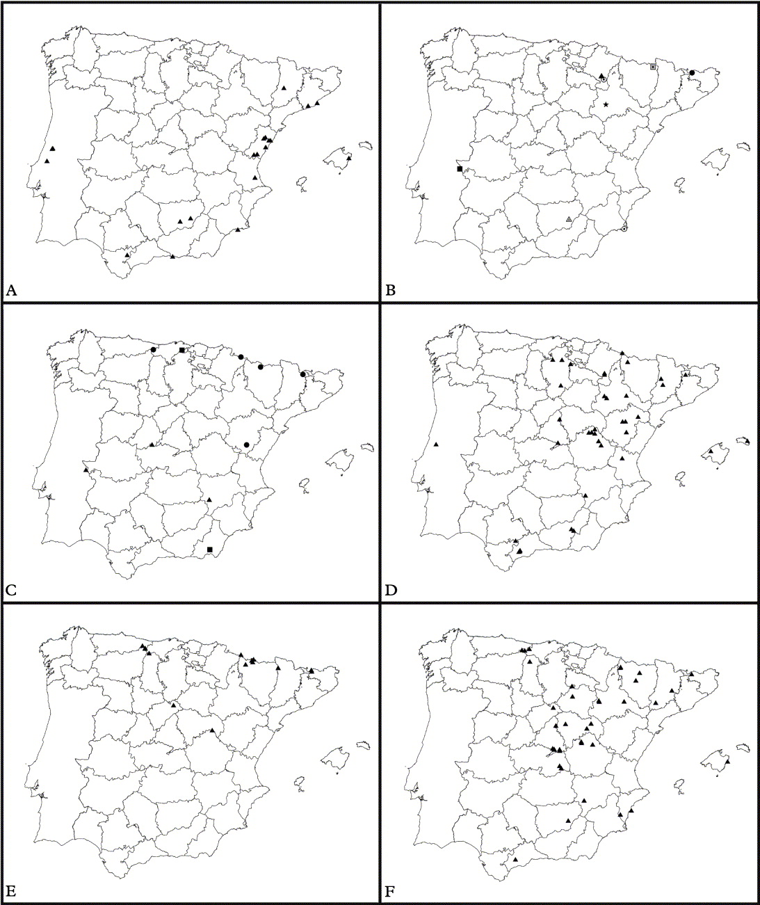
Distribution of Catapyrenium s. lat. species in Iberia. A, Anthrocarpon virescens; B, Catapyrenium cinereum; C, C. daedaleum; D, C. psoromoides; E, Heteroplacidium acervatum (●); H. compactum (■), H. congestum (○) and H. contumescens (▲); F, H. divisum (▲) and H. phaeocarpoides (■).

Anthracocarpon virescens is widely distributed in the Mediterranean region, being known from Croatia (Breuss Reference Breuss1990a), Greece (Christensen et al. Reference Christensen, Pišút and Sipman1997; Sipman & Raus Reference Sipman and Raus1999), Italy (Nimis & Martellos Reference Nimis and Martellos2004), Spain (Breuss Reference Breuss1990a), Tunisia (Breuss Reference Breuss1994a) and Turkey (Breuss & John Reference Breuss and John2004; Tufan et al. Reference Tufan, Sümbül and Özdemir2005), growing from sea level to 1400 m altitude.
Catapyrenium Flot
Bot. Zeitung 8: 361 (1850); type: C. cinereum (Pers.) Körb.
The thallus is composed of small squamules, 1–4 mm wide, forming a more or less continuous thallus; there is a cinereum-type upper cortex, i.e. thin (5–40 µm), poorly delimited from the algal layer, paraplectenchymatous, with small, roundish-subangular cells 4–10 µm diam. (Fig. 2B). The algal layer is not clearly delimited above and below, being more or less diffuse; the algal cells are small (2·5–10 µm). The asci are clavate, 8-spored, with ascospores biseriate, simple, colourless, 12–24 × 5–9 µm. Occasionally, we have found some pseudoseptate ascospores (see Prieto et al. Reference Prieto, Martínez, Aragón, Otálora and Lutzoni2010). Pycnidia absent. There are seven species world-wide, after the recent exclusion of C. dactylinum from the genus (Prieto et al. Reference Prieto, Martínez, Aragón, Otálora and Lutzoni2010). Four species are present in Europe, three of which are here reported from the Iberian Peninsula.
Key to Catapyrenium species
1 Squamules with dark margins; lower cortex well-developed, paraplectenchymatous, medulla composed of spherical hyphae; ascospores 14–24 × 6–9 µm ... ... C. cinereum
Squamules without dark margins; lower cortex absent; medulla loose, composed of a mixture of elongated and spherical hyphae ... 2
2(1) On soil; ascospores 15–24 × 5–9 µm ... C. daedaleum
On bark; ascospores 12–18 × 5–7 µm ... C. psoromoides
Catapyrenium cinereum (Pers.) Körb
Syst. Lich. Germ.: 325 (1855).—Endocarpon cinereum Pers., Nueue Ann. Bot. 1: 28 (1794); type: Germania [‘Reperi hanc speciem prope Schanzfeld: circa die alte kirche & prope montem Meisner’] (H-ACH nº 851 – type?!).
Thallus squamulose; squamules tightly contiguous, forming a continuous thallus, small, (0·5) 1–3 mm wide, finely lobulate; upper surface whitish, rarely greenish grey or brownish, pruinose, with dark margins; lower surface black, with dark rhizohyphae. Anatomy: thallus (100) 198 ± 45 (312) µm thick, with or without epinecral layer, up to 30 µm when present; upper cortex (5) 18 ± 7 (35) µm thick, poorly delimited from the algal layer, with small, roundish-subangular cells (5) 6·7 ± 0·9 (8) µm diam. Algal layer 75–175 µm thick, distributed over almost the entire thallus, with scattered cells or in clusters, algal cells (3) 5·3 ± 1 (8) µm diam. Medulla up to 125 µm thick, composed of globular cells 3–12 µm diam.; lower cortex paraplectenchymatous, blackish, (7) 19 ± 5 (30) µm thick, with roundish-angular cells in two or three layers, (7) 8·6 ± 1·3 (12) µm diam. Rhizohyphae thin, (3) 3·8±0·6 (5) µm, brown to black.
Perithecia up to 350 µm wide, subglobose; exciple initially pale, later brown to black, up to 30 µm diam. Asci clavate, c. 65–70 × 16–22 µm (Breuss Reference Breuss1993), ascospores biseriate, simple (occasionally pseudoseptate), (14) 19·4 ± 1·9 (24) × (6) 7·6 ± 0·9 (9) µm, l/b ratio (1·7) 2·6 ± 0·4 (3·3).
Pycnidia absent.
Notes. The type was not located with certainty, but a specimen in H-ACH (nº 851) named as Endocarpon tephroides from ‘Germania’ and annotated as ‘Endocarpon cinereum Pers.’ probably represents the type.
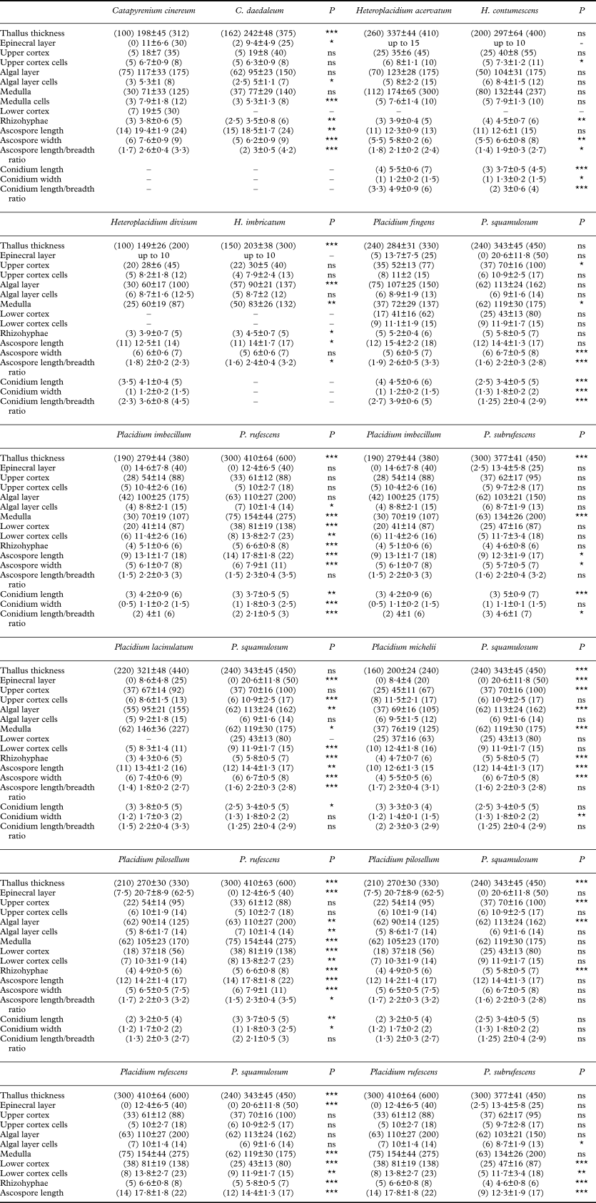
Catapyrenium cinereum, C. daedaleum and C. psoromoides, are the only members of Catapyrenium s. str. present in the Iberian Peninsula. These three species have very similar anatomical characters; however, C. cinereum is easily recognized by the usually whitish, dark rimmed squamules, and is the only one with a lower cortex clearly delimited and a medulla composed of globular cells. Catapyrenium daedaleum also differs in the longer and wider ascospores, and in thallus thickness (thinner in C. cinereum); see Table 1 for further differences. Catapyrenium psoromoides is easily differentiated by its corticolous habit.
Results from ANOVA comparing similar species. Measurements are expressed as mean ± standard deviation in µm and ranges in parentheses

*** = P <0·001; ** = P <0·01; * = P <0·05; ns = no significant differences.
Ecology. Catapyrenium cinereum grows predominantly in alpine grasslands, over calcifereous soils or in fissures, frequently mixed with mosses, but is also found in Juniperus thurifera L. forests. The species occurs at high altitudes, from 1270 to 2350 m, together with Catapyrenium daedaleum and Involucropyrenium waltheri.
Distribution. Catapyrenium cinereum is found in montane areas of the Iberian Peninsula, mainly in northern Spain, but with occurrences in the central and southern mountains (Fig. 3B).
Records from Andorra, Granada, La Rioja (Casares & Llimona Reference Casares and Llimona1984; Azuaga & Gómez-Bolea Reference Azuaga and Gómez-Bolea1998; Burgaz et al. Reference Aragón, Martínez, Burgaz, Belinchón, Prieto, Otálora, García, López, Amo, Ramírez and Asensio2007) and Portugal (Sampaio Reference Sampaio1924; Jansen Reference Jansen1993) could not be revised and are not included in the map (Fig. 4B).
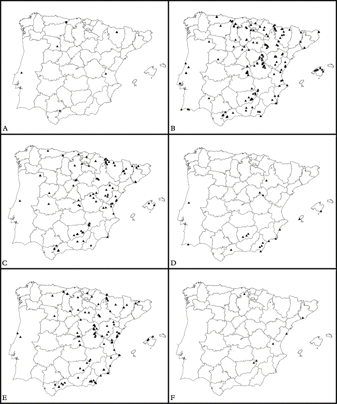
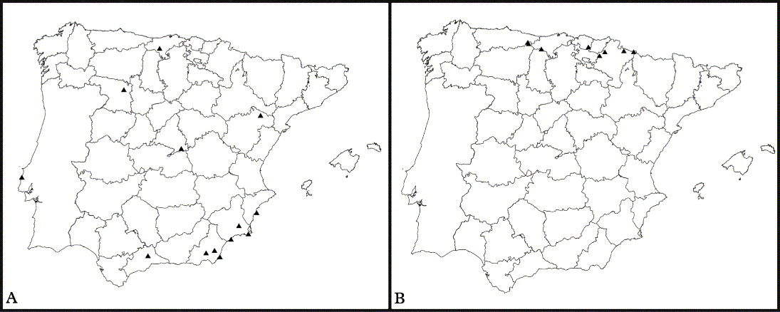
Distribution of Catapyrenium s. lat. species in Iberia. A, Heteroplacidium imbricatum; B, Involucropyrenium llimonae (▲), I. nuriense (●), I. pusillum (■), I. tremniacense(○), I. waltheri (□), Neocatapyrenium cladonioideum (△) and N. latzelii (★); C, Placidium adami-borosi (■), P. boccanum (▲) and P. fingens (●); D, P. imbecillum; E, P. lachneum; F, P. lacinulatum.

Catapyrenium cinereum is an arctic-alpine taxon, found in Africa, Asia, Australia, Europe, New Zealand and North and South America (Breuss Reference Breuss1993, Reference Breuss and McCarthy2001a, Reference Breuss, Nash, Ryan, Gries and Bungartz2002a; Harada Reference Harada1993; Egea Reference Egea1996).
Catapyrenium daedaleum (Kremp.) Stein
In Cohn, Krypt.-Fl. Schlesien (Breslau) 2(2): 312 (1879).—Endocarpon daedaleum Kremp., Flora 38: 66 (1855); type: BRD, Bayern, Berchtesgadener Alpen, Steinthal und Vorderberg zwischen Hochkalter und Kammerlinghorn, c. 5800–6000′, 1854, Rauchenberger (M—lectotype); BRD, Karwendel (bei Mittenwald), 5009′, 1849, Krempelhuber (WU—syntype).
Thallus squamulose; squamules tightly contiguous, adnate or with slightly raised margins, forming a continuous thallus, small, 1–4 mm wide, finely crenulate; upper surface faintly pruinose, greenish grey to brownish, never darker at margins; lower surface dark, with dark rhizohyphae. Anatomy: thallus (162) 242 ± 48 (375) µm thick; upper cortex (5) 19 ± 8 (40) µm thick, poorly delimited from the algal layer, with small, roundish-subangular cells of (5) 6·3 ± 0·9 (8) µm diam.; epinecral layer up to 25 µm. Algal layer not clearly delimited, diffuse, c. 62–150 µm thick, with algal cells of (2·5) 5 ±1·1 (7) µm diam. Medulla up to c. 140 µm thick, composed mainly of elongated cells of (3) 5·3 ± 1·3 (8) µm diam., loose, becoming darker below; lower cortex lacking. Rhizohyphae dark, (2·5) 3·5 ± 0·8 (6) µm.
Perithecia up to 300 µm wide, subglobose; exciple up to 30 µm, hyaline to brown, darker at the ostiole. Asci clavate, c. 75–85 × 17–20 µm (Breuss Reference Breuss1993); ascospores biseriate, simple (occasionally pseudoseptate), (15) 18·5 ± 1·7 (24) × (5) 6·2 ± 0·9 (9) µm, l/b ratio (2) 3 ± 0·5 (4·2).
Pycnidia absent.
Notes. Many herbarium specimens identified as C. daedaleum were found to be C. cinereum. Both species can sometimes be confused because of their similar thallus morphology, but the lack of a lower cortex in C. daedaeleum is a good diagnostic character (see above for further details).
Ecology. Catapyrenium daedaleum lives predominantly in alpine grasslands, on calcareous soils and in rock fissures, mixed with bryophytes; it has also been collected in Fagus sylvatica L. and Juniperus thurifera forests. The species was collected from 1000 to 2300 m altitude, occasionally with Catapyrenium cinereum and Involucropyrenium waltheri.
Distribution. Catapyrenium daedaleum presents now a wider distribution than previously known in the Iberian Peninsula (Aragón Reference Aragón2003). The species has a similar distribution to C. cinereum, as both species occur in mountainous areas; nevertheless, C. daedaleum is more common in the Mediterranean region than C. cinereum (Fig. 3C). It has been recently recorded in Portugal for the first time by Paz-Bermúdez et al. (Reference Paz-Bermúdez, López de Silanes, Terrón, Arroyo, Atienza, Brime, Burgaz, Carvalho, Figueras and Llop2009).
According to Breuss (Reference Breuss1993) and Nimis & Martellos (Reference Nimis and Martellos2004) the species has a more pronounced arctic-alpine distribution than C. cinereum, although in the Iberian Peninsula we have observed the opposite pattern, C. daedaleum being more common in the southern mountains than C. cinereum.
C. daedaleum has an arctic-alpine distribution, known from Asia (Aptroot et al. Reference Aptroot, Ferraro, Lai, Sipman and Sparrius2003), Europe (Breuss Reference Breuss1990a), North America (Goward et al. Reference Goward, McCune and Meidinger1994; revised in this study, hb. COLO) and South America (Breuss Reference Breuss1993).
Catapyrenium psoromoides (Borrer) R. Sant
In Hawksworth, James & Coppins Lichenologist 12: 106 (1980).—Verrucaria psoromoides Borrer, in Hooker & Sowerby, Engl. Bot. [Suppl. 1, tab. 2612, Fig. 1] (1831); type: England, on elm at Hurstpierpoint and on ash at Beeding, Sussex, Borrer (K—holotype).
Thallus squamulose; composed of tightly contiguous to slightly overlapping squamules forming a continuous thallus; squamules adnate and lobate, up to 4 mm wide; upper surface greyish brown to brown (Fig. 1B), pruinose (mostly on external parts of the lobes); lower surface with blackish-brown rhizohyphae. Anatomy: thallus (140) 211 ± 58 (330) µm thick; upper cortex (5) 16·8 ± 8·2 (32·5) µm thick, paraplectenchymatous, with roundish-subangular cells (4) 6·9 ± 1·5 (10) µm diam.; epinecral layer thin, up to 15 µm. Algal layer not clearly delimited, diffuse, c. 45–112 µm thick, with cells (4) 6·4 ± 1·5 (10) µm diam. Medulla loose, (50) 72 ± 18 (175) µm thick, composed mainly of elongated hyphae of (3) 5·2 ± 1·4 (8) µm diam., with some spherical cells and interhyphal spaces; lower cortex lacking. Rhizohyphae brown to black, (3) 3·4 ± 0·5 (4) µm.
Perithecia pyriform, up to 200 µm wide; exciple hyaline to pale brown, darker on the ostiole, up to c. 25 µm thick; asci clavate, 55–65 × 13-15 µm (Breuss Reference Breuss1990a), ascospores biseriate, hyaline, simple (occasionally pseudoseptate), (12) 15 ± 1·6 (18) × (5) 5·6 ± 0·6 (7) µm, l/b ratio (2·1) 2·7 ± 0·4 (3·6).
Pycnidia absent.
Notes. Catapyrenium psoromoides is the only corticolous species of Catapyrenium s. str. It is very similar to C. daedaleum from which it differs in its significantly smaller ascospores (15 ± 1·6 × 5·6 ± 0·6 µm in C. psoromoides versus 18·5 ± 1·7 × 6·2 ± 0·9 µm in C. daedaleum) and in its ecology and distribution (C. daedaleum is terricolous and arctic-alpine whereas C. psoromoides is corticolous and from temperate climates).
Ecology. Catapyrenium psoromoides is a nitrophilous species growing on ancient trees in open forests (Aragón & Sarrión Reference Aragón and Sarrión2003); we have found it on Fraxinus angustifolia Vahl, Juniperus thurifera, Olea europaea L. and on different species of Quercus (Q. faginea Lam., Q. faginea subsp. broteroi (Cout.) A. Camus, Q. ilex subsp. ballota (Desf.) Samp., Q. pyrenaica Willd. or Q. robur L.). The species has been collected from 500 to 1400 m altitude.
Although C. psoromoides has been found growing only on trees in the Iberian Peninsula, in other parts of Europe the species also occurs on mosses (Breuss Reference Breuss1990a).
Distribution. We recorded few specimens only of C. psoromoides in the Iberian Peninsula, maybe because the species can be overlooked due to its small size. Based on its habitat preferences, we suspect the species probably has a wider distribution. Despite this, new data extend the distributional area of the species in the Iberian Peninsula (Fig. 3D). Records from Navarra (Etayo & Breuss Reference Etayo and Breuss1996), Palencia (López de Silanes et al. Reference López de Silanes, Terrón and Etayo1998) and Zaragoza (Etayo et al. Reference Etayo, Aguirre and Diederich1993) could not be localized and are not included on the map.
Catapyrenium psoromoides is known from temperate Asia, eastern Africa, Europe, New Zealand and SW North America (Breuss Reference Breuss, Nash, Ryan, Gries and Bungartz2002a).
Heteroplacidium Breuss
Ann. Naturhist. Mus. Wien, Ser. B, Bot. Zool. 98 (Suppl.): 40 (1996); type: H. imbricatum (Nyl.) Breuss.
The thallus is crustose-areolate to squamulose, composed of small squamules or areoles (1–3 mm), with a thallus anatomy entirely paraplectenchymatous or subparaplectenchymatous (Fig. 2C). Perithecia are conspicuous and generally have the exciple turning brown when mature. Asci clavate with biseriate ascospores, pycnidia are laminal of Dermatocarpon-type and with oblong-ellipsoidal to cylindrical conidia. Differences from Placidium are mainly based on squamule size, thallus anatomy and asci. Differences between species are few because of the weak differentiation of tissues and consequently lack of diagnostic characters.
The species grow on soil and rock (although some parasitic species have been recently included in the genus), living in warm-temperate regions.
Until now, ten species have been reported worldwide, six of which were present in Europe. As a result of this study the number of species rises to eight in Europe, seven of which are present in the Iberian Peninsula.
Key to Heteroplacidium species
1 Thallus areolate to subsquamulose, epilithic, parasitic or not ... H. compactum
[Conidia bacilliform (5–7 µm long), ascospores 11–18 × 8–10 µm, medulla (sub)paraplectenchymatous]
Thallus squamulose, epilithic or not, never parasitic ... 2
2(1) Squamules densely imbricate ... 3
Squamules not densely imbricate ... 4
3(2) Thallus 100–200 µm thick, ascospores 11–14 × 6–7 µm ... H. divisum
Thallus 150–300 µm thick, ascospores 11–17 × 5–7 µm ... H. imbricatum
4(2) Squamules very small, 0·5–1 mm ... H. phaeocarpoides
[ascospores 11–13 × 8–10 µm, conidia oblong-ellipsoidal]
Squamules up to 3 mm ... 5
5(4) Conidia bacilliform, 4–7 × 1–1·5 µm, ascospores 11–13 × 5–6 µm, rhizohyphae 3–5 µm wide ... H. acervatum
Conidia oblong-ellipsoid to subcylindrical, 3–4·5 × 1–1·8 µm, ascospores 11–15 × 5·5–8 µm long, rhizohyphae up to 6·5 µm wide ... 6
6(5) Ascospores 12–15 × 8–10 µm, rhizohyphae 5–6·5 µm wide ... H. congestum
Ascospores 11–15 × 5–5–8 µm, rhizohyphae 4–6 µm wide ... H. contumescens
Heteroplacidium acervatum (Breuss) Breuss
Ann. Naturhist. Mus. Wien, Ser. B, Bot. Zool. 98 (Suppl.): 40 (1996).—Catapyrenium acervatum Breuss, Linzer biol. Beitr. 28: 525 (1996); type: España, Islas Baleares, Mallorca, Serra de Llevant, Puig de sa Font bei Son Cervera, 200–270 m, 15/04/1987, O. Breuss (W—holotype; LI—isotype!).
Thallus epilithic, composed of tightly contiguous to sometimes overlapping squamules, forming compact cushion-like aggregations; squamules 0·3–3 mm wide, irregularly rounded to slightly lobate, convex; upper surface pale brown to brown, matt, greyish towards the margins; lower surface brown, with bundles of rhizohyphae forming a stipe-like organ. Anatomy: thallus (260) 337 ± 44 (410) µm thick; upper cortex (25) 35 ± 6 (45) µm thick, paraplectenchymatous, with small roundish-angular cells (6) 8 ± 1·1 (10) µm diam.; epinecral layer thin or lacking, up to 15 µm when present. Algal layer 70–175 µm, with cells of (5) 8 ±2·2 (15) µm diam. Medulla (112) 174 ± 65 (300) µm thick, composed of spherical hyphae of (5) 7·6 ± 1·4 (10) µm diam., the lower ones turning brownish; lower cortex not clearly delimited from the medulla. Rhizohyphae hyaline, (3) 3·9± 0·4 (5) µm, aggregated in central parts of the squamules giving the aspect of a stipe.
Perithecia (sub)globose, up to 350 µm wide, exciple up to 30 µm, pale except at the ostiole which is brown. Asci clavate, 50–60 × 11–15 µm; with biseriate ascospores, simple, (11) 12·3 ± 0·9 (13) × (5·5) 5·8 ±0·2 (6) µm, l/b ratio (1·8) 2·1 ± 0·2 (2·4).
Pycnidia laminal; conidia bacilliform, (4) 5·5 ± 0·6 (7) × (1) 1 ± 0·2 (1·5) µm, l/b ratio (3·3) 4·9 ± 0·9 (6).
Notes. This species is characterized by its growth form of compact cushions of aggregated squamules; although we collected three specimens which did not show this typical form. Because their anatomical characters are similar to H. acervatum as well as molecular data (M. Prieto et al. unpublished), we classify these three specimens as H. acervatum. A few specimens from Murcia (MUB) with the typical growth form of compact cushions of aggregated squamules and previously classified as H. contumescens belong to H. acervatum.
Heteroplacidium acervatum and H. contumescens, can be differentited mainly by the shape of conidia (cylindrical, 5·5 ± 0·6 µm long in H. acervatum and shortly cylindrical to oblong, 3·7 ± 0·5 µm in H. contumescens), and width of the ascospores (5·8 ± 0·2 µm in H. acervatum versus 6·6 ± 0·8 µm in H. contumescens); moreover, the rhizohyphae are thinner in H. acervatum (3·9 ± 0·4 µm versus 4·5 ± 0·7 µm in H. contumescens) (but see Table 1 for further differences). The cushion-like growth of H. acervatum is very characteristic, but not always present, as mentioned above.
Ecology. Heteroplacidium acervatum grows directly on calcareous rocks or over a slight accumulation of soil; it has been collected in xeric areas dominated by Chamaerops humilis L., from 200 to 400 m altitude.
Distribution. Until now the species has been found in very few localities in the Balearic Islands (Breuss Reference Breuss1996b); we have found it sparsely distributed in Murcia province, located in south-eastern Spain (Fig. 3E), and is recorded here for the first time in the Iberian Peninsula and also continental Europe.
Heteroplacidium compactum (A. Massal.) Gueidan & Cl. Roux
Bull. Ass. Fr. Lichénologie 33(1): 25 (2008).—Placidium compactum A. Massal., Misc. Lichenol.: 62 (1856); type: Germany, Arnold.
Thallus epilithic, areolate to subsquamulose, areoles up to 2 mm diam., angular to rounded, flat to slightly convex; upper surface medium to dark brown, dull to somewhat shiny. Anatomy: thallus up to 600 µm thick; upper cortex 10–25 µm thick, with cells 5–8 µm diam., the uppermost layer with a brown tinge; epinecral layer up to 20 µm thick. Algal layer 100–200 µm thick, with cells 8–15 µm diam. Medulla of globular cells of 7–10 µm thick; lower cortex not clearly delimited. Rhizohyphae 4–6 µm.
Perithecia nearly spherical, 250–400 µm wide, with pale exciple, darkening with age. Asci clavate 55–70 × 13–20 µm; ascospores 11–18 × 8–10 µm.
Pycnidia laminal; conidia bacilliform, 5–7 × c. 1·5 (2) µm.
Notes. Recently, Verrucaria compacta and V. fuscula have been combined into the genus Heteroplacidum (Gueidan et al. Reference Gueidan, Roux and Lutzoni2007; Roux Reference Roux2008). The two species have been considered to be the same taxon by some authors (Fröberg Reference Fröberg1989; Santesson Reference Santesson1993), with H. compactum the autotrophic form of H. fusculum; or they have been retained as different species by other authors (Clauzade & Roux Reference Clauzade and Roux1985; Breuss Reference Breuss1994b). Breuss (Reference Breuss1994b) conducted a detailed investigation and concluded that the main differences between the two taxa were as follows. Heteroplacidium compactum has bacilliform conidia (5–7 µm long), longer ascospores (11–18 µm), a cellular texture in the medulla, and is either autotrophic or is parasitic on various crustose lichens. In contrast, H. fusculum has shortly cylindrical conidia (3–5 µm long), a medulla with some cylindrical hyphae, shorter ascospores (9–14 µm) with a thick wall (1 µm), and is parasitic on Aspicilia calcarea (L.) Körb., or later becoming autotrophic.
Only three specimens we revised corresponded clearly to H. compactum with bacilliform conidia and long ascospores, and were either parasitic or non-parasitic. The remainder showed characters intermediate between H. compactum and H. fusculum; for example they were parasitic on Aspicilia calcarea with long ascospores but with either shortly cylindrical or longer, bacilliform conidia. Nevertheless, all the specimens living on A. calcarea had a thick ascospore wall. These observations lead us to doubt the distinction between these two species, but more material is necessary in order to clarify this question.
Few measurements could be made, due to the small number of specimens available; consequently, the description above is based on published data (Breuss Reference Breuss1994b).
Ecology. The species has been collected from acid rocks and lava; between 30 and 1000 m altitude.
Distribution. Heteroplacidium compactum is very sparsely distributed in the Iberian Peninsula, occurring only in eastern Spain (Fig. 3E). The species has been previously reported from Asia, Europe and northern Africa (Breuss Reference Breuss1994b).
Heteroplacidium congestum (Breuss & McCune) Breuss
Ann. Naturhist. Mus. Wien, Ser. B, Bot. Zool. 98 (Suppl.): 40 (1996).—Catapyrenium congestum Breuss & McCune, Bryologist 97: 365 (Reference Breuss and McCune1994); type: USA, Idaho, Owihee Co, Atriplex confertifolia-Artemisia spinescens flats, Castle Creek Road, 4·5 km NW of junction with Mudflat Road, 1095 m, vi 1991, McCune (OSC—holotype; LI —isotype!).
Thallus densely squamulose or subverruculose; squamules small, 0·5–2 mm wide, tightly contiguous and scarcely overlapping, rounded to slightly lobate, flat to slightly convex; upper surface medium to blackish brown, matt; lower surface pale. Anatomy: thallus 250–400 µm thick, upper cortex 20–40 µm, of roundish angular cells 5–9 µm diam., epinecral layer of 10–20 µm. Algal layer 70–100 µm, with cells of 6–13 µm diam. Medulla composed of globular cells of 6–10 µm diam.; lower cortex hardly discernible, of more closely packed cells. Rhizohyphae colourless, thick-walled, 5–6·5 µm diam.
Perithecia subglobose, up to 400 (500) µm wide; exciple pale when young, dark brown at maturity. Asci clavate, 65–85 × 20–25 µm; ascospores biseriate, broadly ellipsoid to subglobose, 12–15 × 8–10 µm.
Pycnidia laminal, conidia oblong ellipsoid to subcylindrical, 3–4 × 1·5–1·8 µm.
Notes. The combination of densely aggregated (but not overlapping) small squamules, thick walled rhizohyphae and broadly ellipsoidal ascospores distinguish this species from H. acervatum and H. contumescens, which have narrower ascospores and thinner rhizohyphae.
The material collected in the Iberian Peninsula was too sparse for detailed examination, so details of morphology and anatomy are based on Breuss & McCune (Reference Breuss and McCune1994).
Ecology. Heteroplacidium congestum has been collected on calcareous soils in dry open habitats together with Placidium squamulosum. The species was found at 380 m altitude.
Distribution. The species is recorded here for the first time in the Iberian Peninsula (Fig. 3E), constituting the first record from Europe; it was previously known only from North America, where it was considered to be endemic.
Heteroplacidium contumescens (Nyl.) Breuss
Ann. Naturhist. Mus. Wien, Ser. B, Bot. Zool. 98 (Suppl.): 40 (1996).—Endocarpon contumescens Nyl., Flora 61: 341 (1878); type: Algeria, Biskra, 1878, Norrlin (H-NYL!—lectotype; H-NYL!—isotypes).
(Fig. 3E)
Thallus squamulose; squamules 1–3 mm wide, scattered or contiguous to hardly overlapping, rounded to slightly lobate, flat or with deflexed to revolute margins; upper surface pale to medium brown, matt; lower surface pale to brown. Anatomy: thallus (200) 297 ± 64 (400) µm thick, upper cortex (25) 40 ± 8 (55) µm thick, paraplectenchymatous, with small roundish-angular cells (5) 7·3 ± 1·2 (11) µm diam.; epinecral layer thin or lacking, up to 10 µm when present. Algal layer (50) 104 ± 31 (175) µm, with cells (6) 8·4 ± 1·5 (12) µm diam. Medulla (80) 132 ± 44 (237) µm, composed of spherical hyphae of (5) 7·9 ± 1·3 (10) µm diam., the lower ones turning brownish; lower cortex not clearly delimited from the medulla. Rhizohyphae colourless, (4) 4·5 ±0·7 (6) µm.
Perithecia globose to subglobose, up to 400 µm wide; exciple up to 30 µm, pale or darkening at maturity. Asci clavate, 60–70 × 15–18 µm; ascospores biseriate, simple, (11) 12·6 ± 1 (15) × (5·5) 6·6 ± 0·8 (8) µm, l/b ratio (1·4) 1·9 ± 0·3 (2·7).
Pycnidia laminal; conidia oblong-ellipsoid to subcylindrical, (3) 3·7 ± 0·5 (4·5) × (1) 1·3 ± 0·2 (1·5) µm, l/b ratio (2) 3 ± 0·6 (4).
Notes. Differences between H. contumescens and H. acervatum have been previously discussed under that species and are shown in Table 1. Some herbarium specimens revised in this study were previously identified as H. imbricatum or H. divisum, but both these species are easily distinguishable from H. contumescens by their imbricate squamules. The species could also be confused with H. compactum or H. fusculum, but these are areolate-subsquamulose and with wider ascospores than H. contumescens, and are parasitic or free-living.
Ecology. Heteroplacidium contumescens has been collected on sandy soils or directly on rock with an accumulation of soil; the species can live on both calcareous or acid substrata and even on volcanic rocks; it was collected predominantly in dry habitats from 200 to 700 m altitude.
Distribution. The species occurs mainly in coastal areas of south-eastern Spain, with some occurrences in central regions of the Iberian Peninsula. We have collected it for the first time in Portugal (Fig. 3E).
Most of the samples studied here are from herbaria and were collected from 1975 to 1990; during our field trips very few specimens of H. contumescens were collected possibly because of habitat destruction and modification that has affected the south-east Mediterranean coast. For this reason, we believe that H. contumescens should be treated as an endangered species in the Iberian Peninsula, or at least a detailed population study of this species should be carried out.
Heteroplacidium contumescens is a rare species with scattered occurrences in Australia, central Asia and the Mediterraneanregion (Breuss Reference Breuss and McCarthy2001b).
Heteroplacidium divisum (Zahlbr.) Breuss
Ann. Naturhist. Mus. Wien, Ser. B, Bot. Zool. 98 (Suppl.): 40 (1996).—Dermatocarpon divisum Zahlbr., Österr. Bot. Z. 59: 349 (1909); type: Jugoslawien, Dalmatien, Ragusa (Dubrovnik), Mokosica, c. 200 m, 21 ix 1908, A. Latzel (W!—lectotype; PRM—isotype).
Thallus composed of densely imbricate squamules; squamules small and thin, 1–3 mm wide and c. 0·2 mm thick, flat to undulate, entire to crenulate (Fig. 1C), with margins free and without rhizohyphae; upper surface greyish or greenish to pale or medium brown, somewhat pruinose; lower surface whitish, usually blackening, attached by rhizohyphae accumulated at the centre of the squamules. Anatomy: thallus (100) 149 ± 26 (200) µm wide, upper cortex (20) 28 ± 6 (45) µm thick, with roundish angular cells (5) 8·2 ± 1·8 (12) µm diam.; epinecral layer very thin or lacking. Algal layer (30) 60 ± 17 (100) µm thick, with cells (6) 8·7 ± 1·6 (12·5) µm diam. Medulla (25) 60 ± 19 (87) µm thick, with many globular cells (6) 7·9 ± 2·0 (15) µm diam.; lower cortex not clearly delimited from the medulla. Rhizohyphae colourless to slightly brownish, (3) 3·9 ± 0·7 (5) µm, usually concentrated on the middle of the squamule, giving the appearance of a stipe.
Perithecia (sub)globose, up to 500 µm wide; exciple pale to brown, darkened towards the ostiole, up to 25 µm thick. Asci clavate, with biseriate ascospores, simple, (11) 12·5 ± 1 (14) × (6) 6 ± 0·6 (7) µm, l/b ratio (1·8) 2 ± 0·2 (2·3).
Pycnidia laminal; conidia subcylindrical, (3·5) 4·1 ± 0·4 (5) × (1) 1·2 ± 0·2 (1·5) µm, l/b ratio (2·3) 3·6 ± 0·8 (5).
Notes. Heteroplacidium divisum and H. imbricatum are the only species in the genus with imbricate squamules. Of the two, H. divisum has significantly thinner squamules (149 ± 26 µm versus 203 ± 38 µm in H. imbricatum) and consequently the algal layer and medulla are also thinner; in addition, ascospores are shorter in H.divisum (12·5 ± 1 µm in H. divisum versus 14 ±1·7 µm in H. imbricatum) (see Table 1 for further differences). Some herbarium specimens belonging to H. imbricatum have previously been erroneously identified as H. divisum.
Ecology. Heteroplacidium divisum has been collected on calcareous rocks, in fissures and cavities. Most specimens were collected in semiarid or dry regions and only the specimen from the Balearic Islands was found in a very humid place on damp rocks; from 100 to 600 m altitude.
Distribution. The species is very scarce in the Iberian Peninsula, with scattered occurrences in the south-east coast of Spain and the Balearic Islands (Fig. 3F). Heteroplacidium divisum is very rare worldwide, distributed mainly in Mediterranean regions, occurring in Croatia, France and Turkey (Breuss Reference Breuss1990a, Reference Breussb, Reference Breuss1998). A specimen revised by us from LI represents a new record from Italy.
Heteroplacidium imbricatum (Nyl.) Breuss
Ann. Naturhist. Mus. Wien, Ser. B, Bot. Zool. 98 (Suppl.): 40 (1996).—Endocarpon imbricatum Nyl., Bot. Not. 1853: 161; type: Frankreich, Dept. Hérault, ad Mireval prope Monspelium (H-NYL!—lectotype, H-NYL!—isotype).
Thallus squamulose, composed of densely imbricate squamules; squamules 1–3 mm wide, rounded to lobate or crenulated, slightly curved to concave or convex, sometimes curved up with free margins and without rhizohyphae; upper surface greyish or greenish brown to brown, somewhat pruinose at margins; lower surface whitish to pale brown, attached by rhizohyphae accumulated in the centre of the squamule. Anatomy: thallus (150) 203 ± 38 (300) µm thick; upper cortex (22) 30 ± 5 (40) µm thick, with roundish angular cells of (4) 7·9 ± 2·4 (13) µm diam.; epinecral layer very thin or lacking, up to 10 µm when present. Algal layer (57) 90 ± 21 (137) µm thick, with cells (5) 8·7 ± 2 (12) µm diam. Medulla (50) 83 ± 26 (132) µm thick, composed of globular cells (4) 7·6 ± 1·7 (11) µm diam.; lower cortex not clearly delimited from the medulla. Rhizohyphae colourless, (3) 4·5 ± 0·7 (5) µm, accumulated on the middle of the squamule, giving the appearance of a stipe.
Perithecia (sub)globose, up to 350 µm wide, exciple pale to brown, darkening towards the ostiole, up to 25 µm thick (Fig. 2D). Asci clavate; ascospores biseriate, simple, (11) 14 ± 1·7 (17) × (5) 6 ± 0·6 (7) µm, l/b ratio (1·6) 2·4 ± 0·4 (3·2).
Pycnidia laminal; conidia subcylindrical, 4–5 × c. 1 µm.
Notes. This species has often been confused with members of the genus Placidium, from which it differs mainly in the habitus, with squamules distinctly imbricate, and smaller than in Placidium; moreover, Heteroplacidium has an entirely (sub)paraplectenchymatous thallus (Fig. 2C).
Specimens reported from the provinces Zamora (Terrón et al. Reference Terrón, Burgaz and Álvarez2000) and Asturias (Aragón et al. Reference Aragón, Martínez, Burgaz, Belinchón, Prieto, Otálora, García, López, Amo, Ramírez and Asensio2007) belong to Placidium tenellum and Placidium sp., respectively. One record from Almería (Egea & Llimona Reference Egea and Llimona1981) proved to be H. contumescens.
Ecology. Heteroplacidium imbricatum occurs mainly on calcareous rocks with some soil accumulation, and in fissures or cavities, frequently sharing a habitat with Placidium rufescens; from 100 to 850 m altitude.
Distribution. Heteroplacidium imbricatum is a common species in coastal areas of Mediterranean Spain, with some records from the more interior provinces and the Balearic Islands (Fig. 4A). New data reported here extends the distributional area of the species in both Spain and Portugal. On the other hand, records from Alicante and Madrid (Barreno & Merino Reference Barreno and Merino1981; Alonso et al. Reference Alonso, Egea and Moreno1989) could not be verified and the species has not been collected by us in these provinces.
The species is not very common worldwide, known only from southern Europe, Macaronesia and northern Africa (Breuss Reference Breuss1988).
Heteroplacidium phaeocarpoides (Nyl.) Breuss
Ann. Naturhist. Mus. Wien, Ser. B, Bot. Zool. 98 (Suppl.): 40 (1996).—Endocarpon phaeocarpoides Nyl., Flora 62: 358 (1879); type: Frankreich, Dépt. Bouches-du-Rhone, in regione Massiliensi, Le Ciotat, 06/1879, A. Taxis (H-NYL!—holotype).
Thallus squamulose; squamules very small (0·5–1 mm), scattered to contiguous, flat; upper surface greyish brown, matt, pruinose. Anatomy: thallus 150–250 µm thick, upper cortex 20–30 µm thick, paraplectenchymatous, with cells 7–11 µm diam.; epinecral layer thin, c. 10 µm. Algal layer occupying almost the entire thallus, algal cells 8–19 µm diam. Medulla undifferentiated; lower cortex of roundish cells. Rhizohyphae colourless, 4–5 µm.
Perithecia up to 450 µm wide, globose; exciple pale. Asci oblong-clavate, 60–70 × 16–20 µm; ascospores biseriate, simple, 11–13 × 8–10 µm.
Pycnidia laminal; conidia oblong-ellipsoid, 2·5–3·5 × 1–1·5 µm.
Notes. The species has been reported once from Spain (Breuss Reference Breuss1990b); however, this specimen was not found, and no further specimens have been found during field work. The measurements are based on the reference cited.
Ecology. Heteroplacidium phaeocarpoides was reported as growing on sandy soils at 350 m altitude in a semi-arid region (Breuss Reference Breuss1990b).
Distribution. Only one record has been reported from Almería (Breuss Reference Breuss1990b) (Fig. 3F). Although this specimen has not been revised by us we have included it in the map as it constitutes the only one from this region.
The species is known from France, Spain, Tunisia (Breuss Reference Breuss1994a), Macaronesia (Boom & Etayo Reference van den Boom and Etayo2006), North Africa (Breuss Reference Breuss1994a) and Ukraine (M. Prieto et al., unpublished, revised by us from herbarium LI).
Involucropyrenium Breuss
Ann. Naturhist. Mus. Wien, Ser. B, Bot. Zool. 98 (Suppl.): 38 (1996); type: I. waltheri (Kremp.) Breuss.
The thallus is composed of small squamules (0·1–3 mm), thin (50–300 µm), and with a thallus anatomy similar to the genus Catapyrenium (i.e. upper cortex of cinereum-type, medulla not clearly defined, algal layer occupying almost the entire thallus, clavate asci, biseriate ascospores, etc). The main diagnostic characters for this genus are the position of the perithecia, situated between the squamules (see Fig. 1D) and the presence of an involucrellum (complete, dimidiate or apical) (Fig. 2E); pycnidia have not been observed except in I. squamulosum (Brand & Van den Boom) Breuss, and were described as unilocular (Endocarpon-type) (Roux Reference Roux2005).
The species occur on calcareous and gypsiferous soils, in rock fissures or directly on limestones, also on artificial substrata in ruderal conditions, such as old bricks or mortar. They occupy semi-arid to alpine and temperate environments.
The genus is distributed mainly in Europe, with a single species present in North America or Asia (I. waltheri). It comprises eight species, five of which are reported here from the Iberian Peninsula. Two species (i.e. I. llimonae and I. nuriense) are known only from this region.
The genus must be regarded as poorly known, since several species are known only from their type localities, or in no more than a handful of sites, and are probably often confused with species of Verrucaria.
Key to Involucropyrenium species
1 Involucrellum partial, not enclosing the entire perithecium ... 2
Involucrellum entire, enclosing the perithecium ... 3
2(1) Thallus epilithic, of densely imbricate squamules; upper cortex not paraplectenchymatous, with cells of irregular shape; involucrellum dimidiate, only reaching half-way down the perithecia; exciple colourless, ascospores 11–14 × 7–9·5 µm ... ... I. nuriense
Thallus consisting of more or less dispersed squamules; upper cortex paraplectenchymatous, with roundish-angular cells; involucrellum apical; exciple brown to black; ascospores 13–18 × 6–8 µm ... I. tremniacense
3(1) Rhizohyphae dark, hypothallus well developed ... I. waltheri
[Ascospores 15–23 × 7.5–11 µm. Artic-alpine distribution]
Rhizohyphae colourless, without hypothallus ... 4
4(3) Thallus consisting of dispersed squamules with a very characteristic shape, more or less irregular to digitate-coralloid; involucrellum dark brown, 30-60 µm thick; ascospores 14–22 × 7–11 µm ... I. llimonae
Thallus of more crowded squamules, sometimes overlapping; involucrellum black, 25–35 µm, thicker in the upper part; ascospores 19–24 × 9–14 µm ... ... I. pusillum
Involucropyrenium llimonae (Etayo, Nav.-Ros. & Breuss) Breuss
Beitr. Naturk. Oberösterreichs 13: 213 (2004).—Catapyrenium llimonae Etayo, Nav.-Ros. & Breuss, Candollea 51: 140 (Reference Navarro-Rosinés, Etayo and Breuss1996); type: España, Navarra, Rada, la Bárdena Blanca, sobre suelos yesífero-arcillosos, 300–400 m, 28 v 1990 J. Etayo (LI!—holotype).
Thallus squamulose; squamules small, (0·1) 0·3–0·5 (0·7) mm, dispersed, irregularly shaped to digitate-coralloid; upper surface brown. Anatomy: thallus (50) 70–150 µm thick; upper cortex 10-20 µm thick, paraplectenchymatous, with roundish-subangular cells (3) 4–7 µm; epinecral layer thin 10–15 µm. Algal layer occupying almost the entire thallus, with cells of 6–10 µm diam. Medulla and lower cortex absent. Rhizohyphae colourless, 3–5 µm.
Perithecia dispersed to aggregated, developed between the squamules (Fig. 1D), globose to slightly conical, 0·2–0·4 mm. Exciple colourless to brown, 15–20 µm with a complete dark brown involucrellum, 30–60 µm thick. Asci clavate, 55–80 × 15–21 µm; ascospores biseriate, broadly ellipsoid, (14) 16–18·2–21 (22) × (7) 9–9·7–11 µm (Navarro-Rosinés et al. Reference Navarro-Rosinés, Etayo and Breuss1996).
Pycnidia not seen.
Notes. This species is easily recognized by the irregular to digitate-coralloid squamules. Involucropyrenium pusillum is a similar species but with a thicker involucrellum and larger ascospores (19–24 × 9–14 µm). Two further species, I. terrigenum (Zschacke) Breuss and I. sbarbaronis (Servít) Breuss (neither of which has been found in the Iberian Peninsula) have smaller ascospores than I. llimonae (15–20 × 6–8 µm in I. sbarbaronis; 10–12 × 7–8·5 µm in I. terrigenum) and a nearly crustose thallus rather than clearly dispersed squamules as in I. llimonae. Only one specimen has been found up to now in the Iberian Peninsula; for this reason we have included the measurements based on Navarro-Rosinés et al. (Reference Navarro-Rosinés, Etayo and Breuss1996).
Ecology. Involucropyrenium llimonae has been collected on very eroded and weathered gypsiferous and clayey soils, at the base of shrubs.
Distribution. This species has been found only in the type locality, situated in the northern part of Spain, in a semi-arid environment at 300–400 m altitude (Fig. 4B).
Involucropyrenium nuriense (Nav.-Ros. & Breuss) Breuss
Beitr. Naturk. Oberösterreichs 13: 214 (2004).—Catapyrenium nuriense Nav.-Ros. & Breuss, Candollea 51: 144 (Reference Navarro-Rosinés, Etayo and Breuss1996); type: España, Cataluña, Prov. Girona, Ripollés, Queralbs, Vall de Núria, Forat de l'Embut, cerca del Pla dels Eugassers, 2300–2400 m, paredes verticales de roca calcárea orientados al norte, 1 viii 1984, P. Navarro-Rosinés (BCC!—holotype).
Thallus epilithic, almost continuous, 0·5–2 cm diam.; squamules densely imbricate, (0·5) 1–2 (3) mm wide, irregularly shaped to sublobulate. Anatomy: thallus 150–200 µm thick; upper cortex 30–40 µm thick, not clearly paraplectenchymatous, with cells irregularly arranged and elongated, (3) 5–7·5 (8·5) × (1·5) 2–4 µm; epinecral layer absent or very thin (6–12 µm). Algal layer 100–150 µm, with algal cells 7–10 µm diam. Medulla not differentiated; lower cortex 15 µm thick, dark brown, cells with thick walls. Rhizohyphae colourless, 4 µm thick.
Perithecia globose, 0·2–0·5 (0·7) mm, dispersed to aggregated, developed in the marginal region of the squamules, rarely in the centre; exciple colourless, 20–30 µm thick; involucrellum black, dimidiate (only reaching to the middle of the perithecia); periphyses 30–40 × 1·5 µm. Asci clavate 55–65 × 15–20 µm; ascospores biseriate, ovoid, broadly ellipsoid or subglobose, 11–12·5–14 × (7) 8·2–9 (9·5) µm (Navarro-Rosinés et al. Reference Navarro-Rosinés, Etayo and Breuss1996).
Pycnidia not seen.
Notes. Involucropyrenium nuriense is the only species in the genus without a clearly paraplectenchymatous upper cortex. Moreover, the involucrellum is dimidiate, not entire, a character that approaches I. tremniacense, although this species has an apical involucrellum. The two species also differ in ascospore size (11–14 × 7–9·5 µm in I. nuriense versus 13–18 × 6–8 µm in I. tremniacense) and in their ecology, I. tremniacense being a terricolous species growing exclusively in the Mediterranean region (see below).
Only one specimen has been found in the Iberian Peninsula and for this reason we have included the measurements based on Navarro-Rosinés et al. (Reference Navarro-Rosinés, Etayo and Breuss1996) in the description.
Ecology. Involucropyrenium nuriense is a saxicolous species and has been found at very high altitudes (2300–2400 m), on vertical limestone facing N-NE.
Distribution. The species is known only from the type locality, collected in the eastern Pyrenees, north east of Spain (Fig. 4B).
Involucropyrenium pusillum Breuss & Türk
Beitr. Naturk. Oberösterreichs 13: 214 (2004); type: Österreich, Oberösterreich, Puglalm SE vom Hengstpass, 920 m, auf Moosen über Kalkblöcken, 5 x 1987, S. Wagner (LI!—holotype).
Thallus squamulose; squamules small, 0·2–0·5 mm wide, greenish brown, matt, contiguous to aggregated and often overlapping, slightly lobulate, closely adnate to the substratum or with slightly raised margins. Anatomy: thallus 100–150 µm thick, almost entirely (sub)paraplectenchymatous; upper cortex not always clearly delimited, up to 20 µm thick, with cells 3–8 µm diam.; epinecral layer lacking or very thin, up to 10 µm when present. Algal layer occupying almost the entire thallus, with algal cells 5–8 µm diam. Medulla and lower cortex not differentiated. Rhizohyphae hyaline, thin, 2·5–3 µm diam.
Perithecia developed between the squamules, sometimes in groups, nearly globose, 0·25–0·40 mm diam. Exciple hyaline to brown, c. 25 µm thick, ostiole paler. Involucrellum entire, black, 25–35 µm thick, thicker in the upper part. Periphyses 20–30 × 1–1·5 µm. Asci clavate, 75–85 × 20–25 µm; ascospores (19) 21·2 ± 1·7 (24) × (9) 12·2 ± 1·3 (14) µm.
Pycnidia absent.
Notes. The ascospores of the Iberian material are slightly wider than in the description of I. pusillum in Breuss & Türk (Reference Breuss and Türk2004), although the other characteristics agree with this species. Involucropyrenium pusillum apparently looks like I. squamulosum, but the involucrellum in I. pusillum completely encloses the exciple, whereas in I. squamulosum it only reaches the base of the perithecium. Moreover, the ascospores are larger in I. squamulosum (24–27 µm in I. squamulosum versus 19–24 µm in I. pusillum). Other species in the genus with an entire involucrellum are I. waltheri, clearly distinguishable by its hypothallus, and I. llimonae with smaller ascospores and a thicker involucrellum. Involucropyrenium terrigenum and I. sbarbaronis also develop an entire involucrellum, but in both cases the ascospores are shorter and narrower (10–12 × 7–8·5 in I. terrigenum and 15–20 × 6–8 in I. sbarbaronis); however, these two species have not been found in the Iberian Peninsula.
Ecology. This species has been collected on wall mortar, at 850 m altitude, growing with Placidium boccanum. Breuss & Türk (Reference Breuss and Türk2004) reported it only from the type locality, growing on mosses above limestone at 920 m altitude. Although the habitat in which we collected the species agrees with that of I. squamulosum, a saxicolous species common on artificial substrata, the measurements of the ascospores and the anatomy of the involucrellum correspond with I. pusillum.
Distribution. We have collected I. pusillum in Portugal (Fig. 4B), widening considerably the distribution of the species previously known only from the type locality in Austria.
Involucropyrenium tremniacense (A. Massal.) Breuss
Ann. Naturhist. Mus. Wien, Ser. B, Bot. Zool. 98 (Suppl.): 38 (1996).—Catapyrenium tremniacense A. Massal., Lotos 6: 79 (1856); type: Italien, Veneto, Tregnano, 1855, M. Brojo (M—lectotype; BM, O, S, W!—isotypes).
Thallus squamulose to subcrustose; squamules 0·3–1·5 (2) mm, roundish to slightly lobulate, flattened to slightly convex, epruinose, closely adpressed to the substratum; upper surface beige to pale brown. Anatomy: thallus 100–150 µm thick; upper cortex paraplectenchymatous, 10–25 µm thick, with roundish-angular cells 6–9 µm diam.; epinecral layer 20–50 µm. Algal layer 60–120 µm thick, with cells 6–10 µm diam. Medulla and lower cortex not differentiated. Rhizohyphae colourless, c. 4 µm thick.
Perithecia frequent, developed between the squamules, globose, up to 350 µm diam.; exciple brown to black, with an apical involucrellum. Asci clavate; ascospores biseriate, ellipsoid, 13–17 (18) × 6–7 (8) µm.
Notes. This species is characterized by the combination of incomplete involucrellum and thallus of dispersed squamules (or subcrustose). Additional differences from other species in the genus are discussed above.
Ecology. Involucropyrenium tremniacense was found in open grassland on calcareous substrata in semi-arid regions, from 90 – c. 400 m altitude.
Distribution. The species has been found in two distant localities in Spain, one located in the north and the other in the south-east (Fig. 4B).
The worldwide range of the species reaches south-central Europe and the Mediterranean region (Breuss Reference Breuss1990a; Nimis & Martellos Reference Nimis and Martellos2004).
Involucropyrenium waltheri (Kremp.) Breuss
Ann. Naturhist. Mus. Wien, Ser. B, Bot. Zool. 98 (Suppl.): 38 (1996).—Verrucaria waltheri Kremp., Flora 38: 69 (1855); type: BRD, Bayern, Karwendel, 26 viii 1850, Krempelhuber (M—lectotype; WU—isotype).
Thallus squamulose, with a nearly crustose appearance; squamules less than 1·5 mm diam., brown to grey, matt, flattened to slightly convex, contiguous, forming a crust; margins entire to crenate-lobulate, surrounded by a dark hypothallus; lower surface dark, with 4–5 µm thick, dark rhizohyphae. Anatomy: thallus (70) 100–200 (300) µm thick; with a paraplectenchymatous upper cortex, 10–25 µm thick; epinecral layer thin or absent, up to 25 µm when present. Algal layer distributed over nearly the entire thallus, with algal cells 6–11 µm. Medulla not clearly differentiated; lower cortex composed of polygonal cells, dark; rhizohyphae dark brown, 4–5 µm thick.
Perithecia globose, often aggregated; exciple brown to black with an entire carbonaceous involucrellum, developed in the hypothallus between the squamules. Asci clavate, with biseriate ascospores (15) 17–21 (23) × (7·5) 8–10 (11) µm.
Notes. This species is easily recognized by its nearly crustose thallus, with brown to grey squamules and perithecia developed from a black hypothallus. It is the only species in the genus with dark rhizohyphae. Sometimes it can resemble Catapyrenium cinereum, from which it differs in the perithecia that are not immersed in the squamules and by the presence of the involucrellum.
Ecology. Involucropyrenium waltheri was collected on calcareous soils, together with C. cinereum, between bryophytes, in alpine grasslands at 2300 m.
Distribution. This species was erroneously cited in Spain by Aragón et al. (Reference Aragón, Rico and Belinchón2006), but the revised samples (hb. Aragón 3663/97 and 2299/97) corresponded to young specimens of Endocarpon sp. Consequently, the only record in the Iberian Peninsula was found during this study in the Pyrenees on alpine grassland (Fig. 5B).
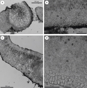
Anatomy of Placidium species. A, cross-section of a marginal Dermatocarpon-type pycnidium of Placidium semaforonense; B–D, cross-sections of the thalli; B, Placidium squamulosum showing the medulla (m) composed of globular cells (r = rhizohyphae); C, Placidium lachneum showing the prosoplectenchymatous medulla (m) and the lower cortex of anticlinally arranged cells (lc); D, Placidium velebiticum, showing the prosoplectenchymatous medulla composed of elongated cells and the lower cortex clearly delimited.

The species has an arctic-alpine distribution, occurring mainly in north and central Europe (Breuss Reference Breuss1990a) with some occurrences in Asia (Sun et al. Reference Sun, Ren, Hau-Jie and Zun-Tian2008) and North America (Breuss & McCune Reference Breuss and McCune1994).
Neocatapyrenium H. Harada
Nat. Hist. Res. 2: 129 (1993); type: N. cladonioideum (Vain.) Harada.
The thallus has a cushion-like growth, composed of imbricate squamules; thallus anatomy is similar to Placidium, but the genus is characterized by Endocarpon-type pycnidia with cylindrical conidia (Fig. 2F), the lack or reduction of rhizohyphae and the attachment to the substratum by rhizines or by the basal end of the squamule. Perithecia have colourless exciples and asci are clavate with biseriate ascospores. There are five species reported world-wide, three of which are present in Europe. In the Iberian Peninsula, we have found two species. All species are rare and have limited distributions (Breuss Reference Breuss2005).
Key to Neocatapyrenium species
1 Squamules fixed to the substratum by elongated basal ends ... ... N. cladonioideum
Squamules fixed to the substratum by pale rhizines ... N. latzelii
Neocatapyrenium cladonioideum (Vain.) H. Harada
Nat. Hist. Res. 2 (2): 129 (1993).—Siphula cladonioides Vain., Bot. Mag. (Tokyo) 35: 47 (1921); type: Japan, Honshu, Gunma-ken (as Prov. Kozuke), Mt. Myôgi, on rock, 19 iv 1916, A. Yasuda 142 (TUR—holotype).
Thallus squamulose, with a cushion-like growth; squamules lobate, 2–5 mm wide, convex, imbricate, attached to the substratum by basal ends only; upper surface flesh coloured to beige, matt or a little glossy, epruinose; lower surface pale brown (Fig. 1E); rhizohyphae and rhizines lacking. Anatomy: thallus (360) 407 ± 53 (480) µm thick, upper cortex (35) 43 ± 5 (50) µm, paraplectenchymatous, with cells (7) 7·6 ± 0·9 (9) µm diam.; epinecral layer very thin or lacking, up to c. 12 µm thick when present. Algal layer (50) 67 ± 15 (87) µm, with algal cells (7) 10 ± 1·9 (12) µm diam. Medulla prosoplectenchymatous, (175) 206 ± 44 (287) µm thick, with elongated hyphae of 3–5 µm diam., with some spherical cells in the uppermost and lowermost parts of the medulla; lower cortex clearly delimited from the medulla, (42) 55 ± 8 (62) µm thick, with rounded to angular cells of (5) 7·4 ± 1·1 (9) µm, the lowermost brownish.
Perithecia pyriform to globose, up to 400 µm wide; exciple 30–40 µm pale, sometimes brownish in the upper part. Asci clavate; ascospores biseriate, simple, (15) 15·9 ± 1·2 (18) × (6) 6·9 ± 0·4 (7·5) µm, l/b ratio (2) 2·3 ± 0·3 (2·6).
Pycnidia laminal, immersed, inconspicuous, Endocarpon-type; conidia bacilliform, 5–7 × c.1 µm.
Notes. Anatomically, the specimen collected in Spain is very similar to the specimens described by Harada (Reference Harada1993), although the squamules are thicker and wider, and more lobate rather than sublinear as Harada described; in addition our specimen has a thin epinecral layer whereas the species was described as lacking this layer. We did not feel these differences were sufficient to justify describing a new species, so we include this specimen as N. cladonioideum. The species is easily recognizable by its cushion-like growth with imbricate squamules.
Ecology. Neocatapyrenium cladonioideum has been collected on calcareous rocks and soils (specimens from Asia are reported from lava or granitic rocks). It has been found together with Heteroplacidium imbricatum and Placidium pilosellum, at 850 m altitude.
Distribution. The record reported here from the Segura mountains, south-east Spain (Fig. 4B), together with N. latzelii (see below), constitute the first records of this genus for the Iberian Peninsula.
Neocatapyrenium cladonioideum was previously known only from Asia, being here for the first time reported from Europe, and having a different ecology to specimens from Asia.
Neocatapyrenium latzelii (Zahlbr.) Breuss
Ann. Naturhist. Mus. Wien, Ser. B, Bot. Zool. 98 (Suppl.): 41 (1996).—Dermatocarpon latzelii Zahlbr., Österr. Botan. Z. 68: 70 (1919); type: Jugoslawien, Dalmatien, Slano, Feldmauern, 3m, 4 i 1909, A. Latzel nº 799 (W!—holotype).
Thallus squamulose, with a cushion-like growth; squamules lobate, 3–7 mm wide, imbricate; attached to the substratum only by pale rhizines, rhizohyphae lacking; upper surface pale to dark brown, matt; lower surface pale brown. Anatomy: thallus 300–500 µm thick, upper cortex 50–70 µm thick, paraplectenchymatous, with cells 5–8 µm diam.; epinecral layer thin, up 10–15 µm thick. Algal layer up to 100 µm thick, with cells 7–12 µm diam. Medulla prosoplectenchymatous, with elongated hyphae of 3–5 µm diam., with some spherical cells in the uppermost and lowermost parts of the medulla; lower cortex clearly delimited from the medulla, with rounded to angular cells 7–10 µm.
Perithecia pyriform to globose, up to 550 µm wide, with a pale exciple. Asci clavate; ascospores biseriate, simple, 13–17 × 6·5–8·5 µm.
Pycnidia laminal, immersed, inconspicuous, Endocarpon-type; conidia bacilliform, 4·5–8·5 × 1–1·5 µm.
Notes. The species is easily identified by its cushion-like growth form, imbricate squamules, the lack of rhizohyphae and the presence of pale rhizines. Only one specimen has been found in the Iberian Peninsula, so we have included measurements based on the literature (Breuss Reference Breuss1990a).
Ecology. In the Iberian Peninsula the species was collected in cavities and fissures of calcareous rocks, in dry habitats, at c. 450 m altitude.
Distribution. Neocatapyrenium latzelii has been previously recorded in Croatia and Greece (Breuss Reference Breuss1990a; Sipman & Raus Reference Sipman and Raus2002). This record constitutes the first from Spain, and, together with N. cladonioideum, the only representatives of the genus in the Iberian Peninsula (Fig. 4B).
Placidium A. Massal
Symmicta 75 (1855); type: P. michelii A. Massal.
Thallus squamulose, with squamules (1) 2–8 (15) mm wide, upper cortex 20–100 µm thick, with cells 5–17 µm diam.; medulla and lower cortex usually well developed (Figs. 3B, 3C, 3D). Asci are usually cylindrical at least when young, but clavate in some species with rhizines. Pycnidia laminal or marginal, Dermatocarpon-type, with oblong-ellipsoidal to cylindrical or bacilliform conidia (Fig 3A). Rhizines present or not. Placidium species inhabit soil, rocks and fissures, ledges and cavities, rarely trees [e.g. P. arboreum (Schweinitz ex Michener) Lendemer and P. corticola (Räsäsen) Breuss]; usually over calcareous substrata, although there are some species occurring on gypsiferous or acid substrata or even on artificial substrata.
The genus comprises 32 species worldwide, 16 of which have been reported from Europe. In this study, we report 14 species occurring in the Iberian Peninsula and the Balearic Islands.
Key to Placidium species
1 Rhizines present (rhizohyphae also present), few to many per squamule ... 2
Rhizines absent ... 3
2(1) Pycnidia laminal; medulla composed of globular cells, ascospores 11–16 × 6–9 µm ... P. lacinulatum
Pycnidia marginal; medulla prosoplectenchymatous, ascospores 13–22 × 7–12 µm ... P. semaforonense
3(1) Medulla composed of globular cells ... 4
Medulla prosoplectenchymatous ... 11
4(3) Pycnidia laminal ... 5
Pycnidia marginal ... 10
5(4) Thallus thin, < 250 µm; perithecial wall black, thin, <20 µm thick ... P. michelii
[Lower side black; conidia oblong-ellipsoidal, 3–4 × 1·2–1·5 µm;ascospores 10–15 × 4–6 µm, rhizohyphae 4–6 µm]
Thallus thicker, >250 µm; perithecial wall pale, >20 µm thick ... 6
6(5) Conidia oblong-ellipsoid, 2·5–5 × 1·2–2 µm, l/b ratio 1·25–2·9 ... 7
Conidia cylindrical to bacilliform, more than 4 µm long × 1–2 µm, l/b ratio 2·5– 5 ... 9
7(6) Mainly epilithic, on mortar or calcareous rock; squamules with black margins, lower side black throughout ... P. boccanum
[rhizohyphae 4–6 µm; ascospores 11–15 × 5–8 µm]
Mainly terricolous, on soils, rock ledges and in fissures; squamules without black margins, lower side pale to brown, rarely blackish ... 8
8(7) Squamules up to 6 mm; lower cortex cells 11–15 µm, rhizohyphae 5–7 µm; ascospores 12–17 × 6–8 µm ... P. squamulosum
Squamules up to 3·5 mm; lower cortex cells 7–12 µm, rhizohyphae 3–5 µm; ascospores 11–15 × 5–7·5 µm ... P. tenellum
9(6) Thallus 240–330 µm, rhizohyphae 5–6 µm; conidia 4–6 × 1–1·5 µm; ascospores 12– 18 × 5–7 µm ... P. fingens
Thallus 350–550 µm, rhizohyphae 6–8 µm; conidia 5–8 × 1·5–2 µm; ascospores 15–24 × 7·5–11·5 µm ... P. norvegicum
10(4) Thallus 300–600 µm thick, rhizohyphae thick, 5–8 µm; ascospores 14–22 × 6–11 µm ... P. rufescens
Thallus 210–330 µm thick, rhizohyphae thin, 4–6 µm; ascospores 12–17 × 5–7·5 µm ... P. pilosellum
11(3) Pycnidia laminal, or laminal and marginal in the same squamule ... P. velebiticum
[including P. pyrenaicum]
Pycnidia marginal only ... 12
12(11) Lower cortex of anticlinally arranged angular cells ... 13
Lower cortex of irregularly arranged angular cells ... 15
13(12) Lower cortex without black pigment; rhizohyphae thin 5–6 µm; conidia oblong-ellipsoidal ... P. velebiticum
Lower cortex with black pigment; rhizohyphae thick, of 6–8 µm thick; conidia bacilliform ... 14
14(13) Squamules > 6 mm, with a thin margin; ascospores 14–17 × 6–7·5 µm; conidia 4·5–6·5 × 1–1·5 µm ... P. adami-borosi
Squamules < 6 mm, with a thick margin; ascospores 12–18 × 6–8 µm; conidia 5–7 × 1–2 µm ... P. lachneum
15(12) Squamules large, up to 12 mm wide, almost foliose; conidia oblong-ellipsoid, 2·5–5 × 1–2·5 µm, l/b ratio 1·2–4 ... 16
Squamules up to 6 mm wide; conidia cylindrical to bacilliform, 3–7 × 0·5–1·5 µm, l/b ratio 2–7 ... 17
16(15) Thallus 300–600 µm thick; rhizohyphae thick, 5–8 µm; ascospores 14–22 × 6–11 µm ... P. rufescens
Thallus 270–360 µm thick, rhizohyphae thin, 5–6 µm; ascospores 13–17 × 6–7 µm ... P. velebiticum
17(15) Thallus thin, 190–380 µm thick, medulla 30–107·5 µm; conidia 3–6 × 0·5–1·5 µm ... P. imbecillum
Thallus 300–450 µm thick, medulla 63–200 µm; conidia 3–7 × 1–1·5 µm ... ... P. subrufescens
Placidium adami-borosi Szatala
Ann. Mus. Nat. Hungar. 7: 271 (1956); type: Hungaria, comit. Heves, in rupibus andesit. Orient. supra vallem Csatorna-völgy montis Remetebérc prope Mátraufüred, c. 750 m, 5 vi 1951, Á. Boros (BP—holotype!; hb Vězda— topotype).
Thallus squamulose; squamules 3–10 mm wide, scattered to contiguous, mainly overlapping, with upturned margins, flat to concave, roundish to slightly lobate, the margins sometimes irregularly lobate and thin; upper surface pale to medium brown, sometimes with a reddish tinge, matt or a little glossy; lower surface pale to brown. Anatomy: thallus 400–700 µm thick; upper cortex 50–80 µm, paraplectenchymatous, with cells of 8–13 µm; epinecral layer up to 15 µm thick. Algal layer 100–130 µm, with cells of 8–14 µm diam. Medulla 150–250 µm thick, prosoplectenchymatous, with filamentous hyphae of 3–5 µm diam.; lower cortex clearly delimited from the medulla, 50–60 µm thick, composed of angular cells, of 12–17 (20) µm wide, conglutinated in vertical rows, the lowermost cells with black pigment (Fig. 3C). Rhizohyphae thick, of 6–8 µm diam., colourless.
Perithecia broadly pyriform, up to 550 µm wide; exciple pale, 30–35 µm. Asci cylindrical, 80–90 × 14–18 µm, ascospores uniseriate, simple, 14–17 × 6–7·5 µm.
Pycnidia marginal, inconspicuous; conidia bacilliform, (4·5) 5·8 ± 0·8 (7) × (1) 1·3 ± 0·2 (1·5) µm) (conidia measurements are based on the Iberian specimens).
Notes. Only two specimens have been found in the Iberian Peninsula; thus, description of the species is based on previous studies (Breuss Reference Breuss1990a).
This species is almost identical to P. lachneum from which it differs mainly in the diameter of the squamules [3–10 mm in P. adami-borosi and 2–7 (10) mm in P. lachneum] and thickness of the margin (thin in P. adami-borosi and thick in P. lachneum); the lower cortex is not so evident in the margins in P. adami-borosi. The most important difference reported is the ecology and distribution. Placidium adami-borosi grows at lower altitudes in southern Europe and west-central Asia, whilst P. lachneum is reported as an arctic-alpine lichen distributed in Asia, northern Europe, North and South America, at higher altitudes (Breuss Reference Breuss, Nash, Ryan, Gries and Bungartz2002b). The minor morphological differences could be explained by the plasticity of the species in response to their different habitats. Moreover, data from a preliminary molecular analysis (M. Prieto et al. unpublished) do not support P. adami-borosi and P. lachneum as distinct species. However, at present we have decided to maintain P. adami-borosi as an independent species pending a forthcoming wider study.
Ecology. Placidium adami-borosi has been collected from 1300 to 1650 m altitude in cavities among mosses, or in crevices, on calcareous substrata. In Europe the species has been reported growing on calcareous and siliceous substrata, from 620 to 1800 m.
Distribution. Placidium adami-borosi has been collected in widely separated localities in northern and southern Spain (Fig. 4C).
The species is distributed in Europe, mainly in the Mediterranean region, and Asia. It has been reported from Bulgaria (Breuss Reference Breuss1990a), Cyprus (Breuss Reference Breuss1990a; Litterski & Mayrhofer Reference Litterski and Mayrhofer1998), France (Breuss Reference Breuss1990a), Greece (Spribille et al. Reference Spribille, Schultz, Breuss and Bergmeier2006), Hungary (Breuss Reference Breuss1990a), Italy (Breuss Reference Breuss1990a, Reference Breussb; Nimis & Martellos Reference Nimis and Martellos2004), Spain (Breuss Reference Breuss1990a) and Tajikistan (Kudratov & Mayrhofer Reference Kudratov and Mayrhofer2002).
Placidium boccanum (Servít) Breuss
Ann. Naturhist. Mus. Wien, Ser. B, Bot. Zool. 98 (Suppl.): 38 (1996).—Dermatocarpon boccanum Servít, Rozpr. Českoslov. Akad. Věd. 65 (3): 23 (1955); type: Jugoslawien, Hrvatska, Dalmatia, Hercegnovi, inter Trebesin et Kameno, 250 m, calc., 1929, M. Servít (PRM!—holotype).
Thallus squamulose, mainly epilithic; squamules up to 4 mm wide, scattered to contiguous, rarely somewhat overlapping, adpressed to the substratum or with slightly upturned margins, roundish to slightly lobate, flat to fairly convex; upper surface pale to dark brown, matt or a little glossy, with a black margin; lower surface black throughout. Anatomy: thallus (210) 313 ± 66 (400) µm thick, upper cortex (50) 75 ± 12 (87) µm thick, paraplectenchymatous, with cells (6) 9 ± 2·1 (13) µm diam.; epinecral layer up to 35 µm thick. Algal layer (75) 95 ± 14 (112) µm thick, with cells (10) 12·5 ± 1·6 (15) µm diam. Medulla (37) 109 ± 52 (162) µm thick, composed of globose cells of 9–13 µm diam.; lower cortex not clearly delimited from the medulla, (25) 29 ± 4 (33) µm thick, composed of more densely aggregated rounded to angular cells of 10-15 µm, the lowermost with dark pigment. Rhizohyphae (4) 4·9 ± 0·6 (6) µm thick, colourless.
Perithecia pyriform, up to 500 µm wide; exciple pale, 30–35 µm thick. Asci cylindrical, (60) 66 ± 5 (73) × (10) 11·2 ± 1·2 (13) µm; ascospores uniseriate, simple, (11) 13·2±1·1 (15) × (5) 6·4 ± 0·7 (8) µm, l/b ratio (1·7) 2·1 ± 0·3 (2·8); periphyses 18–40 × 2–3 µm.
Pycnidia laminal, immersed; conidia oblong-ellipsoid, 3–4 × c. 1·5 µm.
Notes. Placidiun boccanum is characterized by its epilithic growth, being, together with P. rufescens, the only species of the genus occurring directly on rocks. The species is anatomically very similar to P. squamulosum, which also has a medulla composed of globose cells, laminal pycnidia and oblong-ellipsoidal conidia. However, in addition to its epilithic habit, P. boccanum differs from P. squamulosum in the black margins of the squamules, and the lower surface that is continuously black, while the lower surface in P. squamulosum is usually pale to dark brown, rarely black. They also differ in the size of rhizohyphae (4·9 ± 0·6 µm in P. boccanum versus 5·8 ± 0·5 µm in P. squamulosum) and ascospores (13·2 ± 1·1 µm long in P. boccanum versus 14·4 ± 1·3 µm in P. squamulosum).
The differentiation from P. tenellum is more difficult, because both species have relatively thin and small squamules, but they differ mainly in the thinner rhizohyphae of P. tenellum (4 ± 0·6 µm versus 4·9 ± 0·6 µm thick in P. boccanum). The presence of the black margin in P. boccanum and the ecology can also be useful as a distinguishing feature.
Another similar species, with laminal pycnidia, oblong-ellipsoidal conidia and a medulla composed of globular cells is P. michelii, which is clearly distinguished from P. boccanum by its dark and thin exciple, thinner thallus and upper cortex (313 ±66 µm thick and 75 ± 12 µm thick, respectively, in P. boccanum versus 200 ± 24 and 45 ± 11 µm thick in P. michelii); moreover, the ascospores are wider in P. boccanum (6·4 ± 0·7 µm versus 5·5 ± 0·5 µm in P. michelii).
Placidium fingens and P. norvegicum have larger ascospores and bacilliform conidia (see below).
Ecology. Placidium boccanum lives on mortar, rocks and consolidated soils, on both acid and basic substrata. Only three specimens have been found in the Iberian Peninsula; one of them living on mortar of a castle wall, the second was collected on a rock in oak forest of Quercus ilex subsp. ballota, and the third was found on acid soil which was very consolidated due to a high proportion of small grains of mica and quartz.
Although in Europe P. boccanum has been collected at low altitudes (70–250 m), in the Iberian Peninsula the altitudinal range increases from 500–1450 m.
Distribution. Placidium boccanum is a very rare species in the Iberian Peninsula. The record from Portugal was previously reported by Carvalho (Reference Carvalho1997), whilst in Spain it has been found for the first time in two scattered localities (Fig. 4C), situated in central and south-western Spain.
In Europe, P. boccanum has been little collected in the west and south (Breuss Reference Breuss1990a; Purvis et al. Reference Purvis, Coppins, Hawksworth, James and Moore1992), thus showing a mediterranean distribution and reaching the Macaronesian Region (Breuss Reference Breuss1996c). It has been recently recorded in Turkey (Halici & Aksoy Reference Halici and Aksoy2009).
Placidium fingens (Breuss) Breuss
Ann. Naturhist. Mus. Wien, Ser. B, Bot. Zool. 98 (Suppl.): 38 (1996).—Catapyrenium fingens Breuss, Stapfia 23: 79 (Reference Seaward and Arvidsson1990); type: Islas Canarias, Tenerife, Las Cañadas, La Fortaleza, 2050–2150 m, auf bemooster Erde, 21vii1987, O. Breuss (LI!—holotype).
Thallus squamulose; squamules up to 6 mm wide, thin, c. 0·25–0·35 mm thick, scattered to contiguous, fully adpressed to the substratum with slightly raised margins, lobate; upper surface pale to medium brown, matt; lower surface pale to brownish, sometimes with hyphal outgrows in the margins of the squamules. Anatomy: thallus (240) 284 ± 31 (330) µm thick, upper cortex (35) 52 ± 13 (77) µm thick, paraplectenchymatous, with cells (8) 11 ± 2 (15) µm diam.; epinecral layer thin, up to 25 µm. Algal layer (75) 107 ± 25 (150) µm thick, with cells (6) 8·9 ± 1·9 (13) µm diam. Medulla (37) 72 ± 29 (137) µm thick, composed of globular cells 7–13 µm diam. Lower cortex not clearly delimited from the medulla, (17·5) 41·4 ± 16·1 (62·5) µm thick, with rounded to angular cells of (9) 11·1 ± 1·9 (15) µm, the lowermost with dark pigment. Rhizohyphae (5) 5·2 ± 0·4 (6) µm thick, colourless.
Perithecia broadly pyriform, up to 500 µm wide, with a pale exciple. Asci cylindrical; ascospores uniseriate, simple, (12) 15·4 ± 2·2 (18) × (5) 6 ± 0·5 (7) µm, l/b ratio (1·9) 2·6 ± 0·5 (3·3).
Pycnidia laminal, immersed; conidia cylindrical to bacilliform, (4) 4·5 ± 0·6 (6) × (1) 1·2 ± 0·2 (1·5) µm, l/b ratio (2·7) 3·9 ± 0·6 (5).
Notes. Anatomically, North American populations of P. fingens are distinct from the Iberian ones; American specimens have smaller ascospores (11–15 × 5–6·5 µm) and longer conidia (5–7 µm). In addition, the ecology is rather different, as some of the specimens from North America are from bark, whereas European specimens do not occur on trees. It is not clear whether these two populations represent different species as only a small amount of material was available; a more detailed analysis is needed.
In the Iberian Peninsula, Placidium fingens is characterized by its bacilliform conidia 4–6 × 1–1·5 µm, which are thus significantly different in length, breadth and length/breadth ratio from the remaining species of Placidium with laminal pycnidia. Although this difference is significant it is not always easy to observe. Consequently, Iberian populations are difficult to distinguish from P. squamulosum and, because conidium shape is one of the main diagnostic characters, it is necessary to have specimens with fully developed pycnidia for correct identification. However, the upper cortex and medulla are significantly thicker in P. squamulosum. Ascospores are narrower in P. fingens and, consequently, the length/breadth ratio is also different (see Table 1).
Other characters distinguish P. fingens from the remaining species with laminal pycnidia. Placidium boccanum can be easily distinguished by its epilithic habit (see previous description for further details), P. michelii has a dark exciple and thinner squamules, the ascospores are significantly shorter (12·6 ± 1·3 µm in P. michelii versus 15·4 ± 2·2 µm in P. fingens) and the length/breadth ratio is also significantly smaller. Placidium tenellum has smaller squamules (up to 6 mm in P. fingens versus up to 3·5 mm in P. tenellum), significantly thinner rhizohyphae (5·2 ± 0·4 µm in P. fingens versus 4 ± 0·6 µm in P. tenellum), and significantly shorter ascospores than P. fingens (15·4 ± 2·2 µm in P. fingens versus 13·2 ± 1·1 µm in P. tenellum).
The only other species with laminal pycnidia and bacilliform conidia is P. norvegicum, which can be distinguished by thallus thickness (0·35–0·55 mm in P. norvegicum versus 0·24–0·33 mm in P. fingens), conidia length (5–8 µm in P. norvegicum versus 4–6 in P. fingens) and ascospore size (15–24 × 7·5–11·5 µm in P. norvegicum versus 12–18 × 5–7 µm in P. fingens). The growth form of P. norvegicum and the ecology are also important distinguishing characters (see below).
Placidium fingens has an external morphology similar to P. pilosellum, as some specimens of both taxa have rhizohyphae on the margins of the squamules, but distinguishing between these two species is very easy when pycnidia are present, as P. pilosellum has marginal pycnidia.
Ecology. In the Iberian Peninsula P. fingens was found mainly in mountainous areas with occurrences in more xeric and lower lands; the species has been found on calcareous soils, shelves and fissures, from 976 to 2000 m altitude. However, in North America the species has been reported growing on soil and bark in warm, dry places.
Distribution. The species was described from the Macaronesian region and cited by Seaward & Arvidsson (Reference Seaward and Arvidsson1997) in southern Spain, the latter constituting the first record from Spain (and from Europe). Unfortunately, a voucher for this record could not be examined.
In the Iberian Peninsula, it is sparsely distributed and we have not found it in Portugal. Placidium fingens extends along the northern part of Spain, being rarer to the south (Fig. 4C). These records represent the northern limit of this species in the European continent.
Placidium fingens is also distributed in SW North America (Breuss Reference Breuss, Nash, Ryan, Gries and Bungartz2002b), occurring in dry and warm places. One specimen from Argentina has been found (LI 297524) demonstrating its presence in South America; this specimen is closer to the Iberian than North American specimens.
Placidium imbecillum (Breuss) Breuss
Ann. Naturhist. Mus. Wien, Ser. B, Bot. Zool. 98 (Suppl.): 38 (1996).—Catapyrenium imbecillum Breuss, Stapfia 23: 80 (1990); type: Österreich, Steiermark, Totes Gebirge, am Vorderen Lahngangsee, 1500 m, 11 vii 1982, O. Breuss (hb. Breuss nº 2644—holotype).
(Fig. 1F)
Thallus squamulose; squamules up to 5 mm wide, contiguous to densely aggregated and sometimes slightly overlapping, thin (c. 0·2–0·4 mm); roundish to lobate, with wavy and raised margins. Upper surface light to medium brown, matt or a little glossy; lower surface pale, upturned margins without rhizohyphae (Fig. 1F). Anatomy: thallus (190) 279 ± 44 (380) µm thick, upper cortex (28) 54 ± 14 (88) µm thick, paraplectenchymatous, with cells of (5) 10·4 ± 2·6 (16) µm diam.; epinecral layer sometimes lacking, up to 40 µm when present. Algal layer (42) 100 ± 25 (175) µm thick, with cells (4) 8·8 ± 2·1 (15) µm diam. Medulla (30) 70 ± 19 (107) µm thick, prosoplectenchymatous, with filamentous hyphae of 3–5 µm diam.; lower cortex clearly delimited from the medulla, (20) 41·4 ± 13·8 (87·5) µm thick, with roundish to angular cells of (6) 11·4 ± 2·6 (16) µm. Rhizohyphae (4) 5·1 ± 0·6 (6) µm thick, colourless.
Perithecia pyriform, up to 400 µm wide, with a pale exciple, up to 30 µm thick. Asci cylindrical, 70–80 × 12–15 µm (Breuss Reference Breuss1990a); ascospores uniseriate, simple, (9) 13·1 ± 1·7 (18) × (5) 6·1 ± 0·7 (8) µm, l/b ratio (1·5) 2·2 ± 0·3 (3).
Pycnidia marginal, relatively inconspicuous, Dermatocarpon-type; conidia cylindrical to bacilliform, (3) 4·2 ± 0·9 (6) × (0·5) 1·1 ± 0·2 (1·5) µm, l/b ratio (2) 4 ± 1 (6).
Notes. The species is characterized by its relatively thin thallus, with a medulla that is significantly thinner (up to 100 µm) than in the other species with marginal pycnidia, and by its bacilliform conidia. Some specimens have hyphal outgrowths on the margins as in P. pilosellum. Placidium imbecillum could be confused with other species with marginal pycnidia and a prosoplectenchymatous medulla (e.g., P. lachneum, P. rufescens and P. subrufescens). Placidium lachneum and P. rufescens differ in their thicker thalli, medulla and lower cortex, and in the longer and broader ascospores (see Table 1 for differences with P. rufescens). Placidium lachneum presents a very typical lower cortex composed of anticlinally arranged cells. The differentiation from P. subrufescens is more difficult, since both species are very similar and share the same conidial shape. Our analysis shows that they differ mainly in thickness of thallus and medulla; also the ascospores are longer and wider in P. imbecillum and the conidia are shorter (see Table 1).
The medulla of P. imbecillum is very thin, and sometimes it is difficult to identify the medulla type. Furthermore, in samples without pycnida it could be confused with species with medulla composed of globular cells, and these must be differentiated on features of thallus, rhizohyphae and ascospores.
Ecology. Placidium imbecillum is very common in the Iberian Peninsula, living on calcareous soils, rock fissures or ledges in Juniperus thurifera and Quercus ilex forests and in shrublands dominated by Buxus sempervirens, Lavandula latifolia, Rosmarinus officinalis and Thymus sp., or with Herniaria fruticosa L. and Quercus coccifera L. on gypsiferous soils. One specimen has been collected in subalpine grasslands. Often it grows together with Anthracocarpon virescens and Placidiopsis cinerascens.
Distribution. Placidium imbecillum has previously been reported only once in the Iberian Peninsula (Breuss Reference Breuss1996c). Data from our study demonstrates that P. imbecillum is relatively abundant, with a wide distribution, living predominantly in central and north-eastern parts of the Iberian Peninsula. We have found some specimens in the south and in the Balearic Islands and also in Portugal (Fig. 4D), the latter constituting the first record from Portugal.
The species is distributed in Central and Mediterranean Europe (Breuss Reference Breuss1990a, Reference Breussb, Reference Breuss1994a) being recorded from Austria, Croatia, Greece, Italy, Morocco, Slovenia, Spain and Switzerland (Breuss Reference Breuss1996c; Egea Reference Egea1996; Mrak et al. Reference Mrak, Mayrhofer and Batic2004; Spribille et al. Reference Spribille, Schultz, Breuss and Bergmeier2006). It is also found in North America (McCune & Rosentreter Reference McCune and Rosentreter2007).
Placidium lachneum (Ach.) B. de Lesd
Ann. Cryptog. Exot. 5: 100 (1932).—Lichen lachneus Ach., Lich. Svec. Prodr.: 140 (1798); type: Suecia (H-ACH! —lectotype).
(Fig. 5C)
Thallus squamulose; squamules up to 7 mm wide, scattered to contiguous or slightly overlapping, thick (c. 0·25–0·6 mm), roundish to lobate or crenate, adpressed to the substratum or with raised margins free from thesubstratum. Upper surface dark brown to red-brown, matt; lower surface black throughout, upturned margins without rhizohyphae. Anatomy: thallus (240) 374 ± 77 (600) µm thick, upper cortex (25) 59 ± 18 (95) µm thick, paraplectenchymatous, with cells (6) 11·2 ± 2·7 (16) µm diam.; epinecral layer very thin or lacking, up to c. 15 µm when present. Algal layer (80) 113 ± 23 (162) µm thick, with cells (5) 9·6 ± 2·7 (18) µm diam. Medulla (50) 147 ± 49 (325) µm, prosoplectenchymatous, with elongated hyphae of 3–5 µm diam.; lower cortex clearly delimited from the medulla, (30) 63 ± 25 (125) µm thick, composed of angular cells, (8) 15 ± 4·1 (28) µm × (7) 12·7 ± 4·5 (30) µm, conglutinated in vertical rows, the lowermost cells with black pigment (Fig. 3C). Rhizohyphae thick, (5) 6·4 ± 0·7 (8) µm diam., colourless or dark in the proximal zone.
Perithecia broadly pyriform, up to 550 µm wide, with a pale exciple c. 30 µm. Asci cylindrical, 70–90 × 12–18 µm (Breuss Reference Breuss1990a); ascospores uniseriate, simple, (12) 15·3 ± 1·4 (18) × (6) 7·2 ± 0·7 (8) µm, l/b ratio (1·5) 2·1 ± 0·3 (2·7).
Pycnidia marginal, relatively prominent; conidia bacilliform, (4·5) 5·7 ± 0·6 (7) × (1) 1·6 ± 0·4 (2) µm, l/b ratio (3) 3·8 ± 1·1 (6).
Notes. Two further varieties apart from the type were described by Breuss (Reference Breuss1990a). One of them, P. lachneum var. globiferum (Breuss) Breuss, is characterized by the presence of pycnidia substantially larger than in the type variety, and by auriculiform squamules. This variety is reported from Russia (Breuss Reference Breuss1990a). The other variety, P. lachneum var. oleosum (Breuss) Breuss, is characterized by the presence of oil drops in the lower cortex and even in the medulla, and pycnidia being rare or sometimes completely lacking. The distribution is similar to the type variety (arctic-alpine), although it is less common (Breuss Reference Breuss1990a). However, because the reported differences are small, we have considered all specimens to belong to the same variety. Most Iberian samples possess oil drops in the lower cortex, and we have observed this feature in other species of the genus, including P. squamulosum and P. pilosellum.
The species is easily distinguished by the lower cortex anatomy, only shared by P. velebiticum. Although P. velebiticum shares with P. lachneum a prosoplectenchymatous medulla with marginal pycnidia, the lower cortex is not black pigmented and its cells are not always in vertical rows. The main difference between them is the shape of conidia, which are bacilliform in P. lachneum and oblong-ellipsoid in P. velebiticum. Rhizohyphae and ascospores are significantly thinner in P. velebiticum (rhizohyphae 6·4 ±0·7 µm and ascospore width 7·2 ± 0·7 in P. lachneum versus 5·3 ± 0·5 µm and 6·4 ± 0·5, respectively in P. velebiticum). Placidium lachneum is, together with P. rufescens, P. semaforonense and P. subrufescens, one of the species with the thickest thallus of the group; moreover, all of them have marginal pycnidia and a prosoplectenchymatous medulla. Nevertheless, P. lachneum is the only species that has long bacilliform conidia. Differences with P. adami-borosi have been discussed above.
Ecology. The species has been reported from both basic or acid substrata, although in the Iberian Peninsula it has been found mainly on calcareous substrata. It grows on soil, between mosses or in fissures, in alpine grasslands; we have also collected it in Juniperus thurifera forests. In most cases it is accompanied by Catapyrenium cinereum, sharing the same ecological preferences. It has been collected predominantly from high altitudes, 1200 m to 2350 m.
Distribution. Placidium lachneum is distributed predominantly in northern Spain, mainly in the Pyrenees and Picos de Europa mountains; it is less common, in the Mediterranean region (Fig. 4E). Records from Granada (Casares & Llimona Reference Casares and Llimona1984; Breuss Reference Breuss1990a) could not be confirmed.
In the Iberian Peninsula the species has often been wrongly identified; many specimens have proved to be Placidium squamulosum and P. rufescens.
Placidium lachneum has an arctic-alpine distribution, in northern and montane regions of Europe, Asia, North and South America and in the north of Africa (Breuss Reference Breuss, Nash, Ryan, Gries and Bungartz2002b).
Placidium lacinulatum (Ach.) Breuss
Ann. Naturhist. Mus. Wien, Ser. B, Bot. Zool. 98 (Suppl.): 39 (1996).—Endocarpon hepaticum var. lacinulatum Ach., Lich. Univ.: 299 (1810); type: Helvetia (H-ACH sub Endocarpon hepaticum var. “laciniatum”—lectotype).
Thallus squamulose; squamules up to 8 mm wide, scattered to contiguous, rarely overlapping, closely adpressed to the substratum or with slightly raised margins, roundish to deeply lobate, flat to concave or convex. Upper surface pale to dark brown, matt, epruinose or slightly pruinose; lower surface pale to dark, rhizinate, rhizines few to many per squamule, pale or brown, simple or branched. Anatomy: thallus (220) 321 ± 48 (440) µm thick, upper cortex (37) 67 ± 14 (92) µm thick, paraplectenchymatous, cells (6) 8·6 ± 1·5 (13) µm diam.; epinecral layer thin or lacking, up to 25 µm when present. Algal layer (55) 95 ± 21 (155) µm thick, cells (5) 9·2 ± 1·8 (15) µm. Medulla (62) 146 ± 36 (227) µm, composed of globular cells (6) 9·5 ± 1·9 (13) µm diam., lower cortex not clearly delimited from the medulla, of more densely aggregated cells of (5) 8·3 ± 1·4 (11) µm. Rhizohyphae (3) 4·3 ± 0·6 (5) µm, colourless. Rhizines pale, few to many per squamule, simple to branched; up to 0·3 mm thick, and more than 10 mm long (although it is very difficult to remove the entire rhizine intact), but easily broken in dry conditions and sometimes inconspicuous.
Perithecia broadly pyriform to subglobose, up to 600 µm wide, with a pale to brown exciple. Asci cylindrical to clavate, (55) 73·23 ± 10·9 (90) × (13) 15 ± 1·7 (18) µm; ascospores uniseriate to biseriate, simple, (11) 13·4 ± 1·2 (16) × (6) 7·4 ± 0·6 (9) µm, l/b ratio (1·4) 1. ± 0·2 (2·7).
Pycnidia laminal, immersed; conidia oblong-ellipsoid, (3) 3·8 ± 0·5 (5) × (1·2) 1·7 ± 0·3 (2) µm, l/b ratio (1·5) 2·2 ± 0·4 (3·3).
Notes. Both the algal layer and especially the medulla are much thicker than described by Breuss (Reference Breuss2002b). However, the lower cortex is often difficult to distinguish, so that our medulla measurements apparently refer to both medulla and lower cortex together.
Breuss (Reference Breuss1990a) distinguished two varieties, var. lacinulatum and var. latisporum, mainly on the basis of ascospore size. Ten years later a new variety was described by Breuss (Reference Breuss2000), var. erythrostratum, which has a reddish colouring of the lower part of the medulla and wider spores than in the type variety. Breuss (Reference Breuss, Nash, Ryan, Gries and Bungartz2002b) included var. atrans (later published in Lendemer Reference Lendemer2004), characterized by dark brown to blackish exciple and rhizines and subcylindrical conidia.
The infraspecific classification of P. lacinulatum is still not clear. Breuss (Reference Breuss, Nash, Ryan, Gries and Bungartz2002b) reported that intermediates are found and from our point of view a complete wordwide phylogenetic and taxonomic study based on all species of Placidium with rhizines (e.g. P. umbrinum, P. pseudorufescens, etc) as well as the varieties of P. lacinulatum. (i.e. P. lacinulatum var. atrans, var. erythrostratum, var. lacinulatum and var. latisporum) would be necessary in order to solve the relationships at the species level.
In the Iberian Peninsula most specimens corresponded to var. lacinulatum, although ascospores were wider than in the original description. This could be explained by the recombination of different varieties which are not reproductively isolated. For this reason we consider all the specimens as the same variety for the time being.
The species can be easily distinguished by its rhizines, the deeply lobed squamules and the presence of laminal pycnidia. In cases when rhizines are inconspicuous or individuals are young, the species could be confused with P. squamulosum. However, this has thicker epinecral and algal layers, longer and narrower ascospores and thicker rhizohyphae (see Table 1).
Placidium semaforonense, another rhizinate Placidium species in the Iberian Peninsula, has a prosoplectenchymatous medulla and marginal pycnidia. Moreover, the ascospores are longer and wider than in P. lacinulatum (see below).
Ecology. Placidium lacinulatum has been found in a wide range of habitats, mainly in mediterranean calcareous or gypsiferous shrublands (e.g. associated with Helianthemum squamatum (L.) Dum. Cours., Reseda suffruticosa Loefl. ex Koelp. and Bupleurum rigidum L.); however, the species also occurs rarely in alpine-subalpine grasslands, reaching 2350 m altitude, or in the thermomediterranean belt, with some records at sea level. Placidium lacinulatum lives on soils, fissures and ledges, showing a preference for fine grained and bare soils. Sometimes it was mixed with mosses and associated with Anthracocarpon virescens, Endocarpon loscosii Müll. Arg. and Placidiopsis cinerascens.
Distribution. The species is very common in Spain; it has not been found in mainland Portugal, but we have confirmed a specimen (in ABL) from Porto Santo in the Azores.
Until now, P. lacinulatum has been recorded mainly from central and southern Spain (Breuss Reference Breuss1990a; Llimona & Hladun Reference Llimona and Hladun2001). Nevertheless, new data show that the species lives predominantly in central and north-eastern Spain, in continental regions, with occurrences in mountainous areas such as the Pyrenees, Picos de Europa, Sierra de Segura, Sierra de Alcaraz and Sierra de las Nieves (Fig. 4F). Although we have found some specimens in coastal areas, it is not very common and there are reported records that we could not revise from Cádiz, Granada (this belonging to var. latisporum, Breuss Reference Breuss1990a), Murcia and Valencia provinces.
Placidium lacinulatum is widespread, being distributed in America, Africa, Asia and Europe, showing clear preferences for Mediterranean to mild temperate regions (Breuss Reference Breuss, Nash, Ryan, Gries and Bungartz2002b).
Placidium michelii A. Massal
Sched. Crit. Lich. 5: 100 (1856); type: Massalongo, Lich. exs. Ital. 161 (W!—lectotype).
Thallus squamulose; squamules up to 5 mm, rather thin, up to c. 0·25 mm, scattered to contiguous, adpressed to the substratum, roundish to slightly lobed. Upper surface pale to dark chestnut brown, matt; lower surface black throughout. Anatomy: thallus thin (160) 200 ± 24 (240) µm thick, upper cortex (25) 45 ± 11 (67) µm thick, paraplectenchymatous, cells (8) 11·5 ± 2·1 (17) µm diam.; epinecral layer thin or lacking, up to 20 µm when present. Algal layer (37) 69 ± 16 (105) µm thick, cells (6) 9·5 ± 1·5 (12) µm diam. Medulla (37) 76 ± 19 (125) µm thick composed of globular cells 6–13 µm diam.; lower cortex not clearly delimited from the medulla, (25) 37 ± 16 (63) µm, but with more densely aggregated, rounded to angular cells, (10) 12·4 ± 1·8 (16) µm diam., the lowermost with dark brown pigment. Rhizohyphae (4) 4·7 ± 0·7 (6) µm thick, colourless.
Perithecia pyriform, up to 350 µm wide, with a thin, 15–25 µm, dark brown exciple. Asci cylindrical, c. 60–70 × 10–13 µm (Breuss Reference Breuss1990a); ascospores uniseriate, simple, (10) 12·6 ± 1·3 (15) × (4) 5·5 ± 0·5 (6) µm; l/b ratio (1·7) 2·3 ± 0·4 (3·1).
Pycnidia laminal, immersed; conidia oblong-ellipsoid, (3) 3·3 ± 0·3 (4) × (1·2) 1·4 ± 0·1 (1·5) µm, l/b ratio (2) 2·3 ± 0·3 (2·9).
Notes. The thin thallus (160–240 µm) clearly separates P. michelii from the remaining species of the genus; the dark and thin exciple is also a good diagnostic character when perithecia are present. Some similar species (e.g., P. fingens, P. squamulosum and P. tenellum), living in dry habitats, could be mistaken for P. michelii as they may have some overmature perithecia with a darker colour than usual.
Placidium squamulosum differs in its thicker thallus and thallus layers (see Table 1), thicker rhizohyphae and conidia, and longer and wider ascospores. Placidium tenellum also has a relatively thin thallus, but thicker than in P. michelii, and also differs in the thinner rhizohyphae and broader conidia.
Differences from P. boccanum and P. fingens are discussed above.
Ecology. Predominantly in dry, open habitats in forests (Juniperus thurifera and Quercus ilex) or shrublands (Buxus sempervirens, Lavandula latifolia, Rosmarinus officinalis), on calcareous soils, in rock crevices and on rock ledges and showing a preference for sandy soils. It has been found associated with Anthracocarpon virescens, Placidiopsis cinerascens, Placidium pilosellum and P. squamulosum; from c. 450–1100 m altitude.
Distribution. Placidium michelii was recorded in the Iberian Peninsula by Colmeiro (Reference Colmeiro1889) and in the early 20th century by Sampaio (Reference Sampaio1921). Unfortunately, these specimens have not been revised by us. Two further records (LEB 4395 and 4984) from Zamora province proved to be Placidium squamulosum; in these specimens, the perithecia are dark possibly because they are overmature. However, another specimen from the same locality is P. michelii (MACB 69463). The species was also cited in La Rioja (Burgaz et al. Reference Aragón, Martínez, Burgaz, Belinchón, Prieto, Otálora, García, López, Amo, Ramírez and Asensio2007), but these specimens have not been revised.
The species is sparsely distributed and rather rare in the Iberian Peninsula. The records are too scattered to suggest any particular distribution pattern (Fig. 6A). Nevertheless, our records constitute an increase in the known distribution of this rare species in the area studied and include the first record from Portugal.
Distribution of Placidium species in Iberia. A, P. michelii (▲) and P. norvegicum (•); B, P. pilosellum; C, P. rufescens; D, P. semaforonense; E, P. squamulosum; F, P. subrufescens.

Placidium michelii is widely distributed but scattered in Asia, Europe and North America, and unknown in the Southern Hemisphere (Breuss Reference Breuss, Nash, Ryan, Gries and Bungartz2002b).
Placidium norvegicum (Breuss) Breuss
Ann. Naturhist. Mus. Wien, Ser. B, Bot. Zool. 98 (Suppl.): 39 (1996).—Catapyrenium norvegicum Breuss, Plant Syst. Evol. 159: 100 (1988); type: Norvegia, Troms, in jugo inter Kilpisjärvi-Lyngen, 1967 J. P. Norrlin (H!—holotype).
Thallus squamulose; squamules 3–6 mm, upper surface medium to dark brown, matt, rarely somewhat glossy towards the margins. Squamules adpressed to the substratum; contiguous but not overlapping; lower surface black. Anatomy: thallus rather thick, 350–550 µm, upper cortex 40–60 µm, paraplectenchymatous; epinecral layer thin. Algal layer 100–130 µm, with cells 8–14 µm diam. Medulla thick, composed of globular cells up to 15 µm. Lower cortex clearly delimited, with roundish to polygonal cells 11–15 µm, with brown pigment between the cells. Rhizohyphae hyaline but brownish in the proximal zone, 6–8 µm thick.
Perithecia broadly pyriform, up to 600 µm wide, with colourless to pale brown exciple, only darkening around the ostiole. Asci cylindrical, 90–100 × 15–23 µm, ascospores uniseriate, simple, (15) 17–22 (24) × (7·5) 8–10·5 (11·5) µm. Periphyses 40–50 (60) × 2–3 µm.
Pycnidia laminal, immersed; conidia bacilliform, (5) 6·1 ± 1·0 (8) × (1·5) 1·8 ± 0·2 (2) µm, l/w ratio (2·5) 3·6 ± 0·7 (5·3) (conidia measurements are based on the Iberian specimen).
Notes. Only one sterile specimen was found in the Iberian Peninsula; thus the description of the species is based on Breuss (Reference Breuss1990a).
The species is characterized by its thallus shape (forming rosettes rather than with isolated squamules), the large and wide ascospores and the bacilliform conidia. It could be confused with P. rufescens, because both species have a thick thallus; however, P. rufescens has a prosoplectenchymatous medulla and marginal pycnidia.
Ecology. On calcareous soil in alpine grasslands, at 2180 m, together with Catapyrenium cinereum and C. daedaleum.
Distribution. Placidium norvegicum is recorded from a single locality in northern Spain (Fig. 6A).
The species shows an arctic-alpine distribution in the boreal zone of Europe and North America, also in the Alps over 2000 m (Breuss Reference Breuss1990a). This record represents the southern limit of the species.
Placidium pilosellum (Breuss) Breuss
Ann. Naturhist. Mus. Wien, Ser. B, Bot. Zool. 98 (Suppl.): 39 (1996). —Catapyrenium pilosellum Breuss, Stapfia 23: 98 (1990); type: Ireland, Clare, limestone pavement near Crusheen, crevice of limestone, grid ref. R/3788, 17 ix 1977, A. M. Burnet (hb. Seaward nº 102259—holotype).
(Fig. 6B)
Thallus squamulose; squamules up to 6 mm wide, scattered to contiguous and partly overlapping, comparatively thin (0·2–c. 0·35 mm); roundish to lobate, flat and fully adpressed to the substratum or ± wavy and with margins free from the substratum, with thin hairs on the margin. Upper surface yellowish to medium or dark brown, usually with an orange tinge, matt; lower surface usually pale throughout, rarely dark. Anatomy: thallus (210) 270 ± 30 (330) µm thick, upper cortex (22) 54 ± 14 (95) µm thick, paraplectenchymatous, cells (6) 10 ± 1·9 (14) µm diam.; epinecral layer thick, up to c. 65 µm. Algal layer (62) 90 ± 14 (125) µm thick, cells (5) 8·6 ± 1·7 (14) µm diam. Medulla (62) 105 ± 23 (170) µm thick, composed of globular cells (7) 10.3 ± 1.9 (16) µm; lower cortex hardly discernible from the medulla, (18) 37 ± 18 (56) µm, with more densely aggregated cells of (7) 10·3 ± 1·9 (14) µm diam., the lowermost sometimes with a brown tinge. Rhizohyphae (4) 4·9 ± 0·5 (6) µm, colourless.
Perithecia pyriform, up to 500 µm wide, with a pale exciple, up to 35 µm thick. Asci cylindrical, 70–90 × 10–15 µm (Breuss Reference Breuss, Nash, Ryan, Gries and Bungartz2002b); ascospores uniseriate, simple, (12) 14·2 ± 1·4 (17) × (5) 6·5 ± 0·5 (7·5) µm, l/b ratio (1·7) 2·2 ± 0·3 (3·2).
Pycnidia marginal; conidia oblong-ellipsoid, (2) 3·2 ± 0·5 (4) × (1·2) 1·7 ± 0·2 (2) µm, l/b ratio (1·3) 2 ± 0·3 (2·7).
Notes. The species is characterized by a medulla made up of globular cells, marginal pycnidia, oblong-ellipsoid conidia and a thin thallus and rhizohyphae. Hyphal outgrowths are mostly but not always present on the lobe margin; they are not unique to this species having also been observed in P. fingens (with laminal pycnidia and bacilliform conidia) and P. imbecillum (with a thinner thallus, prosoplectenchymatous medulla, differentiated lower cortex and bacilliform conidia). When the medulla is hardly discernible Placidium pilosellum could also be confused with P. rufescens (with much thicker thallus and rhizohyphae, and larger ascospores) (see Table 1).
Placidium pilosellum and P. squamulosum are the two most abundant species in the Iberian Peninsula. Placidium squamulosum has laminal pycnidia; when pycnidia are absent the species can be distinguished on thickness of thallus, upper cortex, algal layer and rhizohyphae thickness (see Table 1).
We found one specimen having both marginal and laminal pycnidia in the same squamule (a characteristic never reported until now); its anatomical features and phylogenetic analyses (M. Prieto et al. unpublished) led us to conclude that the specimen corresponded to P. pilosellum.
Ecology. Placidium pilosellum lives in a wide range of ecological conditions, predominantly at low altitudes, in dry open habitats dominated by Mediterranean shrubs (e.g., Buxus sempervirens, Lavandula latifolia, Rosmarinus officinalis or Thymus sp). Occasionally, the species was found in forested areas, such as Juniperus thurifera, Pinus halepensis and Quercus ilex forests. We have also collected it in alpine grasslands between rock outcrops. The species lives on calcareous and gypsiferous soils, in fissures and on ledges and also on sandy soils from sea level to 2300 m.
Distribution. Placidium pilosellum is the most frequent species of Placidium in the Iberian Peninsula being distributed mainly along the eastern half of Spain, where calcareous substrata dominate, although we have also collected it in the west on calcareous outcrops. The species is also very common in the Balearic Islands. The new records, most of them new at the provincial level, greatly extend the distribution of the species in Spain.
Until now the species was known from Portugal in one scattered locality only in Tras-os-Montes e Alto Douro province (Paz-Bermúdez et al. Reference Paz-Bermúdez, López de Silanes, Terrón, Arroyo, Atienza, Brime, Burgaz, Carvalho, Figueras and Llop2009), but new data expand its distribution to Algarve, Beira Litoral and Estremadura provinces (Fig. 6B).
Placidium pilosellum occurs in Australia, Europe, the north of Africa, North America and SW Asia (Breuss Reference Breuss, Nash, Ryan, Gries and Bungartz2002b), and has been recently recorded in South America (Prieto et al. Reference Prieto, Aragón and Martínez2008), thus demonstrating the cosmopolitan behaviour of the species.
Placidium rufescens (Ach.) A. Massal
Sched. Crit. Lich. 6: 114 (1956).—Endocarpon rufescens Ach., Lich. Univ.: 304 (1810); type: Helvetia (H-ACH—lectotype).
Thallus squamulose to almost foliose; squamules large, up to 15 mm wide and up to 0·6 mm thick, densely aggregated and overlapping; roundish to lobate, with broad lobes and undulate margins free from the substratum, without rhizohyphae in marginal areas; upper surface medium to dark brown, red-brown or chestnut-brown, matt or glossy; lower surface pale brown at the margins, turning black toward the centre. Anatomy: thallus (300) 410 ± 64 (600) µm thick, upper cortex (33) 61 ± 12 (88) µm, paraplectenchymatous, with cells (5) 10 ± 2·7 (18) µm diam.; epinecral layer sometimes lacking, up to 40 µm when present. Algal layer (63) 110 ± 27 (200) µm, cells (7) 10 ± 1·4 (14) µm diam. Medulla predominantly prosoplectenchymatous, (75) 154 ± 44 (275) µm thick, with elongated cells 3–5 µm diam., spherical cells usually few, but occasionally numerous; lower cortex clearly delimited from the medulla, (38) 81 ± 19 (138) µm thick, with rounded to angular cells (8) 13·8 ± 2·7 (23) µm diam. Rhizohyphae thick, (5) 6·6 ± 0·8 (8) µm, colourless.
Perithecia pyriform, up to 550 µm wide, with a pale exciple up to 30 µm thick. Asci cylindrical, 80–100 × 12–21 µm (Breuss Reference Breuss1990a); ascospores uniseriate ascospores, simple, (14) 17·8 ± 1·8 (22) × (6) 7·9 ± 1 (11) µm, l/b ratio (1·5) 2·3 ± 0·4 (3·5).
Pycnidia marginal, relatively prominent and abundant; conidia oblong-ellipsoid, (3) 3·7 ± 0·5 (5) × (1) 1·8 ± 0·3 (2·5) µm, l/b ratio (2) 2·1 ± 0·5 (3).
Notes. Placidium rufescens is easily recognized by its large and thick squamules, large ascospores and thick rhizohyphae; the marginal pycnidia are quite evident, with oblong-ellipsoid conidia. The medulla is typically prosoplectenchymatous, but in some specimens the prosoplectenchymatous nature is obscured by numerous globular cells. It is the only species of Placidium growing directly on rock, apart from P. boccanum.
Placidium squamulosum differs in the dimensions of ascospores, rhizohyphae, lower cortex cells and thallus layers (Table 1), laminal pycnidia and medulla of globular cells. Placidium subrufescens shares a prosoplectenchymatous medulla and marginal pycnidia, but differs in the smaller ascospores and bacilliform conidia (Table 1). Placidium pilosellum and P. imbecillum also have marginal pycnidia; for differences, see above and Table 1. Placidium velebiticum differs by the thinner thallus and thallus layers, and by the thinner rhizohyphae (see Table 1).
Ecology. Placidium rufescens occupies a wide range of habitats, predominantly in canyons, on rocky ground and on vertical rocks, in Mediterranean scrublands (e.g., Buxus sempervirens, Lavandula latifolia, Rosmarinus officinalis or Thymus sp.) and forests of Abies pinsapo Boiss., Juniperus thurifera, Pinus halepensis, Quercus ilex or Q. pyrenaica. Also in alpine grasslands, commonly living directly on rocks but also on mortar, in fissures and cavities and on soil. Occasionally, we have collected it on gypsiferous soils, from sea level to 2100 m.
Distribution. Placidium rufescens is very common in the Iberian Peninsula, being widely distributed in the eastern half of Spain on calcareous soils with some occurrences on calcareous outcrops in the west, and in the Balearic Islands. In Portugal it was previously cited by Paz-Bermúdez et al. (Reference Paz-Bermúdez, López de Silanes, Terrón, Arroyo, Atienza, Brime, Burgaz, Carvalho, Figueras and Llop2009). The new records extend the distributional area of this species, being cited for the first time in many provinces (Fig. 6C).
It is a widespread species cited in Asia, Europe, north of Africa and North America (Breuss Reference Breuss, Nash, Ryan, Gries and Bungartz2002b).
Placidium semaforonense (Breuss) Breuss
Ann. Naturhist. Mus. Wien, Ser. B, Bot. Zool. 98 (Suppl.): 39 (1996).—Catapyrenium semaforonense Breuss, Stapfia 23: 112 (1990); type: Kanarische Inseln, Tenerife, Macizo de Anaga, SW-Hang des Semáforo bei Igueste de San Andrés, 80–150 m, 10 vii 1986 (hb. Breuss nº 4290—holotype).
(Fig. 5A)
Thallus squamulose; squamules up to 6 mm wide, scattered to contiguous, not overlapping, with the margins free from the substratum and raised, sometimes up-turned and undulate; roundish to lobate, flat to convex or concave. Upper surface pale to dark brown, matt, often pruinose at margins; lower surface pale or darkening, rhizinate, with few to many rhizines per squamule, pale, simple or branched, sometimes anastomosing. Anatomy: thallus (300) 378 ± 58 (550) µm thick, upper cortex (50) 79 ± 17 (112) µm thick, paraplectenchymatous, cells (5) 8·9 ± 1·7 (12) µm diam.; epinecral layer thin or lacking, up to 20 µm when present. Algal layer (75) 124 ± 29 (200) µm thick, cells (7) 9·1 ± 1·6 (12) µm diam. Medulla (87) 175 ± 50 (375) µm thick, prosoplectenchymatous in the upper part and with more spherical cells towards the base, the globose cells 7–13 µm diam. and the elongated ones 3–5 µm diam. Lower cortex not clearly delimited from the medulla, (25) 42 ± 16 (70) µm thick, of more densely aggregated cells (6) 8·8 ± 1·4 (11) µm diam. Rhizohyphae (3) 4·2 ± 0·6 (5) µm thick, colourless; rhizines pale, few to many per squamule, simple to branched; up to 0·35 mm thick, and more than 10 mm long (although is very difficult to dig up the entire rhizine), easily breaking in dry conditions and sometimes inconspicuous.
Perithecia broadly pyriform to globose, up to 500 µm wide, with a pale exciple, up to 40 µm thick. Asci cylindrical to clavate; ascospores uniseriate to biseriate, simple, (13) 16·1 ± 1·8 (22) × (7) 9 ± 1 (12) µm, l/b ratio (1·4) 1·8 ± 0·2 (2·6).
Pycnidia marginal; conidia oblong-ellipsoid, (3) 3·8 ± 0·5 (5) × (1·2) 1·8 ± 0·3 (2) µm, l/b ratio (1·5) 2·1 ± 0·4 (3·3).
Notes. The species is easily recognized by the rhizines and the lobulate squamules with marginal pycnidia. It could be confused with P. rufescens with a prosoplectenchymatous medulla and marginal pycnidia, but P. rufescens does not have rhizines (for more differences see notes under P. rufescens).
The differences with P. lacinulatum are discussed above.
Ecology. Placidium semaforonense grows predominantly in xeric areas, such as dry mediterranean scrublands with Chamaerops humilis, Lavandula latifolia, Stipa tenacissima L. and Tetraclinis articulata (Vahl) Mast. at low altitudes from sea level to 1300 m altitude. The species was collected mainly from calcareous soils and shelves, with some occurrences on gypsiferous substrata. It prefers sandy soils although in the type locality it was collected on volcanic rocks.
Distribution. Until now P. semaforonense has been little collected in the Iberian Peninsula, always on the east coast, and with few records in Portugal. New data extend its distributional area to more continental habitats and to the centre of Spain, although it is more abundant in coastal areas (Fig. 6D). It is not very frequent in Spain, being mainly distributed in Asia, the Macaronesian region, Mediterranean Europe, and with some records from Africa (Breuss Reference Breuss2004).
Placidium squamulosum (Ach.) Breuss
Ann. Naturhist. Mus. Wien, Ser. B, Bot. Zool. 98 (Suppl.): 39 (1996).—Endocarpon squamulosum Ach., Method. Lich.: 126 (1803); type: Germania, Sprengel (H-ACH—lectotype).
(Fig. 5B)
Thallus squamulose; squamules up to 6 mm, scattered or contiguous to densely aggregated or slightly overlapping, nearly completely adnate to the substratum, sometimes with slightly raised margins; roundish to lobate. Upper surface pale to dark brown, matt; lower surface pale to brown, rarely blackish. Anatomy: thallus (240) 343 ± 45 (450) µm thick, upper cortex (37) 70 ± 16 (100) µm thick, paraplectenchymatous, cells (6) 10·9 ± 2·5 (17) µm diam.; epinecral layer sometimes lacking, up to 50 µm when present. Algal layer (62) 113 ± 24 (162) µm thick, cells (6) 9 ± 1·6 (14) diam. Medulla (62) 119 ± 30 (175) µm thick, composed of globular cells of 7–15 µm diam. (Fig. 3B); lower cortex not clearly delimited from the medulla, (25) 43 ± 12 (80) µm, made up of more densely aggregated rounded to angular cells, (9) 11·9 ± 1·7 (15) µm wide. Rhizohyphae (5) 5·8 ± 0·5 (7) µm thick, colourless.
Perithecia broadly pyriform, up to 650 µm wide, with a pale exciple. Asci cylindrical, 70–90 × 10–15 µm (Breuss Reference Breuss, Nash, Ryan, Gries and Bungartz2002b); ascospores uniseriate, simple, (12) 14·4 ± 1·3 (17) × (6) 6·7 ± 0·5 (8) µm; l/b ratio (1·6) 2·2 ± 0·3 (2·8). Periphyses 30–40 × 3–4 µm (Breuss Reference Breuss, Nash, Ryan, Gries and Bungartz2002b).
Pycnidia laminal, immersed; conidia oblong-ellipsoidal, (2·5) 3·4 ± 0·5 (5) × (1·3) 1·8 ± 0·2 (2) µm, l/b ratio (1·25) 2 ± 0·4 (2·9).
Notes. The species could be confused with P. tenellum, which differs in the small squamules, the thinner thallus, algal layer and medulla thickness, smaller lower cortical cells, thinner rhizohyphae and smaller ascospores (see Table 1).
The differences between P. semaforense and other species with laminal pycnidia are discussed above.
Ecology. Placidium squamulosum, together with P. pilosellum, have the widest ecological amplitude in the Iberian Peninsula, growing mainly in open habitats, in calcareous or gypsiferous shrublands (with e.g., Buxus sempervirens, Chamaerops humilis, Lavandula latifolia, Quercus coccifera, Stipa tenacissima and Tetraclinis articulata,) and forests (with e.g., Juniperus thurifera, Pinus halepensis, Quercus ilex and Q. pyrenaica). We also have collected specimens in the coastal regions and alpine grasslands, and occasionally in more humid places such as Fagus sylvatica forests, growing in rock fissures. It was collected from sea level to 2350 m altitude from calcareous and gypsiferous soils, in rock cavities and fissures and on ledges.
Distribution. Placidium squamulosum is found throughout the Iberian Peninsula but especially in the Mediterranean region and the eastern half of the Peninsula, extending to parts of central and northern Spain and the Balearic Islands (Fig. 6E).
The species is thought to be the most common member of the genus worldwide, and the most cosmopolitan species (Breuss Reference Breuss1993).
Placidium subrufescens (Breuss) Breuss
Catapyrenium subrufescens Breuss, Linzer biol. Beitr. 20 (2): 832 (1988); type: Kanarische Inseln, Tenerife, Montaña de la Crucita, c. 1450 m, Breuss, 12 vii 1983 (LI!—holotype).
Thallus squamulose; squamules up to 6 mm wide, contiguous to slightly overlapping; roundish to lobate, with undulate margins raised from the substratum. Upper surface light to medium brown, matt or a little glossy; lower surface pale, margins without rhizohyphae. Anatomy: thallus (300) 377 ± 41 (450) µm thick, upper cortex (37) 62 ± 17 (95) µm thick, paraplectenchymatous, cells (5) 9·7 ± 2·8 (17) µm; epinecral layer thin, up to 25 µm. Algal layer (62) 103 ± 21 (150) µm thick, cells (6) 8·7 ±1·9 (13) µm diam. Medulla (63) 134 ± 26 (200) µm thick, prosoplectenchymatous, with elongated cells, 3–5 µm long; lower cortex clearly delimited from the medulla, (25) 47 ± 16 (87) µm thick, with rounded to angular cells of (5) 11·7 ± 3·4 (18) µm. Rhizohyphae (4) 4·6 ± 0·8 (6) µm thick, colourless.
Perithecia pyriform, up to 500 µm wide, with a pale exciple. Asci cylindrical, 65–80 × 10–12 µm (Breuss Reference Breuss1990a); ascospores uniseriate, simple, (9) 12·3 ± 1·9 (17) × (5) 5·7 ± 0·5 (7) µm, l/b ratio (1·6) 2·2 ± 0·4 (3·2).
Pycnidia marginal, scattered, comparatively small and inconspicuous; conidia bacilliform, (3) 5 ± 0·9 (7) × (1) 1·1 ± 0·1 (1·5) µm, l/b ratio (3) 4·6 ± 1 (7).
Notes. The species could be confused with P. imbecillum and P. rufescens, which share the prosoplectenchymatous medulla and marginal pycnidia; the differences from both species have been previously discussed (see notes on P. imbecillum and P. rufescens for details).
Ecology. In the Iberian Peninsula the species lives predominantly in coastal areas, in shrublands dominated by Chamaerops humilis, or more interior regions in Juniperus thurifera and Quercus ilex forests; it has been collected on calcareous soils, and in rock fissures and on ledges, from c. 150 m to 1300 m.
Distribution. Scattered in the Iberian Peninsula, on the east coast , the south-east and in the north of Spain (Fig. 6F). Recorded here for the first time from the Iberian Peninsula, but previously known from the Canary Islands and Greece (Breuss Reference Breuss1990a; Sipman & Raus Reference Sipman and Raus2002).
Placidium tenellum (Breuss) Breuss
Ann. Naturhist. Mus. Wien, Ser. B, Bot. Zool. 98 (Suppl.): 39 (1996).—Catapyrenium tenellum Breuss, Stapfia 23: 126 (1990); type: Cyprus, 4 km. N Gecitköy, on calcareous soil, 200 m, vii 1987, P. Jacobsen (LI!—holotype).
Thallus squamulose; squamules up to 3·5 mm, scattered to contiguous, roundish or shallowly lobate; upper surface medium to dark brown, matt; lower surface pale to black. Anatomy: thallus (200) 283 ± 50 (400) µm thick, upper cortex (45) 66 ± 14 (100) µm, paraplectenchymatous, with cells of (8) 11·5 ± 2·2 (15) µm diam.; epinecral layer up to c. 40 µm. Algal layer (40) 93 ± 24 (155) µm thick, cells (6) 9·1 ± 1·5 (12) diam. Medulla (47) 91 ± 24 (150) µm, composed of globular cells, 7–12 µm diam. Lower cortex not clearly delimited from the medulla, (25) 35 ± 17 (62) µm, made up of more densely aggregated cells, roundish to angular, (7) 9·9 ± 2 (12) µm diam. Rhizohyphae thin, (3) 4 ± 0·6 (5) µm, colourless.
Perithecia pyriform, up to 500 µm wide, with a pale exciple. Asci cylindrical, 65–75 × 10–13 (Breuss Reference Breuss1990a) µm; ascospores uniseriate, simple, (11) 13·2 ± 1·1 (15) ×(5) 5·9 ± 0·7 (7·5) µm, l/b ratio (1·6) 2·3 ± 0·3 (3). Periphyses 30–35 × 3·5 µm (Breuss Reference Breuss1990a).
Pycnidia laminal, immersed; conidia oblong-ellipsoid, (2·5) 3·2 ± 0·5 (4) × (1·5) 1·8 ± 0·2 (2) µm, l/b ratio (1·3) 1·8 ± 0·4 (2·7).
Notes. Characterized by the small squamules and thin rhizophyphae. Another diagnostic character is the size of the cells in the lower cortex. These characters distinguish P. tenellum from P. squamulosum (Table 1). The differences between P. tenellum and other species with laminal pycnidia have been discussed above.
Ecology. Placidium tenellum is relatively common in coastal areas in shrublands dominated by Chamaerops humilis and Tetraclinis articulata, but rarer towards the interior; it was collected mainly on calcareous and sandy soils, with some occurrences in gypsiferous soils, usually on bare substrata. One herbarium specimen was labelled as growing on acid soil, but we have not found the species on this type of substratum. It often occurs with Endocarpon pusillum, Placidium lacinulatum and P. semaforonense and from sea level to 1000 m altitude, although more frequently at low altitude.
Distribution. Relatively frequent in the Iberian Peninsula, occurring mainly on the Mediterranean coast, with some records from central and north Spain (Fig. 7A). Placidium tenellum was previously cited in Spain in an unpublished work (Navarro-Rosinés Reference Navarro-Rosinés1992) but this specimen could not be traced. The records shown here are the first published records from the Iberian Peninsula and for both Portugal and Spain.
Distribution of Placidium species in Iberia. A, P. tenellum; B, P. velebiticum.

Placidium tenellum has a mainly Mediterranean distribution with occurrences in the Irano-Turanian Region and Southern Africa (Breuss Reference Breuss1995).
Placidium velebiticum (Zahlbr. ex Zschacke) Breuss
Ann. Naturhist. Mus. Wien, Ser. B, Bot. Zool. 98 (Suppl.): 39 (1996).—Dermatocarpon velebiticum Zahlbr. ex Zschacke, In Rabenh., Kriptog.-Flora IX, Abt. I/1: 629 (1934); type: Croatia, in monte Velebit, in ditione “Bunjevaćka” supra Raduć ad saxa umbrosa, c. 1150 m, J. Baumgartner (W!— lectotype).
Placidium pyrenaicum (Breuss & Etayo) Breuss, Ann. Naturhist. Mus. Wien, Ser. B, Bot. Zool. 98 (Suppl.): 39 (1996).—Catapyrenium pyrenaicum Breuss & Etayo, Pl. Syst. Evol. 181: 257 (1992); type: España, Navarra, Baraibar, S. Miguel de Aralar, cerca del monasterio, Carici sylvaticae-Fagetum, 1300 m, J. Etayo, 19 iv 1991 (LI! —holotype and isotypes).
(Fig. 5D)
Thallus squamulose to almost foliose; squamules large, up to 15 mm wide, relatively thin, densely aggregated to overlapping; more or less deeply lobate, loosely adnate, attached to the substratum by the central parts of the squamules with the margins free, flat or up-turned, without rhizohyphae in the margins. Upper surface pale brown, rarely dark, matt; lower surface pale brown at the margins, turning black to the centre. Anatomy: thallus (240) 312 ± 47 (430) µm thick, upper cortex (25) 49 ± 9 (67) µm thick, paraplectenchymatous, cells (7) 10·6 ± 2·5 (16) µm diam.; epinecral layer very thin or lacking, up to 10 µm when present. Algal layer (55) 85 ± 18 (125) µm thick, cells (4) 9 ± 2·4 (13) µm diam. Medulla (70) 137 ± 43 (250) µm thick, prosoplectenchymatous, with elongated cells of 3–5 µm; lower cortex clearly delimited from the medulla, (27) 49 ± 13 (77) µm thick, with rounded to angular cells of (6) 13 ± 3 (20) µm, irregularly arranged or in distinct vertical rows (Fig. 3D). Rhizohyphae (4) 5·4 ± 0·6 (6) µm, colourless to brownish.
Perithecia pyriform, up to 500 µm wide, with a pale exciple. Asci cylindrical, 75–90 × 12–17 µm; ascospores uniseriate, simple, (11) 14 ± 1.3 (17) × (5) 6·3 ± 0·5 (7) µm, l/b ratio (1·7) 2·2 ± 0·2 (2·8).
Pycnidia exclusively marginal or marginal and laminal at the same time; conidia oblong-ellipsoid, (2·5) 3·9 ± 0·7 (5) × (1) 1·7 ±0·4 (2) µm, l/b ratio (1·2) 2·5 ± 0·9 (4·2).
Notes. Placidium pyrenaicum was reported to differ from P. velebiticum mainly by the occurrence of laminal pycnidia (Breuss & Etayo Reference Breuss and Etayo1992), but we have observed both laminal and marginal pycnidia in the same squamule in most specimens of P. pyrenaicum. The size of conidia is not significantly different between pycnidia in the two different positions. Only one other specimen has been previously reported as having laminal and marginal pycnidia at the same time (see P. pilosellum). This is a striking characteristic not previously reported in the group.
Placidium pyrenaicum and P. velebiticum do not show any clear anatomical differences, so that the position of pycnidia is the only distinguishing character; moreover, samples with pycnidia in both positions have been found in the type specimen and isotypes of P. pyrenaicum. Results from molecular analyses showed that DNA sequences are identical in the two species (M. Prieto et al. unpublished). Based on these results it was decided to synonymyze P. pyrenaicum with P. velebiticum.
Differences between P. velebiticum and P. rufescens have been given above (see Table 1).
Ecology. The species has been found in mountainous regions, growing on rocks with soil and mixed with mosses, in fissures or cavities and occasionally on soil. In Spain, the species occurs mainly in shaded situations in Fagus sylvatica or Fraxinus sp. forests, from 900 to 2100 m altitude.
Distribution. Within the Iberian Peninsula, it is confined to northern Spain (Fig. 7B). It has previously been cited as P. velebiticum (Aragón et al. Reference Aragón, Martínez, Burgaz, Belinchón, Prieto, Otálora, García, López, Amo, Ramírez and Asensio2007) and P. pyrenaicum (Breuss & Etayo Reference Breuss and Etayo1992).
The species has a scattered European distribution, in Austria, Croatia, Spain and Switzerland (Breuss & Etayo Reference Breuss and Etayo1992), and has been considered as endangered (Breuss & Etayo Reference Breuss and Etayo1992).
Conclusions
Catapyrenium s. lat. comprises c. 69 species in 8 genera worldwide. Forty species and 6 genera are represented in Europe from which 33 species (c. 80%) in 5 genera are reported here from the Iberian Peninsula and the Balearic Islands.
The study of the genus Catapyrenium s. lat. in the Iberian Peninsula demonstrates the poor knowledge of this group in the region. An example of this is Placidium pilosellum, a cosmopolitan species reported only occasionally from the Iberian Peninsula, but which is actually widespread in the region, with many new records reported here. Even more, P. imbecillum, was previously known from one province in the north of Spain, but is now known to be widespread in the eastern part of the Iberian Peninsula. In summary, eight species are new records for the Iberian Peninsula, three of which are reported here for the first time for Europe. Four species were collected for the first time in Portugal and one in Spain.
Generally, members of the group grow on calcareous substrata (on soils, in rock fissures and ledges or directly on rock) predominantly in open habitats or Mediterranean shrublands; however, some species can live on both acid and calcareous substrata (e.g. Heteroplacidium contumescens) or even on gypsiferous soil (e.g. Anthracocarpon virescens).
Species in this group show distinct distributional patterns; from those mainly distributed in coastal areas such as Heteroplacidium contumescens, H. imbricatum and Placidium semaforonense to species preferentially distributed in montane regions such as Catapyrenium cinereum, C. daedaleum and Placidium lachneum. In the region, only one species is corticolous (C. psoromoides). There are species widely distributed over the eastern part of the Iberian Peninsula, e.g. Placidium rufescens, P. squamulosum and P. pilosellum, the latter being one of the most abundant species in the area. The rarer species such as Placidium boccanum, P. michelii and P. subrufescens can be sparsely distributed or known only from a single collection, for example, Involucropyrenium llimonae, I. nuriense, I. pusillum, I. waltheri, Neocatapyrenium cladonioideum, N. latzelii and Placidium norvegicum.
Although this study has resulted in a considerable expansion of the known distribution of most species, Heteroplacidium contumescens showed the opposite pattern, being less collected now than previously suggesting a contraction of its distribution. This could be due to the habitat destruction suffered in the main distributional area of the species on the south-eastern coast of Spain. For this reason this species should be included in the Red List of endangered species in an attempt to conserve its habitats.
We are grateful to the curators of the herbaria in ABL, ARIZ, ASU, B, BCC, BCN, BM, BP, COLO, GB, H, HAL, L, LEB, LI, LISU, MA, MACB, MAF, NY, PRM, S, SANT, TFC, TUR, VAL, VIT and C. Keller for loans of type material and other significant collections. We also thank O. Breuss for helping in several identifications and for improving this manuscript with his useful comments and A. Orange for the English revision and comments on the manuscript. The first author also thanks R. Belinchón, D. Martínez, I. Morell, M.A.G. Otálora, S. Prieto and J. Torres for their help with field work. This research was supported by a grant from the Universidad Rey Juan Carlos to the first author, and the research projects CGL2004-04795-C04-04 as part of “Flora Liquenológica Ibérica” and CGL2007-66066-C04-04 from the Spanish Ministry of Education and Science.