INTRODUCTION
Ancylostoma duodenale and Necator americanus (Nematoda) are human hookworms which infect 0·5–1·0 billion people in developing countries and are a major cause of iron deficiency anaemia (de Silva et al. 2003). Hookworms are responsible for the loss of more disability adjusted life years than many of the more well-known tropical diseases, including African trypanosomiasis, dengue, Chagas' disease, schistosomiasis and leprosy (Hotez et al. 2004).
Hookworms feed on blood and intestinal tissues (Roche and Layrisse, 1966), utilizing released haemoglobin (Hb) and the subsequent break-down products for nutrition, growth and reproduction. The cascade of proteolytic degradation of Hb in the hookworm gut has been elucidated partially (Williamson et al. 2002, 2003a,b, 2004) but the molecules involved in the process immediately preceding Hb degradation, haemolysis, have yet to be identified. Previous research in our laboratory identified a haemolytic, pore-forming protein from membrane extracts of adult hookworms (Don et al. 2004), but the molecular identity of this protein remains elusive.
Recently, proteins identified in 2 liver flukes, Clonorchis sinensis and Fasciola hepatica, were shown to lyse erythrocytes (Lee et al. 2002; Espino and Hillyer, 2003). These fluke cytolysins share sequence identity with amoebapores, pore-forming peptides from the protozoan parasite, Entamoeba histolytica (see Leippe et al. 1992), and can be grouped within a family of distantly related saposin-like proteins (SAPLIPs) that have 6 conserved cysteine residues forming 3 disulfide bridges (Leippe et al. 1994; Andersson et al. 1995; Zhai and Saier, 2000; Bruhn, 2005). Native and recombinant SAPLIPs from both of the liver flukes lysed red cells in vitro (Lee et al. 2002; Espino and Hillyer, 2003), but the exact mechanisms of lysis were not determined – i.e. pore formation was not reported. Clonorin, the cytolysin from C. sinensis, is expressed exclusively in the gut of juvenile and adult flukes, where it is thought to enhance digestion (Lee et al. 2002). Hookworms ingest and subsequently lyse intact erythrocytes with a pore-forming protein (Don et al. 2004), so, we hypothesized that homologues of these fluke pore-forming proteins might also lyse erythrocytes in the intestine of blood-feeding adult hookworms.
In this study, we describe the identification and characterization of mRNAs encoding 2 SAPLIPs, termed Ac-SLP-1 and Ac-SLP-2, from the dog hookworm, Ancylostoma caninum. Antisera raised to recombinant SLP-1 and SLP-2 (produced in insect cells) were used in immunofluorescence to localize the expression sites of the native proteins to vesicles within the intestines of adult and larval stages of A. caninum.
MATERIALS AND METHODS
Sequence identification and analysis
Clonorin from C. sinensis (GenBank Accessison number AF421960) was used to search by BLASTx the parasitic nematode expressed sequence tag (EST) dataset (WuBlast – http://www.ebi.ac.uk/blast2/parasites.html) to identify hookworm homologues. Full length cDNA sequences were obtained for Ac-slp-1 and slp-2 by PCR using gene-specific primers designed from GC-rich regions of the EST sequences in combination with vector-derived primers which flanked the multiple cloning site of an A. caninum adult worm cDNA library constructed in λ-ZAP Express (Stratagene). Primers Ac1GCF (5′ CTGGACCAGCTGGAGAAGGAG) and Ac2GCR (5′ CGGTGCAGATCTTCTCCGGTGAG) were used to obtain the 3′-end of Ac-slp-1 and the 5′-end of Ac-slp-2, respectively. The presence of predicted signal peptides was assessed using SignalP 3.0 (http://www.cbs.dtu.dk/services/SignalP/). Homologues of the hookworm SAPLIPs were identified by BLASTp searches against GenBank nr (http://www.ncbi.nlm.nih.gov/BLAST/) and the parasite EST dataset (http://www.ebi.ac.uk/blast2/parasites.html). Multiple sequence alignments were carried out using the program ClustalW. The analyses were restricted to sequences containing a full length SAPLIP domain; only sequence between the first and sixth Cys residues were included.
Transcriptional analysis in different developmental stages
Reverse transcription PCR (RT-PCR) was used to determine the life-cycle stages in which mRNAs for slp-1 and slp-2 were transcribed. Total RNAs from adult worms (mixed sexes), first-stage larvae (L1), L3 and serum-stimulated L3 (designated L3SS) (Hawdon and Schad, 1991) were extracted using Trizol (Invitrogen) according to the manufacturer's instructions. The cDNA was generated using a Super SMARTTM PCR cDNA synthesis Kit (Clontech). Primers were designed within GC-rich regions of slp-1 and slp-2 to yield expected products of ~150 base pairs (bp). The following primers were used for slp-1: Ac1RTF – 5′ GCAAGATGGCCGTCAAACTCA; Ac1RTR – 5′ GCCAACGCCTTGCACTCCTTC; and for slp-2: Ac2RTF – 5′ GCCGTTGTGCGAGATGTGTGAGG; Ac2RTR – 5′ CGGTGCAGATCTTCTCCGGTGAG. Primers designed to amplify the A. caninum beta-tubulin mRNA (GenBank Accession number AF077870) were used as a constitutively expressed control: AcTubF – 5′ GGAACAATGGATTCGGTCC; AcTubR – 5′ CAGGGAAGCGAAGGCAG. One ng of cDNA was used as a template for the PCR using the following cycling parameters: initial denaturation at 94 °C for 15 min, followed by 35 cycles (94 °C for 30 sec, 55 °C for 30 sec, 72 °C for 1 min), followed by a final extension at 72 °C for 10 min. RT-PCR amplicons were subjected to electrophoresis through 1% agarose gels, stained with ethidium bromide and photographed upon ultraviolet transillumination.
Expression and purification of recombinant proteins in baculovirus
Recombinant proteins were expressed in a baculovirus shuttle plasmid (pMelBac – Invitrogen), such that they were fused to an N-terminal melittin signal peptide. Primers were designed to amplify the open reading frames (ORFs) of Ac-slp-1 and slp-2 without their predicted signal peptides and containing restriction sites for Sac I (5′) and Nco I (3′); 3′-primers also contained nt encoding a 6×His tag, followed by a stop codon for downstream purification using metal ion affinity chromatography. The following primers were used for slp-1: Ac1pmbF (5′ GCGCGCGAGCTCACTCCAGTAGTGATCAACAAC); Ac1pmbR (5′ GCGCGCCCATGGTTAATGGTGATGGTGATGATGACAAGCATGCAGAGTTGTGCA); and for slp-2: Ac2pmbF (5′ GCGCGCGAGCTCGAGAAGAGAAAAAAGCCGTTG); Ac2pmbR (5′ GCGCGCCCATGGTTAATGGTGATGGTGATGATGAACAG CACAAAGATGTATGTCGGT). PCR products were generated from the A. caninum cDNA library using Pfu polymerase (Stratagene). After gel extraction (Qiagen), amplicons were double-digested with Sac I/Nco I, gel purified and cloned into double-digested pMelBac, followed by transformation into Escherichia coli (Top10 cells) by heat shock at 42 °C. Selected plasmids were sequenced using vector primers to identify inserts and determine their orientation. Sf9 (Spodoptera frugiperda) and Hi5 (Trichoplusia ni) insect cells were maintained according to standard procedures (Invitrogen). Transfection and expression of recombinant protein were performed following the Bac-N-Blue protocol (Invitrogen) with minor modifications (Williamson et al. 2004). Recombinant proteins were purified from culture supernatants by gravity-flow affinity chromatography on Ni-NTA agarose (Qiagen). Supernatants were buffer-exchanged into nickel binding buffer (5 mM imidazole, 0·5 M NaCl, 40 mM Tris-HCl pH 7·9) (Williamson et al. 2004) and applied to Ni-NTA agarose pre-equilibrated in binding buffer. The agarose was washed with increasing concentrations of imidazole (binding buffer containing 10–60 mM imidazole), and protein was eluted in 250 mM imidazole. A second Ni-NTA purification was conducted if necessary to achieve [ges ]90% purity. Purified samples were pooled and then buffer exchanged into phosphate-buffered saline (PBS, pH 7·4). Protein concentrations were determined by colorimetric detection using a bicinchoninic acid (BCA) assay kit (Pierce), and the purified proteins were stored at 4 °C for short periods or at −20 °C for extended periods.
After initial expression, we learned that amoebapores from E. histolytica dimerize via a likely head-to-tail mechanism, requiring a ‘flush’ C-terminus without affinity tags (Lee et al. 2002; Hecht et al. 2004). Therefore, we re-engineered Ac-slp-1 and slp-2 into a pMelBac expression plasmid, modified to contain a C-terminal TEV protease cleavage site, V5 epitope, 6×His tag and stop codon (in that order) between the existing Hind III and Sal I sites. The modified plasmid was constructed in our laboratory by M. Smout and was termed pHotWax. Additional 3′-primers without the nucleotides encoding the 6×His tag were designed to clone Ac-slp-1 and slp-2 into pHotWax; the 5′-primers were the same as those used for cloning into pMelBac. Amplicons were produced (as described above) and double-digested with Sac I/Hind III, cloned into the double-digested pHotWax vector and transformed into E. coli (Top10 cells). The transfection, expression and purification of recombinant proteins in insect cells were as described above. To address potential dimerization of the hookworm SLPs, we conducted Western blots using mouse antisera against the recombinant SLPs to probe soluble extracts of A. caninum (adult worm and L3), under denaturing and non-denaturing conditions (Don et al. 2004). Haemolysis assays using recombinant SLPs were conducted as described previously (Don et al. 2004).
Antibody production and immunolocalization
Serum antibodies against recombinant Ac-SLP-1 and Ac-SLP-2 were raised in female BALB/c mice, as described previously (Tran et al. 2006). Pre-immune sera were taken by tail bleed 2 days prior to the first injection. Ten mice were injected subcutaneously with 25 μg of protein emulsified in Freund's Complete Adjuvant for the first immunization and Freund's Incomplete Adjuvant for 2 subsequent boosts. Injections were given at 2-week intervals, and mice were necropsied 2 weeks after the last boost. Whole blood was collected via cardiac puncture, and serum was separated by incubation at 37 °C for 1 h, centrifuged at 10000 g for 10 min and then stored at −20 °C. Immunolocalization by fluorescence microscopy was performed on paraformaldehyde-fixed sections of adult A. caninum cut using a microtome (7·0 μm). Sections were rehydrated in PBS for 15 min before blocking in PBS-0·1% Tween 20 (PBST)/5% skimmed milk powder (SMP) for 1 h at room temperature (RT). Sections were washed in PBST 3×5 min, probed with serum diluted 1[ratio ]25 in PBST/5% SMP for 1 h at RT, followed by 3×5 min washes with PBST. Sections were reacted firstly with α-mouse-Cy3 (Jackson ImmunoResearch) diluted 1[ratio ]500 in PBST/5% SMP for 1 h at RT, washed 3×5 min in PBST and then reacted with 4′,6-diamidino-2-phenylindole (DAPI) (Sigma), diluted 1[ratio ]50 000 in PBS for 30 min at RT. Sections were then washed 3×5 min in PBS, mounted, a cover-slip applied, allowed to air dry for about 1 h and the edges sealed with nail polish. Slides were examined using a fluorescence microscope (Axioskop2, Zeiss) and then stored at 4 °C after viewing.
A. caninum L3 were fixed and processed for microscopy as described previously (Lustigman et al. 1992). Briefly, the L3 were fixed for 1 h at RT in 0·25% glutaraldehyde in 0·1 M phosphate buffer, pH 7·4, containing 1% sucrose. They were then washed in the same buffer, treated with 50 mM ammonium chloride to quench any remaining aldehydes, dehydrated and embedded in LR-White Resin (Electron Microscopy Sciences). Thin sections of embedded parasites were mounted on parlodion-covered nickel grids, blocked in 2% bovine serum albumin (BSA) and probed with mouse anti-SLP-1 and anti-SLP-2 sera (diluted 1[ratio ]50) overnight at 4 °C. Sections were then washed and incubated with goat-anti-mouse IgG(Fc) coupled to gold particles (15 nm) (Amersham Biosciences; 1[ratio ]20 dilution). Pre-immunization serum was used as the control. After staining with aqueous uranyl acetate, sections were examined using a Philips 410 electron microscope (Holland).
RESULTS
cDNAs encoding 2 SAPLIPs from A. caninum
The predicted protein sequence of clonorin (from C. sinensis) was used as the query in a BLASTx search of the nematode EST dataset at WUBlast. Homologues from A. caninum were identified and 2 were selected for further analysis: EST AW488306, termed Ac-slp-1 and EST BM077625, termed Ac-slp-2. The ORF of Ac-slp-1 comprised 104 amino acids (including a signal peptide of 17 residues) with a predicted molecular mass of 11 424 Da and isoelectric point (pI) of 4·69. The ORF of Ac-slp-2 was 98 amino acids (including a signal peptide of 18 residues), with a predicted molecular mass of 11 400 Da and pI of 4·65. The amino acid sequences of both predicted proteins contained features of the SAPLIP family (Bruhn, 2005), including the distribution of hydrophobic amino acids and the 6 cysteine residues involved in disulfide bond formation (Fig. 1). Both proteins contained a His residue situated 2 amino acids N-terminal to the last Cys residue; in amoebapores this His is predicted to be responsible for dimer formation (Andra and Leippe, 1994). SLP-1 has a predicted N-linked glycosylation site between the fourth and fifth Cys residues in the same region as the predicted glycosylation site in FhSAP2 (Fig. 1). Ac-SLP-1 and SLP-2 had positively charged residues in the vicinity of Lys 13 and Lys 17 of mammalian saposin C, 2 residues that are implicated in its anchoring to the cell membrane (Liu et al. 2005). Both A. caninum SAPLIPs had a Pro situated 4 residues N-terminal to the fifth Cys residue, confirming their putative identities as SAPLIPs rather than true saposins which usually contain an Ala in this position.

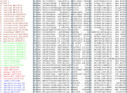
Fig. 1. Sequence alignment of Ac-SLP-1 and SLP-2 with other saposin-like proteins (SAPLIPs). Only the SAPLIP domain from each protein has been used for the alignment. SPB_human – saposin B (GenBank Accession number P07602); Ce spp4 – C. elegans SAPLIP protein family 4 (AAA81416); FhSAP2 – F. hepatica SAP2 (AF286903); Aoah_human – Homo sapiens acyloxyacyl hydrolase (BAD97196); EhA – E. histolytica amoebapore A (M83945); EhB – E. histolytica amoebapore B (CAA54226); countin – Dictyostelium discoideum AX4 countin (XP_643887); Asm_human – H. sapiens acid sphingomyelin phosphodiesterase 1 (BAD93012); Sap_slimemould – D. discoideum saposin A (BAA32237); Sap_C_bovine – Bos taurus saposin C (S21770); Sap_B_human – H. sapiens saposin B (NP_001035930); clonorin – C. sinensis clonorin (AF421960); NKLysin_pig – Sus scrofa NK-Lysin (CAA59720); Ce ZK455.4 – C. elegans hypothetical protein (CAA91493). The 6 conserved cysteine residues are boxed in grey with predicted disulphide bonding patterns shown underneath (1–6, 2–5, 3–4). Blue denotes conserved hydrophobic residues; yellow highlights the conserved proline in saposins or alanine in SAPLIPs and pink indicates the histidine predicted to be involved in dimerization of amoebapores. Red boxes in bovine saposin C denote positively charged residues involved in its anchoring to the cell membrane; positively charged residues of Ac-SLP-1 and SLP-2 in this vicinity are also highlighted in red. Predicted alpha-helices of amoebapore A are shown as solid bars above the alignment. Putative N-linked glycosylation sites are in bold. Gaps have been introduced to improve the alignment.
ESTs encoding SAPLIPs from parasitic nematodes
ESTs encoding a SAPLIP domain were identified from the 4 major clades of animal-parasitic nematodes (clades I, III, IV and V) (Blaxter et al. 1998) (Fig. 2). Again, these nematode SAPLIPs share little sequence identity other than the 6 cysteines and the hydrophobic residues making up the charged surfaces, making it difficult to infer functional inferences based on sequence identities.

Fig. 2. Multiple sequence alignment of parasitic nematode expressed sequence tags encoding SAPLIP domains. GenBank Accession numbers are listed next to the species name. Trichinella spiralis prosaposin (T. spiralis prosap_1–4: GenBank accession number AAR83123) full-length cDNA sequence was included in the analysis – it contains 4 SAPLIP domains; hence, each domain was included as a separate entry. Grey boxes denote the 6 conserved Cys residues. Species are grouped according to the phylogeny of Blaxter et al. (1998) and colour coded as follows: brown – clade V; green – clade IV; red – clade III; blue – clade I.
Transcription in different developmental stages
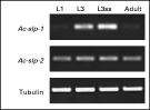
Ac-slp-1 was detected in RNA from all developmental stages of A. caninum, but was most abundant in L3 and L3SS. Ac-slp-2 was detected equally in L1, L3, L3SS and adult stages (Fig. 3). Both transcripts were identified by PCR in the cDNA from adult A. caninum gut tissue extracted by laser capture microdissection (Ranjit et al. 2006) (not shown).

Fig. 3. Transcription profiles of Ancylostoma caninum SAPLIP mRNAs. Reverse transcription (RT)-PCR analysis for mRNA transcription levels of Ac-slp-1 and slp-2 in A. caninum L1, L3, serum stimulated L3 (L3ss) and adult worms. Beta-tubulin mRNA (AF077870) was used as a constitutively expressed positive control. Controls lacking cDNA template did not reveal any bands (not shown).
Expression and analysis of recombinant proteins
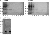
Recombinant Ac-SLP-1 and SLP-2 (in the pMelBac plasmid) were both expressed and secreted by Sf9 and Hi5 insect cells. The yields of purified recombinant proteins were ~0·2 mg/l for Ac-SLP-1 and ~0·3 mg/l for Ac-SLP-2 when expressed in Hi5 cells (Fig. 4A). Recombinant SLP-1 migrated as a doublet, probably representing glycosylated and non-glycosylated forms (SLP-1 has 1 predicted N-linked glycosylation site). No expression of recombinant proteins was achieved using the pHotWax expression vector (where the C-terminal purification tags can be removed by proteolysis with TEV protease), and protein was not detected in either Sf9 or Hi5 insect cells for either Ac-SLP-1 or SLP-2 (not shown), precluding studies aimed at determining whether head-to-tail dimerization occurs in the presence of native termini free of purification tags. Both recombinant proteins were detected by their homologous mouse antiserum on Western blots (Fig. 4B). Antisera to recombinant SLPs were used to probe A. caninum extracts electrophoresed under denaturing and non-denaturing conditions (to address dimerization of native SLPs), but bands were not detected using either method (not shown). The antisera did, however, detect proteins in immunohistochemistry, perhaps reflecting differential accessibility of epitopes when the proteins were electrophoresed in a polyacrylamide gel compared with parasite tissue sections. Therefore, we cannot comment with any degree of certainty on whether these proteins dimerize in their native states.

Fig. 4. Expression and purification of recombinant Ac-SLP-1 and Ac-SLP-2 proteins in baculovirus. (A) Coomassie Brilliant Blue-stained SDS-PAGE gel depicting the purification of recombinant Ac-SLP-1 and SLP-2 by nickel-affinity chromatography. Lane 1, culture supernatant in nickel binding buffer; lane 2, unbound material after initial chromatography; lanes 3–6, washes with increasing concentrations of imidazole (10, 20, 40, 60 mM respectively); lanes 7–10, eluate fractions (1·0 ml) of purified protein in 250 mM imidazole; lane 11, recombinant protein after a secondary round of purification on Ni-NTA agarose to remove contaminating bands seen in lanes 7–10 after primary purification. (B) Western blot showing recognition of recombinant proteins by homologous antisera. Lanes 1 and 2, Ac-SLP-1; lanes 3 and 4, Ac-SLP-2; lanes 1 and 3 probed with normal mouse serum; lanes 2 and 4 probed with mouse anti-SLP-1 and anti-SLP-2, respectively.
Immunolocalization
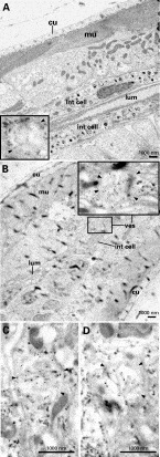
Using specific antisera, both Ac-SLP-1 and SLP-2 were immunolocalized to the intestine of adult A. caninum (Fig. 5). Intense staining was detected in small punctate ‘vesicles’ beneath the brush border epithelium of the intestinal cells. Some fluorescence, albeit weak and dispersed, was also displayed on the microvillar brush border of the intestinal cells. Pre-immunization mouse serum did not bind to any structures in sections. Using electron microscopy and immunogold labelling, Ac-SLP-1 was localized to vesicles within the intestinal cells of the L3 (Fig. 6), supporting the findings for the transcription and expression of these molecules in the intestinal cells of adult A. caninum. Antibodies to Ac-SLP-2 bound to the same structures in the L3 as did anti-SLP-1 sera (not shown), consistent with the punctate vesicles observed in the adult worm.

Fig. 5. Immunostaining of tissue sections from Ancylostoma caninum adult worms using mouse antibodies raised to recombinant SLP-1 or SLP-2 or pre-immunization mouse sera, followed by Cy3-conjugated anti-mouse Ig (red colour). Sections were counterstained with DAPI to highlight the nuclei (blue colour). Both proteins were localized to the intestine, with intense staining seen in small punctate ‘vesicles’ (arrows) and less intense, sporadically distributed staining in the microvillar brush border of the intestinal cells. NMS, normal mouse serum; int, intestinal cell; lum, intestinal lumen.

Fig. 6. Localization of Ac-SLP-1 in Ancylostoma caninum L3 using immunogold electron microscopy. Mouse anti-SLP-1 antibodies were used to localize both proteins in L3. (A) Normal mouse serum. The insert shows unlabelled ‘vesicle’. (B) Mouse anti-SLP-1 antibodies recognized the protein in ‘vesicles’ (arrowheads) within the intestinal cells (boxed). The insert shows a larger magnification of the stained ‘vesicle’. (C and D) Anti-SLP-1 antibody labelled vesicles taken from other regions of the intestinal cells. The same structures were also labelled using anti-SLP-2 antibodies (not shown). cu, cuticle; mu, muscle; lum, intenstinal lumen; int cell, intestinal cell; ves, ‘vesicle’.
Haemolysis
No haemolytic activity was observed for recombinant Ac-SLP-1 or SLP-2 containing C-terminal purification tags (expressed in pMelBac – not shown). Also, no dimer formation of recombinant proteins was detected when proteins were subjected to electrophoresis on native-PAGE gels under non-reducing conditions, despite dimerization of a positive control recombinant protein (thioredoxin peroxidase) (not shown). Insufficient quantities of recombinant proteins were generated by insect cells infected with virus containing the pHotWax plasmid (i.e. where the purification tags could be cleaved with exogenous protease).
DISCUSSION
All members of the SAPLIP family (with the exception of granulysin) share a conserved placement of 6 cysteines, predicted to have a similar disulfide cross-linkage: 1–6; 2–5; 3–4 (Leippe, 1997; Zhai and Saier, 2000; Bruhn, 2005). The disulfide bonds confer remarkable stability to the peptides, particularly against heat and other environmental factors (Kishimoto et al. 1992; Zhai and Saier, 2000; Lee et al. 2002). Apart from the conserved cysteines, there is very little sequence homology among SAPLIPs. Therefore, function may not be reflected in sequence similarity, and is most likely due to similar tertiary structure (Bruhn, 2005). The crystal structure has been elucidated for amoebapore A (Leippe et al. 1992; Hecht et al. 2004) and NK-lysin (Liepinsh et al. 1997), and forms the backbone upon which other SAPLIPs can be modelled. Four or 5 alpha helices are usually present in SAPLIPs, and the surface charge distribution of these proteins is the basis of their membrane binding capacity and ability of some to form pores. The function of SAPLIPs is to bind to lipids in cell membranes (Zhai and Saier, 2000; Bruhn et al. 2003) and then to perform a range of functions, including transporting lipids within and between membranes (e.g. saposins) (Munford et al. 1995), cytolysis (e.g. amoebapores) (Leippe et al. 1991) and antimicrobial activity (e.g. NK lysin) (Leippe, 1995). Both Ac-SLP-1 and SLP-2 contain the 6 conserved cysteines as well as several hydrophobic residues representing the protein core of the SAPLIP superfamily (Bruhn, 2005).
Dimerization is common in SAPLIPs, but the mechanisms by which dimer formation occurs are variable. Amoebapore A forms head-to-tail homodimers which are predicted to insert as pores into lipid membranes and, in turn, facilitate the subsequent lysis of the cells (Hecht et al. 2004). This dimer formation hinges on the presence of a histidine near the C-terminus of the protein (Andra and Leippe, 1994). Similarly, clonorin from C. sinensis has a histidine in the same position as amoebapore A, and is predicted to form dimers when active (Lee et al. 2002). Both Ac-SLP-1 and SLP-2 have a C-terminal histidine between cysteine residues 5 and 6 which might mediate dimer formation. Interestingly, recombinant clonorin is unable to lyse erythocytes when it contains a C-terminal 6×His tag and an enterokinase cleavage site, but does lyse cells after proteolytic removal of the tags, implying that extra residues at the C-terminus interfere with dimer formation (Lee et al. 2002). Recombinant Ac-SLP-1 and SLP-2 produced in pMelBac contained a C-terminal 6×His tag which could not be removed; these proteins were neither haemolytic nor did they dimerize. Therefore, an attempt was made to express them in pHotWax (containing a cleavable 6×His tag), but insufficient yields of recombinant protein prevented the pursuit of dimerization studies.
Both Ac-SLP-1 and SLP-2 are localized to the intestines of adults and L3 of A. caninum, indicating that they could play a role in parasite feeding. Clonorin is haemolytic and localized to the intestine of adult C. sinensis (see Lee et al. 2002), further supporting a role for Ac-SLP-1 and SLP-2 in the lysis of ingested host cells by L3 and adult stages. Both proteins displayed a unique expression site within the intestine of A. caninum, localizing to punctate vesicles/granules within the intestinal cells. A similar pattern of localization has been shown for an intestinal SAPLIP of Fasciola gigantica (see Grams et al. 2006) which is orthologous to a haemolytic protein from F. hepatica (see Espino and Hillyer, 2003). SAPLIPs might be secreted from these ‘vesicles’ and transported to the intestine where they bind to and, subsequently, lyse erythrocytes, releasing haemoglobin into the intestinal lumen where it is degraded by a suite of proteases (Brindley et al. 2001; Williamson et al. 2003b, 2004). A. caninum SAPLIPs have signal peptides, and it is possible that these proteins are secreted across the intestinal epithelium into the lumen upon demand, possibly accounting for the scattered presence of fluorescence on the epithelium. Alternatively, these vesicles might be lysosome-like organelles and the final site of SLP trafficking.
Ac-slp-1 and slp-2 mRNAs are expressed in the intestines of multiple developmental stages of A. caninum, suggesting multiple functions in parasite biology. Ac-slp-1 mRNA is most abundantly expressed in the L3 (determined by non-quantitative methods), both serum-stimulated and non-stimulated; Ac-slp-2 on the other hand is expressed equally in L1, L3 and adult stages. Since the majority of Ac-slp-1 is expressed in the L3 and adult worm, the SLP-1 protein could interact with host cells. SLP-2 protein might perform multiple functions, because it is expressed in L1 as well as the intra-mammalian stages. Alternatively, SLP-2 might lyse multiple cell types in the parasite's intestine – soil bacteria in the L1 and host cells in the L3/adult. SAPLIPs often lyse different cell types (Bruhn, 2005) and, in the absence of functional recombinant proteins (i.e. inability to determine whether dimerization occurs), it would be premature to comment further on the biological functions of these proteins in the gut of A. caninum.
Since both A. caninum SAPLIPs described here are found in pre-parasitic and parasitic developmental stages, they have potential as vaccine candidates. Indeed, vaccination of rabbits with recombinant FhSAP2 achieved protection against challenge infection with F. hepatica (see Espino and Hillyer, 2004), implying that this family of proteins is a valid target for helminth vaccines. Existing hookworm vaccine molecules are restricted in their developmental expression to either the L3 (Bethony et al. 2005) or adult worm (Loukas et al. 2005), prompting the suggestion that a bivalent vaccine may be required to attack both major developmental stages within the host (Hotez et al. 2006). Therefore, a molecule expressed in the gut of different developmental stages might be worthy of pursuit as a single antigen vaccine against hookworm disease. However, prior to vaccine testing, the production of a biologically active recombinant protein (which allows dimer formation, if appropriate) is needed to determine the function(s) of both hookworm SAPLIPs.
We thank Mary Duke, Mai Tran, Mark Pearson, Ben Datu, Michael Smout and Danielle Smyth for helpful discussions and the provision of reagents. We are grateful for advice and support from Drs Peter O'Donoghue and Peter Hotez. T.D. was supported by an Australian Postgraduate Award and by Queensland Institute of Medical Research. A.L. was supported by a Career Development Award from the National Health and Medical Research Council of Australia (NHMRC). The work was supported by grants from the NHMRC and The Sabin Vaccine Institute.