Introduction
Nasal polyps tend to recur despite radical removal by means of functional endoscopic surgery.Reference Larsen and Tos1, Reference Wynn and Har-El2 Certain comorbidities are considered to predispose to massive, recalcitrant nasal polyps, i.e. allergies, allergic rhinitis, bronchial asthma and (especially) Samter's triad. Another ominous prognostic factor is the presence of initial frontal sinus disease.Reference Mendelsohn, Jeremic, Wright and Rotenberg3
In this study, we assessed the replacement of vulnerable nasal vault mucosa with skin as a technique to reduce polyp formation. Skin transfer into the nose has already been reported for the treatment of hereditary haemorrhagic telangiectasia.Reference Lesnik, Ross, Henderson, Joe, Leder and White4–Reference Bridger7 In the current study, the technique was evaluated in a prospective case series.
Materials and methods
Patients were operated upon by the same surgeon (GA) in the Neo Athinaion Hospital, Athens, Greece, between January and December 2011. These patients had been operated upon for polyposis at least twice in the past, with at least one procedure involving functional endoscopic surgery. Every patient considered for nasal dermoplasty received pre-operative treatment with prednisolone and antibiotics. After completion of this medical therapy, patients underwent computed tomography using a navigation system protocol.
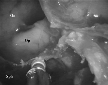
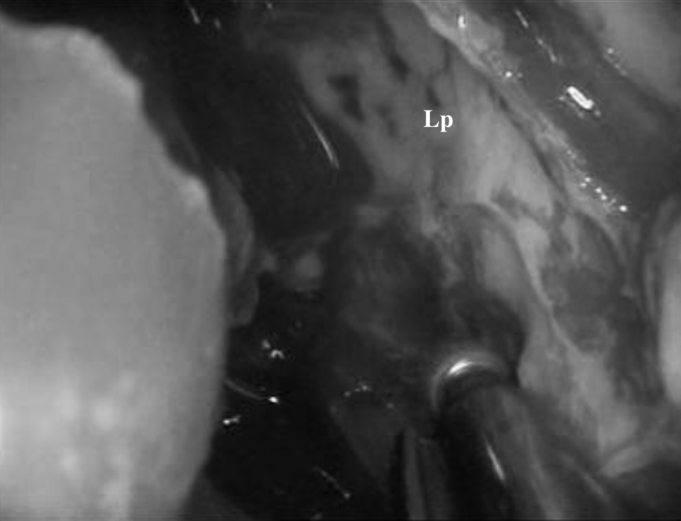
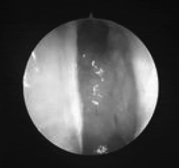
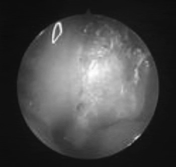
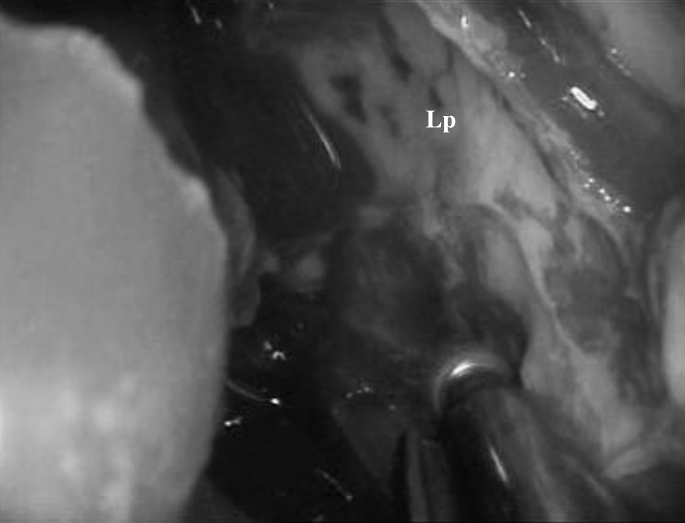
The surgical equipment was arranged as for a conventional endoscopic operation. For dermoplasty, a small area of skin was prepared on the anterior surface of the thigh. Complete ethmoidectomy and opening of the sinuses ostia were performed. Starting from the roof of the sphenoid, the nasal mucosa was removed. For every cell from which the mucosa was removed, the bony partitions were also removed using a 15° angled diamond burr (Figure 1). The aim was to completely flatten the skull base. The same applied for the lamina papyracea (Figure 2). The removal of the mucosa was performed up to the frontal recess.
Endoscopic view showing removal of the bony septa. On = Onodi cell; Op = optic nerve; Sph = sphenoid sinus

Endoscopic view showing flattening of the lamina papyracea (Lp).
After the completion of mucosa removal and skull base flattening, we estimated the amount of skin needed to cover the denuded area. Our intention was to cover the fovea ethmoidalis from the roof of the sphenoid sinus to the base of the posterior wall of the frontal sinus, and to cover the lamina papyracea from the anterior wall of the sphenoid sinus to the area of the lacrimal sac. The rationale was to replace the mucosa of the region that is most responsible for the growth of polyps (i.e. the fovea ethmoidalis). The amount of skin needed was estimated using a piece of paper cut to fit the dimensions of the denuded area. Mean dimensions of 4.5 × 3 cm sufficed for each nostril. A graft thickness of 0.15 mm was used.
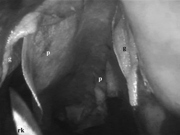
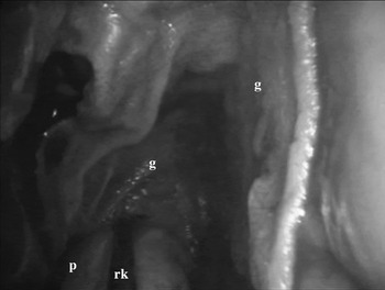
The skin was inserted together with the paper template (Figure 3) and was applied to the aforementioned areas (Figure 4). No further packing was needed. The middle turbinates were stitched to the septum to prevent lateralisation.Reference Musy and Kountakis8
Endoscopic view showing insertion of the skin graft (g) together with the paper template (p). rk = round knife

Endoscopic view showing removal of the paper template (p) with the round knife (rk), leaving the skin graft (g) in situ.

The patients stayed overnight and were discharged from hospital the next day. They were instructed to use saline rinses in the immediate post-operative period and to apply topical corticosteroids after one month had elapsed (to allow complete healing of nasal tissues).
Results
Five male patients were operated upon using the modified nasal dermoplasty technique described above. The coincidence of gender prevalence was probably influenced by aesthetic considerations. Patients' ages ranged from 35 to 60 years. All patients had a history of allergic rhinitis, three had a typical history of coexisting allergic bronchial asthma, and one presented with Samter's triad. All patients had undergone at least two previous operations for polyp removal.
In the current study, patients' surgical procedures lasted between 5 and 6 hours. All patients recovered uneventfully.
Follow up ranged from a minimum of two months to a maximum of one year. One patient did not present for follow up. One case of graft failure occurred. In three cases, graft uptake was successful in both nostrils, albeit with a significant amount of shrinkage (Figure 5).
Endoscopic view showing the skin graft one month after surgery; remnants of hair are seen.

Post-operatively, all four followed-up patients reported that they were in a better condition than they had been at the same post-operative point following previous operations. The need for meticulous preparation of the fovea ethmoidalis, in order to ensure skin graft acceptance, may explain why even the patient whose graft failed considered his surgical outcome a success. Nevertheless, new polyp formation was observed in all four followed-up patients. However, no post-operative polyp formation was observed in the successfully grafted areas; rather, all new polyp formation occurred in non-grafted areas.
Discussion
Five patients suffering from recurrent nasal polyposis, and having undergone multiple previous operations, were operated upon using a modified nasal dermoplasty technique. Preliminary results were promising. In this series, there was a high rate of successful graft uptake and no new polyp formation was noted in the grafted areas. These preliminary results encourage us to apply this method to larger numbers of patients with longer periods of follow up.
Until now, the surgical removal of nasal polyps has had one major drawback. Polyps tend to recur, despite radical removal and intense post-operative follow up and medication. The optimum extent of polyp removal surgery has been a matter of debate. Some authors state that extensive sinus surgery can lead to lower revision rates.Reference Masterson, Tanweer, Bueser and Leong9–Reference Videler, Wreesmann, van der Meulen, Knegt and Fokkens11 Others have compared minimally invasive surgery to radical ethmoidectomy with removal of the mucosa, and found no significant difference in outcomes.Reference Devars du Mayne, Prulière-Escabasse, Zerah-Lancner, Coste and Papon12 In a large study comparing patients undergoing simple nasal polypectomy only versus simple nasal polypectomy plus additional sinus surgery, little difference was detected between the two patient groups regarding recurrence rates.Reference Hopkins, Slack, Lund, Brown, Copley and Browne13 Our suggestion concerns not the extent but, rather, the rationale of the operation.
Our study had two obvious disadvantages: limited patient numbers and restricted follow up. However, the current findings represent only preliminary results for this new technique. Also, the measurement of outcomes could have been improved: patient evaluation could have benefitted from more specific criteria for symptoms and signs, as well as from laboratory tests. A further disadvantage was the need for high-level endoscopic surgery skills in order to successfully apply the modified nasal dermoplasty technique, especially in patients who had undergone previous surgery. Furthermore, the duration and difficulty of the operation were considerably increased by our proposed, extended surgical technique, which added removal of the mucosa, flattening of the skull base, and estimation and insertion of a skin graft. The operations in this study lasted between 5 and 6 hours. The operative time would have been reduced by the presence of a second surgeon, the establishment of standards for the amount of skin needed, and, of course, increased experience. The long duration of the operation necessitated detailed pre-operative diagnostic investigation in order to exclude patients with impaired general health.
Another concern is the safety of the procedure. However, extensive experience with the use of septal dermoplasty in patients with hereditary haemorrhagic telangiectasia has indicated that the procedure is relatively safe, with few complications.
The only serious complication arising from insertion of a skin graft into the nose is crusting due to desquamation.Reference Fiorella, Ross, Henderson and White14 Crusting may lead to nasal obstruction and a foul odour, requiring frequent saline rinsing (Figure 6). Others have noted that graft survival rates are high within the well protected nasal cavity.Reference Tran, Berry and Fee15 A literature search identified no reports of increased rates of squamous cell carcinoma in the nasal cavity.
Endoscopic view for the same patient as in Figure 5, two months after surgery. Desquamated epithelial cells can be seen.

Finally, with the exception of a dermatome, no other special equipment is required for modified nasal dermoplasty, and no prolonged hospitalisation is needed.
Conclusion
We propose a new technique for the surgical management of recalcitrant nasal polyps, namely, nasal dermoplasty of the fovea ethmoidalis and lamina papyracea. Preliminary results show both a high prevalence of graft ‘take’ and strong resistance of the grafted areas to polyp formation. On the other hand, the technique is cumbersome and polyps continue to appear in the remaining, untreated nasal mucosa. We believe that further research is justified in order to evaluate and establish this modified nasal dermoplasty technique.