Total anomalous pulmonary venous connection is rare (1–3% of all CHDs) and encompasses a wide clinical spectrum in presentation, from mild cyanosis to cardiovascular collapse. Reference Husain, Maldonado and Rasch1–Reference Reller, Strickland, Riehle-Colarusso, Mahle and Correa3 With improvement in medical and surgical management, mortality has steadily decreased below 20%, and in some series below 10% in the current surgical era. Reference Husain, Maldonado and Rasch1,Reference Yong, Yaftian and Griffiths2,Reference Kirshbom, Myung and Gaynor4 In the most recent decade, the focus of the literature has been on identifying characteristics associated with recurrent pulmonary venous obstruction and mortality risk after repair of total anomalous pulmonary venous connection. Our own institution previously reported the association of pulmonary venous obstruction at presentation, increased cardiopulmonary bypass and cross-clamp times with mortality, as well as the association of post-repair confluence gradients with the occurrence of pulmonary venous obstruction requiring reintervention. Reference Husain, Maldonado and Rasch1
Long-term morbidity and mortality data (20–40+ years) exist after total anomalous pulmonary venous connection repair; however, these traverse several decades in which significant improvements in surgical and critical care management of total anomalous pulmonary venous connection have occurred. Reference Yong, Yaftian and Griffiths2,Reference Kirshbom, Myung and Gaynor4,Reference Karamlou, Gurofsky and Al Sukhni12 With our updated cohort, we focused on the long-term re-intervention and mortality risk in the most recent two decades. The purpose of this study was to revisit our now larger cohort of patients and review factors associated with surgical and long-term mortality as well as the need for re-intervention
Materials and method
Our study cohort included 81 patients undergoing repair of TAPVC at Christus Santa Rosa Hospital and University Hospital of San Antonio, TX, from January 2002 to January 2018. Our University of Texas Health at San Antonio program moved from one facility to another in 2014. Approval was granted by the institutional review board at all study group hospitals.
Patient demographics and available operative and post-operative records, discharge summaries, and echocardiographic data were reviewed (Tables 1 and 2). In all patients, the diagnosis of TAPVC was confirmed by transthoracic echocardiogram prior to surgery. Pulmonary venous obstruction was defined as any obstruction of the pulmonary venous drainage along the anomalous pathway, resulting in significant pulmonary congestion and haemodynamic compromise. Study points of interest were mortality and pulmonary vein reintervention. Early, or surgical mortality, was defined as death within 30 days of operation, or at any point during the primary hospitalisation during which initial TAPVC repair occurred. Late mortality was defined as death occurring post-discharge and greater than 30 days from the repair at initial hospitalisation. Reintervention was defined as any procedure, operative or trans-catheter, performed to treat pulmonary venous stenosis/obstruction.
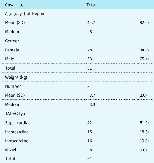
Patient demographics. Demographic variables evaluated in patient cohort.

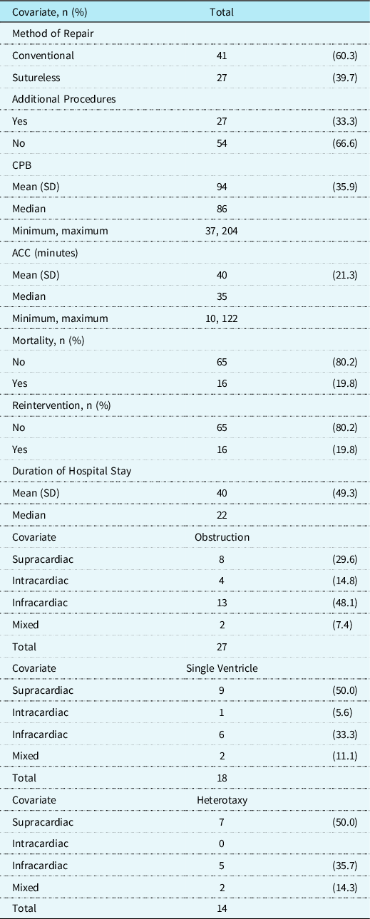
Operative variables evaluated in study cohort.

Our study cohort included interventions performed by six different congenital heart surgeons; with two surgeons performing the majority (76.5%) of the primary repairs. All repairs utilised non-absorbable, polypropylene suture.
Post-repair pulmonary venous confluence gradients were measured by an intraoperative transesophageal echocardiogram. Mean gradients were measured at the level of the pulmonary venous confluence anastomosis to the left atrium. Over the study period, transesophageal echocardiogram recordings were performed by two echocardiographers, with the previously reported cohort all being performed by one.
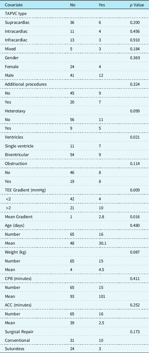
Demographic information including, age and weight at time of operation, gender, single ventricle physiology, heterotaxy, and the presence of pre-operative pulmonary venous obstruction were reviewed. Single ventricle physiology was defined as a cardiac anatomy without the means to provide separate and sufficient systemic and pulmonary circulations. We examined operative variables, including the method of surgical repair, additional procedures at the time of initial palliation, duration of cardiopulmonary bypass and aortic cross clamp time, utilisation of deep hypothermic circulatory arrest, blood products administered, and immediate postoperative transesophageal echocardiogram findings. Additional procedures were identified and defined as any total anomalous pulmonary venous connection repair requiring more than a patent ductus arteriosus ligation or atrial septal defect repair at the time of initial palliation. Variables of interest were then evaluated for possible association with reintervention, mortality, and hospital length of stay (Tables 3–5).
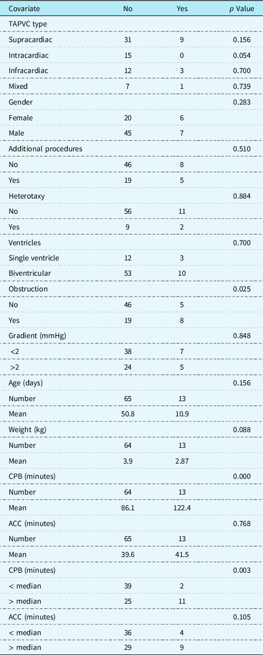
Early mortality. Variables evaluated for association with early mortality after surgical repair.

All mortality. Variables evaluated for association with early or late mortality after surgical repair.

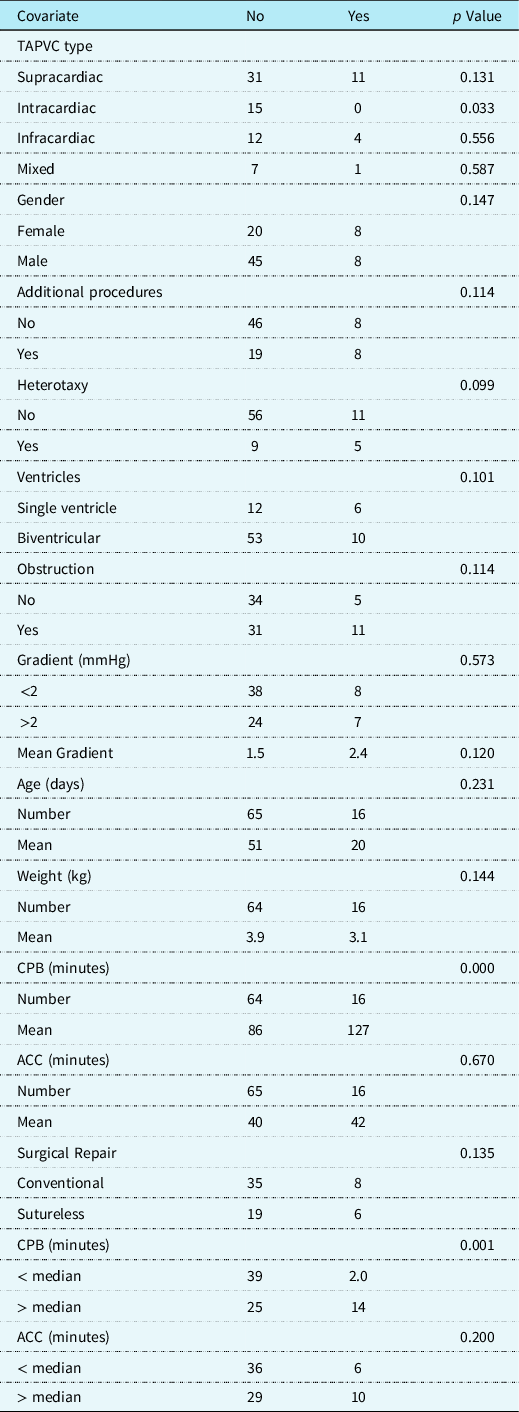
Reintervention. Variables evaluated for association with surgical and/or transcatheter intervention for pulmonary venous confluence obstruction after surgical repair.

Study data were collected and managed using the REDCap© (Research Electronic Data Capture) system. Variables in association with outcomes of interest were investigated by statistical methods, including the difference in means (t-test), Pearson’s chi-squared analysis, and creation of Kaplan–Meier curves using the log-rank test of equality; all utilising the software STATA 14 © StataCorp LLC, College Station, TX, USA.
Results
Patient demographics
Of the 81 patients, 53 (65%) were male and 28 (35%) females. Median age at the time of repair was 8 days (range 0–564 days). Median weight was 3.27 kg (range 2.0 to 16.8 kg). Concerning the anatomic subtypes, 42 (52%) were supracardiac, 15 (19%) intracardiac, 16 (20%) infracardiac, and 8 (9%) had mixed type (Table 1).
Single ventricle physiology was present in 18 (22%) of the 81 patients. Heterotaxy was noted in 14 (17%) patients. At the time of presentation, pulmonary venous obstruction was diagnosed in 27 (33%) of the patients. Among those who presented with obstruction, 13 (48%) were of the infracardiac subtype. Twenty-seven patients (33%) had additional procedures performed at the time of initial total anomalous pulmonary venous connection repair. The most prevalent of these being a right ventricle to pulmonary artery conduit placement (seven patients), pulmonary artery banding (five patients), atrial septectomy (four patients), and coarctation repair (four patients).
Patient follow-up
Total follow-up time ranged from 0 to 17.2 years, with a median of 8.8 months and a mean of 3.5 years. Follow-up information was available for all 81 patients; however, only 64 and 56 patients had followed up at 30 and 60 days, respectively. Median time between initial date of surgery and mortality was 58 days (range 0 to 44.4 months), with 94% occurring within 4.1 months. The median time between the date of surgery and re-intervention was 97 days (range 3 to 3733 days), with 80% occurring within 1.2 years.
Operative technique
Of the 81 initial operations, 41 (51%) were considered “conventional”, 28 (35%) operations utilised the sutureless method. The sutureless method of repair was determined from the operative note, with a technique like those reported. Reference Kilic and Whitman21 The conventional methods of repair included surgical anastomosis between the pulmonary confluence into either the dome of the left atrium, the left atrial appendage, or via a right atriotomy with atrial septostomy and reconstruction. The remaining 12 (14%) were unclassified due to missing or incomplete records. Statistical evaluation of the various methods of repair was not significantly associated with post-operative mortality or re-intervention rates. Cardiopulmonary bypass and aortic cross-clamp times were also evaluated. The median time on cardiopulmonary bypass times was 86 minutes (range of 37 to 204) while median aortic cross-clamp time time was 35 minutes (range of 10 to 122). The use of DHCA was utilised in 16 (20.5%) patients. Blood product use including intraoperative transfusion of packed red blood cells, fresh frozen plasma, platelets, and cryoprecipitate were noted and analysed for possible association with post-operative mortality and re-intervention. They were not found to be significant regarding re-intervention or mortality (Table 2).
Re-intervention for pulmonary venous stenosis
All patients were reviewed for the development of pulmonary venous stenosis at the confluence requiring intervention. There were 16 patients who required re-intervention for significant pulmonary venous obstruction. Of these 16 patients, 5 had multiple reinterventions. Those with single-ventricle anatomy were found to be at significant risk for re-intervention (p = 0.021) (Fig 2). Upon cessation of cardiopulmonary bypass, transesophageal echocardiogram evaluation of the venous confluence identified a post-repair gradient greater than 2 mmHg to be significantly associated with re-intervention (p = 0.009) (Table 5). Kaplan–Meier curves were created in re-intervention analysis (Fig 2). The log-rank test demonstrated the presence of obstruction as a predictor of re-intervention (p = 0.031). A Cox-proportional hazard model was also created, which again found significance in the presence of obstruction and re-intervention rate with a hazard rate of 3.468, and nearly 2.5× higher risk compared to non-obstructed (p = 0.042). Kaplan–Meier curves and Cox proportional hazard models for single ventricle and heterotaxy trended toward significance, but ultimately did not reach a p-value <0.05.
Kaplan Meier Curves for re-intervention in subgroups of ventricular physiology, presence of heterotaxy, and presence of pulmonary venous obstruction at presentation.

Kaplan Meier Curves for mortality in subgroups of ventricular physiology, presence of heterotaxy, and presence of pulmonary venous obstruction at presentation.

Mortality
There were 16 (19.8%) total patient deaths throughout the study period. Thirteen (16.1%) were surgical mortalities, and 3 (3.7%) were late deaths. Of note, all three patients with late mortality had single ventricle anatomy in association with heterotaxy. Among these patients, the latest mortality occurred 1322 days post-initial discharge during a subsequent Fontan palliation. The total anomalous pulmonary venous connection anatomic subtypes were evaluated for associated risk with mortality. The overall mortality in repaired supracardiac subtype was 11/42, intracardiac had no death (0/15), infracardiac 4/16 deaths, and mixed 1/8 died. Intracardiac subtype was found to be negatively correlated with mortality (p = 0.033), indicating a survival benefit. Single and biventricular repair patients were evaluated, and while single ventricle repairs had a higher mortality rate, this was not statistically significant.
Within our cohort, 54 patients had no pulmonary venous obstruction at presentation, and 5/54 (9.82%) died after repair. A total of 27 patients presented with pulmonary venous obstruction and 8/27 (29.6%) died. Those patients presenting with obstruction were at increased risk of early mortality (p = 0.025). Kaplan–Meier curves were created for mortality analysis. The log-rank test of equality demonstrated significance in the presence of obstruction and mortality (p = 0.007) (Fig 2). Cox proportional hazard models were also considered, noting a hazard rate of 3.877 (p = 0.014) for the presence of obstruction, indicating a mortality hazard rate of 287% higher than those without obstruction. Kaplan–Meier curves and Cox proportional hazard models for single ventricle and heterotaxy trended toward significance, but ultimately did not reach a p-value <0.05.
During repair, the length of time on cardiopulmonary bypass, particularly times greater than the median of 86 minutes, were significantly correlated with early and all-mortality (p = 0.003 and p = 0.001, respectively).
Discussion
A growing body of literature pertaining to the management and outcomes of patients with total anomalous pulmonary venous connection has brought to light important factors associated with outcomes. Several characteristics of patient anatomy and surgical management have been previously identified as risk factors for reintervention and mortality after surgical repair of total anomalous pulmonary venous connection. Reference Husain, Maldonado and Rasch1,Reference Kirshbom, Myung and Gaynor4–Reference Caldarone, Najm and Kadletz7,Reference White, Ho and Faerber9–Reference Seale, Uemura and Webber13
Several factors were associated with reintervention for post-operative pulmonary venous obstruction including gender, increased cardiopulmonary bypass, increased aortic cross-clamp times, and post-operative transesophageal echocardiogram confluence gradient ≥2 mmHg. Reference Husain, Maldonado and Rasch1 The initial data published from our institution in 2011 was completed over a relatively short period of five years and follow-up of the younger portion of the cohort was of specific interest. This study contains the original group of 46 patients in addition to 35 patients surgically repaired from 2011 to 2018. Of note, the original study contained 51 patients; however, upon further review of the original patient’s records, 5 were removed in our study due to disqualifying factors.
Reintervention
The incidence of pulmonary venous stenosis (PVS) is cited as occurring in 10–15% of surgically repaired total anomalous pulmonary venous connection and remains a difficult complication to manage in the post-operative period. Reference Vanderlaan and Caldarone15 The location of stenosis can vary, either at the anastomotic site of the confluence, or as an intrinsic disease process affecting the pulmonary veins anywhere throughout their course. More concerning is its progressive nature and mortality rates ranging upwards of 40%. Reference Seale, Uemura and Webber13
The total number of patients requiring reintervention was 16 (19.7%), where 5 (6%) of these patients required more than one intervention. Of these procedures, 18 were surgical and 6 were catheter-based interventions. Additionally, 80% of reinterventions occurred within the first 15 months after initial surgical repair. This is consistent with Seale, et al. who noted that when pulmonary venous obstruction occurs, it tends to do so early, rather than late after repair. Reference Seale, Uemura and Webber13 It is also important to note that four interventions occurred after 15 months and were first time re-interventions. Three were anastomotic in origin, and one intrinsic. Only one went on to have further reintervention.
Among our patients, those with single ventricle physiology were found to be at a statistically higher risk for reintervention compared to those with biventricular physiology. The significant heterogeneity in this population of patients makes it difficult to determine which, if any, specific factors relate to increased risk of reintervention. However, one must certainly consider the anatomic complexity of these patients, as well as lability in physiology as substrate for their high-risk nature.
The evaluation of immediate post-operative confluence gradient by means of transesophageal echocardiogram was found to be a significant predictor of reintervention in our original publication. Once again, significance was found for those with a confluence gradient ≥2 mmHg.
It is well established that there are many factors that affect echo gradients. Physiologically, pulmonary venous flow is dependent on left atrial and ventricular function and compliance. Both are altered with the ensuing haemodynamic lability immediately after cardiopulmonary bypass, external compression from the transesophageal echocardiogram probe, and other factors which makes immediate post-repair gradients potentially difficult to interpret. In fact, surgical programmes have published reports identifying operative echocardiogram measurements suggesting confluence obstruction, when in fact, follow-up evaluation demonstrated a widely patent confluence without obstruction. Reference Herlong, Li, Bengur and Ungerleider18 When an elevated confluence gradient is identified at transesophageal echocardiogram, it is the practice of some programmes to withdraw the transesophageal echocardiogram probe, and re-image with a transthoracic probe. Therefore, we would posit it is incumbent from the surgeon’s perspective to reflect on the quality of the anastomosis to help guide intraoperative decision-making in relation to echo gradients. We believe the measured transesophageal echocardiogram gradient data remains an important factor in a surgeon’s decision-making in whether to consider revising the confluence anastomosis. It is important to note that we do not consider these findings as a certainty to the need for revision, but as a guideline to evaluate the possibility and safety of revising and improving the confluence anastomosis. It also places significant emphasis on the importance of having skilled echocardiographers.
Mortality
Overall, the reported mortality after surgical repair of total anomalous pulmonary venous connection has continually improved with advancing surgical and medical management over recent decades. Early publications demonstrated mortality risk upwards of 20% or more, while more recent reports demonstrate feasibility of mortality rates below 10%. Reference Yong, Yaftian and Griffiths2,Reference St. Louis, Harvey and Menk6–Reference Caldarone, Najm and Kadletz7,Reference Karamlou, Gurofsky and Al Sukhni12 From 2002 to 2018, our programme saw a surgical mortality rate of 16.1% (13/81) and identified three late deaths. Further evaluation revealed 81% (13/16) of overall mortality were neonatal repairs, a finding linked with increased mortality. Reference Yong, Yaftian and Griffiths2
The nature of patient presentation, particularly as a neonate with obstruction, single ventricle physiology, and additional anatomical cardiac anomalies has demonstrated the difficulty in creating equal survival compared to those with simple total anomalous pulmonary venous connection. Amidst advances in the management of total anomalous pulmonary venous connection and the lowering of surgical mortality rates, our programme’s growing experience has made us aware of the intricacies that exist within the growing body of literature. Specifically, the ability to identify the subset of patients within the highest risk categories for reintervention and mortality.
There were a total of three patients who were considered late deaths. Two deaths occurred ithin 4 months, the other at 3 years post-repair. All three patients were single ventricle variants. Overall survival for the entire cohort over the 17.2-year study period was 80.3%, with most of the mortality occurring early. Noting, if one survived the early post-operative period, the late survival in our cohort was excellent at 96% (65/98). This reiterates previously published findings that late survival is excellent for those surviving the initial repair and discharge from the hospital. Reference Yong, Yaftian and Griffiths2,Reference St. Louis, McCracken and Turks14
Within this larger cohort, one new finding identified intracardiac subtype as being protective against mortality. Further evaluation of our intracardiac cohort identified three patients with complex total anomalous pulmonary venous connection, and three presented with obstruction, with the majority being simple and unobstructed. This finding certainly could be related to many of these patients having a relatively benign neonatal course and non-urgent need for repair in simple, non-obstructed forms (Fig 3).
Sub-type Survival. Observed 12-month and late survival for anatomic sub-types of TAPVC after repair.

Limitations
We recognise the limitations of this study, and while we are encouraged by our results, the retrospective nature and small sample size limit our ability to perform and interpret our statistical analyses. Another significant limitation was finding data points of interest in patients from the earlier era, where paper charts have since been destroyed in some instances. Additionally, surgeon preference in certain aspects of operative conduct rather than the lack of standardised surgical management of these patients makes it challenging to generalise our findings. Our study is also limited by difficulties in patient follow-up. Due to the rare nature of this condition, many of our patients come from all over the state of Texas and are lost easily to follow-up after initial repair. For our patients, increased complexity in presentation prevents isolating outcomes to total anomalous pulmonary venous connection, creating uncertainty in our mortality and reintervention rates.
Conclusion
Total anomalous pulmonary venous connection is still a significant problem with significant mortality in the modern era. This study re-evaluated a larger cohort with increased follow-up and confirmed the previously identified factors of confluence obstruction and increased cardiopulmonary bypass times with mortality risk. Intracardiac type was protective against mortality. Immediate post-operative confluence gradients and single ventricle anatomy increase the risk of post-operative pulmonary venous obstruction requiring reintervention. This illustrates the relationship between anatomical complexity and post-operative outcomes in patients with surgically repaired total anomalous pulmonary venous connection. Our institution reports a surgical mortality rate of 16.1%. While this is consistent with the reported literature, we must continually re-evaluate the risk stratification and management of these patients. Our study also sheds light on an expected timeline for re-intervention and mortality which included four patients with first-time reintervention after 15 months, which can help guide providers in surveillance and management.
Acknowledgements
None.
Financial support
This research received no specific grant from any funding agency, commercial, or not-for-profit sectors.
Conflicts of interest
None.