INTRODUCTION
Choline is a vitamin-like compound with two known functions: as choline per se and as a methyl donor, although the two roles overlap. Choline per se is a lipotropic agent playing a major role in lipid metabolism, particularly lipid transport, and is able to prevent or correct excessive fat deposition in the liver. This function is attributable to the presence of choline in the phospolipids (phosphatidylcholine and sphingomyelin) of lipoproteins. Impaired triglyceride secretion in the form of very-low-density-proteins is considered to be a major cause of fatty liver in choline deficiency. As a methyl donor choline, like methionine, is an important source of labile methyl groups for biosynthesis. Actually, the two principal methyl donors in animal metabolism are betaine, a choline metabolite, and S-adenosyl-l-methionine, a metabolite of methionine (Zeisel Reference Zeisel, Shils and Young1988; Pinotti et al. Reference Pinotti, Baldi and Dell'Orto2002). For this reason, it has been suggested that choline, betaine and methionine are closely interrelated metabolically. However, the requirement for choline per se must be met as choline, and betaine can substitute for the methyl donor function of choline only, probably because betaine cannot be reduced to choline (Zeisel Reference Zeisel, Shils and Young1988).
In adult ruminants, choline is extensively degraded in the rumen so dietary choline contributes insignificantly to the choline body pool. Methyl group metabolism is therefore conservative with a relatively low rate of methyl catabolism and an elevated rate of de novo methyl group synthesis via the tetrahydrofolate system (Pinotti et al. Reference Pinotti, Baldi and Dell'Orto2002; Baldi & Pinotti Reference Baldi and Pinotti2006). In dairy ruminants, the output of methylated compounds in milk is high but the dietary availability of choline is still low and precursors from the tetrahydrofolate pathway may be limiting, especially at the onset of lactation (Baldi & Pinotti Reference Baldi and Pinotti2006). Based on those considerations, the effects of rumen-protected choline (RPC) supplementation to transition dairy ruminants have been investigated in several studies (Brüsemeister & Südekum Reference Brüsemeister and Südekum2006; Pinotti et al. Reference Pinotti, Campagnoli, D'Ambrosio, Susca, Innocenti, Rebucci, Fusi, Cheli, Savoini, Dell'Orto and Baldi2008). The latter study investigated the effects of RPC administration on milk production and methyl group metabolism during the periparturient period of dairy goats, finding that RPC supplementation can increase milk yield and milk fat concentration. The milk production response to choline supplementation was obtained without a detrimental effect on plasma metabolites including folate and vitamin B12, indicating that good methyl group status was maintained.
Further studies in dairy cows have also investigated the effects of choline supplementation on hepatic lipid metabolism (Piepenbrink & Overton Reference Piepenbrink and Overton2003) and lipidosis (Grummer Reference Grummer, Joshi and Herdt2006). The results obtained in those studies indicated not only that hepatic fatty acid metabolism and cow performance are responsive to increasing the supply of choline (Piepenbrink & Overton Reference Piepenbrink and Overton2003), but also that choline supplementation can reduce cellular lipid accumulation in the liver (Grummer Reference Grummer, Joshi and Herdt2006). However, data on choline supplementation and its effects on liver lipid accumulation in dairy goats are lacking.
At the mammary gland level in dairy ruminants, choline plays a major role in lipid metabolism, particularly in lipid transport and milk fat secretion. The long-chain fatty acids in milk are obtained from the blood triglycerides of very low density lipoproteins (VLDLs) which arise either from absorbed fat or endogenously via mobilization of adipose fat stores. When dietary or synthetic choline availability is restricted, the rate of choline-containing phospholipid synthesis decreases, affecting lipoprotein lipid transport (Pinotti et al. Reference Pinotti, Baldi and Dell'Orto2002, Reference Pinotti, Baldi, Politis, Rebucci, Sangalli and Dell'Orto2003). Within the mammary tissue many selected enzymes (e.g. lipoprotein lipase (LPL)), transcription factors and nuclear receptors that can be affected by choline availability are involved in lipid uptake and milk fat secretion. Furthermore, choline is actively secreted into mammalian milk. The major choline-containing compounds in bovine milk are unesterified choline, phosphatidylcholine and sphingomyelin (Pinotti et al. Reference Pinotti, Baldi, Politis, Rebucci, Sangalli and Dell'Orto2003). Kinsella (Reference Kinsella1973) reported that a bovine mammary gland yielding 25 litres milk secretes 10±3 g phospholipids per day, corresponding on average to 0·05 of the phospholipids of the mammary tissue. The phospholipids of the membrane of the milk fat globule constitute the major choline-containing component of bovine milk (McPherson & Kitchen Reference Mcpherson and Kitchen1983). The above data suggest that choline is an important metabolite in lactating mammary tissue and that it is used avidly when available (Kinsella Reference Kinsella1973), mainly in lipid metabolism. Thus, choline supplementation can have positive effects on methyl group metabolism (Emmanuel & Kennelly Reference Emmanuel and Kennelly1984; Baldi & Pinotti Reference Baldi and Pinotti2006; Brüsemeister & Südekum Reference Brüsemeister and Südekum2006) and lipid trafficking, particularly lipid transport to extra-hepatic tissues (Piepenbrink & Overton Reference Piepenbrink and Overton2003; Baldi & Pinotti Reference Baldi and Pinotti2006; Cooke et al. Reference Cooke, Silva Del Río, Caraviello, Bertics, Ramos and Grummer2007), including the mammary gland. However, the mechanisms of these effects have not yet been clearly elucidated. In an attempt to shed light on these mechanisms, dairy goats were studied during the first month of lactation, investigating the effects of RPC administration on metabolic profile, selected liver constituents and transcript levels of selected enzymes, transcription factors and nuclear receptors involved in lipid metabolism in the mammary gland.
MATERIALS AND METHODS
Animals, treatment and diet
The animals were kept and cared for in accordance with European Union guidelines (86/609/EEC) approved by the Italian Ministry of Health. The current study was conducted as a parallel experiment to a production trial in which 70 pregnant multiparous Saanen goats were used (Pinotti et al. Reference Pinotti, Campagnoli, D'Ambrosio, Susca, Innocenti, Rebucci, Fusi, Cheli, Savoini, Dell'Orto and Baldi2008). Due to ethical reasons concerning the collection of tissues samples, the experiment was performed using a limited number of animals. In order to have 0·90 power to detect a significant difference at P<0·05, four samples per group were estimated as the minimum required. Eight pregnant multiparous Saanen goats of similar weight (mean 65±3·0 kg) and milk yield in the previous lactation (mean of the first month 2852± 360 g/day) at the Guidobono Cavalchini experimental farm, University of Milan, were assigned to one of two experimental groups: control group (CTR) with no choline supplementation and RPC group given 4 g/day of choline chloride in rumen-protected form (8 g of Sta-Chol Ascor Chimici, Forlì, Italy). The quantity of choline was decided based on experience with dairy cows (Pinotti et al. Reference Pinotti, Baldi, Politis, Rebucci, Sangalli and Dell'Orto2003) and metabolic body weight at the beginning of the experiment. The treatment was administered to each animal individually before the morning feed to ensure complete consumption, starting 4 weeks prior to expected kidding and continuing for 4 weeks after parturition. During this period all goats were fed a basal diet formulated to provide 8·37 and 10·0 MJ of metabolizable energy/kg dry matter (DM), 115 and 143 g of crude proteins (CP)/kg DM, for pre-kidding and lactation phase, respectively. The pre-partum and lactation basal diets were formulated according to the National Research Council (NRC 1981), and fed as total mixed ration (Unifast SpA, Padova, Italy) twice daily (06.30 and 18.30 h). Diet ingredients and composition have been published elsewhere (Pinotti et al. Reference Pinotti, Campagnoli, D'Ambrosio, Susca, Innocenti, Rebucci, Fusi, Cheli, Savoini, Dell'Orto and Baldi2008). Before and after kidding, the animals were housed in contiguous indoor stalls. Each goat delivered two calfs, and after kidding the animals were milked automatically twice a day. Post-kidding dry matter intake (DMI) was assessed weekly for each animal as the difference between feed DM offered and feed DM refused.
Milk and plasma analysis
During the first month of lactation, milk yield and composition were determined weekly. On sampling days, morning (06.30 h) and evening (18.30 h) milk samples from each animal were collected and pooled in proportion to the yield at each milking. The pooled samples were treated with sodium azide and stored at 5°C pending analysis for milk fat and milk protein (Milkoscan, Foss Technology, Denmark). Jugular vein blood samples were taken weekly, before the first meal of the day (06.00 h), and on days 7, 14, 21 and 28 post-partum. Blood samples were collected in heparinized tubes (Venoject; Teruno Europe, Leuven, Belgium) and centrifuged (14 000 g for 15 min at 10 °C) to obtain plasma, which was stored at −20°C pending analysis. Non-esterified fatty acids (NEFA) (acyl CoA synthetase, acyl CoA oxidase method – NEFA Kit, Randox test, UK), cholesterol (Cholesterol oxidase method, Diagnostic Cholesterol Kit, Alfawassermann), and β-hydroxybutyrate (BHBA) (β-hydroxybutyrate dehydrogenase method, RANBUT Kit, Randox) were measured in all plasma samples taken.
Biopsy preparation
On day 28 of lactation, 60 min before the liver and mammary biopsy procedures, the goats were sampled for blood (06·00 h) and treated with oxytocin (2 IU i.v, Izossitocina IZO S.p.A, Brescia, Italy), and then milked out. All goats were fed at the end of biopsy procedure.
Biopsy and analysis of liver samples
Samples of liver parenchyma were obtained from each animal using a blind percutaneous method and a 16G Tru-Cut needle 115 mm long (Urocore, H-S Medical SpA. Pomezia, Rome, Italy). Sedatives were not given so as not to disturb liver metabolism. The biopsy site (11th intercostal space, c. 150 mm below the spine) was shaved, surgically scrubbed and draped with sterile drapes. Five ml of 20 mg/ml lidocaine solution (Lidocaina 2% Fort Dodge, Fort Dodge Animal Health; Aprilia, Latina, Italy) was administered and a 40 mm incision made. A Tru-Cut needle was introduced in a cranio-ventral direction. About 100 mg of liver was collected per animal. The wound was sutured subcutaneously with poliglecaprone 25 monofilament (2–0 USP) (Mnocryl, Ethicon, Johnson & Johnson International, Stevenswulume, Belgium); the skin was closed using individual sutures of polyamide monofilament (0 USP) (Daclon, S.M.I AG, Hünningen, Belgium). The liver samples were frozen in liquid nitrogen and stored at −80°C pending analysis for total lipid and DNA.
Lipids were extracted by the Folch method (Folch et al. Reference Folch, Lees and Sloane-Stanley1957). Briefly, samples were thawed and total lipid extracted in a suitable excess of chloroform:methanol (2:1). After centrifugation, the lower phase was evaporated under a nitrogen stream and weighed. DNA was measured using the Burton (Reference Burton1956) method. Briefly, 100 mg of tissue was homogenized with 7 ml of cold 0·25 mm sucrose and 2 mm MgCl2, and centrifuged at 1000 g for 10 min at 4°C. The pellet was resuspended in 0·2 ml of 9 g/l NaCl solution, washed twice with 1 ml of 0·2 m perchloric acid (PCA) solution and centrifuged at 2000 g for 10 min at 4°C. The pellet was re-suspended in 2 ml 0·5 N PCA at 75 °C for 15 min. After centrifugation at 2000 g for 10 min, 2 ml of diphenylamine reagent (containing 1 g diphenylamine in 50 ml glacial acetic acid, 1 ml conc. H2SO4 and 0·25 ml aqueous acetaldehyde (1:50 v/v)) was added to 1 ml of supernatant. The mixture was then incubated at 75°C for 20 min. Calf thymus DNA (200 μg/ml, Sigma) was used as standard. The blank consisted of 1 ml of 0·5 M PCA solution. Absorbance at 600 nm was measured using a spectrophotometer (Perkin Elmer). DNA tissue content has been used to calculate total lipid/DNA ratio.
Biopsy and analysis of mammary samples
On day 28 of lactation, after liver biopsy mammary tissue was biopsied from each animal. The animals were sedated with 0·08 mg/kg i.v. xylazine chlorohydrate (Rompun, Bayer, Shawnee Mission, Kansas, USA) and then placed in dorsal recumbency. Heart rate and respiration were monitored continuously. The biopsy site – 250 mm distal to the basal part of the right udder − was shaved, surgically scrubbed and draped with sterile drapes. Five ml of 20 mg/ml lidocaine solution (Lidocaina 2% Fort Dodge, Fort Dodge Animal Health; Aprilia, Latina, Italy) was injected at the biopsy site (line block). A 30–40 mm incision was made through the skin, avoiding visible superficial blood vessels. The incision was continued through subcutaneous tissue and fascia. Secretory tissue was exposed and removed using dissecting scissors. Approximately 1 g was removed, rinsed with ml 9 g/l NaCl solution, and inspected to verify tissue homogeneity. It was then frozen in liquid nitrogen and stored at −80°C pending analysis. Haemostasis was achieved by ligation of blood vessels with catgut 3–0 USP (Assugut, Assut Europe S.p.A, Maliano dei Marsi, L'Aquila, Italy). Connective tissue and skin were closed using 3–0 USP poliglecaprone 25 (Mnocryl) and 2–0 USP polyamide (Daclon), respectively. No other medication was given during biopsy or afterwards.
Recovery from the liver and udder biopsies was rapid and uneventful in all cases. In all animals, milk yields returned to preoperative levels within 24 h. The polyamide sutures were removed 13 d later from both liver and mammary biopsy sites.
Transcript levels of LPL, fatty acid synthase (FAS), sterol regulatory binding proteins 1 and 2 (SREBP-1 and SREBP-2), peroxisome proliferator-activated receptor γ (PPARγ) and liver X receptor α (LXRα) were determined in mammary tissue. Total RNA was extracted using PeqGOLD Trifast (Peqlab, Germany) according to the manufacturer's instructions. RNA quality was checked by electrophoresis on denaturating gel; quantity was estimated by measuring optical density at 260 nm; integrity was checked by determining whether the OD260 nm/OD280 nm ratio >1·9. One μg total RNA was used for the synthesis of first strand complementary DNA (cDNA) using 200 units of reverse transcriptase (MMLV-RT, Promega, Madison, WI, USA) and 100 pmol of random primers (Invitrogen, Leek, The Netherlands) following the manufacturers’ protocols.
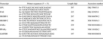
PCR reactions and analyses were performed in a Rotor-Gene 6000 (Corbett Research, Sydney, Australia) using Sensimix No Ref DNA kits (Quantacem, London, UK). Reaction tubes contained 20 ng of cDNA, 1 μl forward primer (5 pmol), 1 μl reverse primer (5 pmol), 5 μl SensiMix (1 mM MgCl2), 0·2 μl 50×SYBR Green I and water to a final volume of 10 μl. Primer sequences for housekeeping (glyceraldehyde-3-phosphate dehydrogenase) and target genes (LPL, FAS, SREBP-1, SREBP-2, PPARγ and LXRα) were designed using published goat nucleic acid sequences. Primer sequences are shown in Table 1. Mixtures underwent the following real-time PCR protocol: a denaturation programme (95°C for 30 s) and a three-phase amplification/annealing programme (95°C for 10 s, 60°C for 10 s, 72°C for 15 s with a single fluorescence acquisition point) for 40 cycles. The specificity and integrity of amplified PCR products were determined by melting curve analysis and subsequent gel electrophoresis separation. Data normalization was achieved using glyceraldehyde-3-phosphate-dehydrogenase (GAPDH) as an internal control gene. This gene was selected as housekeeping gene since its expression did not fluctuate in goat mammary tissues in response to the experimental treatments (R. Bruckmaier, personal communication). Efficiency of amplification for all determined genes was determined by serial dilutions of pooled samples and was found to be 1·9–2·0. Therefore, the normalization could be performed as the difference between the cycle threshold (Ct) values of GAPDH and the target gene for each individual sample. Data are presented as delta Ct values. mRNA expression levels have been normalized to the housekeeping gene GAPDH relative to total RNA and presented as logarithm dualis.
Forward (for) and reverse (rev) primer sequences used for real time PCR amplification of transcripts of LPL, FAS, SREBP-1, SREBP-2, γPPARγ and αLXRα and the housekeeping gene GAPDH

Statistical analysis
Milk and plasma measurements were analysed using the PROC MIXED procedure of SAS (1999). Liver lipid, liver DNA, lipid/DNA ratio and udder transcripts were analysed by the general linear model procedure of SAS. The REPEATED statement was used for variables measured over time (milk yield, milk components and blood metabolites). The random error term used for all mixed models was goat within treatment group. Means were compared with PDIFF option. Differences with P<0·05 were considered significant.
RESULTS
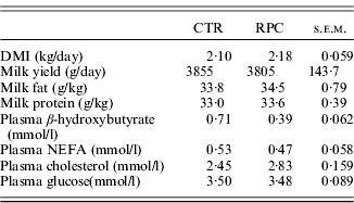
Mean values of DMI, milk yield, milk composition and plasma metabolites from the four determinations (weeks 1−4) after kidding are shown in Table 2. Milk yields were very similar in the CRT and RPC groups, as were all the other milk variables measured over the experimental period.
Mean values of DMI, milk yield, milk composition and plasma metabolites on samples taken weekly during weeks 1–4 of lactation in the CTR (n=16) and RPC (n=16) groups
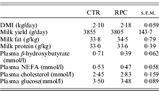
No choline effect was observed on plasma NEFA, cholesterol and glucose. In contrast, plasma β-hydroxybutyrate was significantly lower (P<0·05) in the RPC group than in CRT group. With regard to liver variables, it was found that total liver lipid content was unaffected (P=0·890) by dietary treatment (45·35 and 46·03 mg/g wet tissue in RPC and CRT animals, respectively), while total lipid/DNA ratio was significantly (P<0·05) lower (11·14 v. 15·87) in RPC than CTR goats.
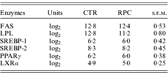
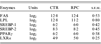
Transcript levels of various proteins and enzymes involved in lipid metabolism in the mammary gland of both CRT and RPC goats are shown in Table 3. Choline supplementation had no effect on the expression of transcripts of LPL, FAS, SREBP-1, SREBP-2, PPARγ and LXRα measured in mammary gland tissues.
mRNA expression levels of selected enzymes, transcription factors and nuclear receptors involved in lipid metabolism in the mammary gland according to treatment group (mammary samples were collected on day 28 of lactation)
DISCUSSION
Previous studies carried out in dairy cows (Baldi & Pinotti Reference Baldi and Pinotti2006; Brüsemeister & Südekum Reference Brüsemeister and Südekum2006) and goats (Pinotti et al. Reference Pinotti, Campagnoli, D'Ambrosio, Susca, Innocenti, Rebucci, Fusi, Cheli, Savoini, Dell'Orto and Baldi2008) have provided evidence that higher choline availability can impact milk production positively, when choline is administered in the rumen-protected form. However, the current work showed that milk yield and composition were similar in both CRT and RPC group during the first month of lactation and that choline supplemented goats were characterized only by lower (P<0·05) levels of plasma β-hydroxybutyrate, compared to the control animals. In a previous study on RPC-supplemented cows (Pinotti et al. Reference Pinotti, Campagnoli, Sangalli, Rebucci, Dell'Orto and Baldi2004), it was found that BHBA plasma concentrations were reduced (not significant) by 30%, while Chung et al. (Reference Chung, Cassidy, Girard, Cavassini and Varga2005) found that RPC reduced plasma NEFA concentrations by 11 and 23%, respectively, in cows receiving doses of 25 and 50 g, supporting a role of choline administration in improving lipid metabolism. Choline serves as methyl donor in carnitine synthesis, which is essential for fatty acid oxidation. At the beginning of lactation in dairy ruminants, lipid mobilization leads to increased liver uptake of NEFA with oxidation to carbon dioxide, or esterification to triglycerides. Less than optimal beta-oxidation of fatty acids induces ketosis, while triglyceride accumulation can lead to fatty liver. Accordingly, liver lipid accumulation was studied in the experimental goats. As a result, even though liver lipid content was closely similar in the two groups, the 35% reduction in lipid/DNA ratio suggests reduced hepatocellular lipid accumulation in RPC-treated animals. Dairy cows receiving choline have also been reported to have reduced hepatocellular lipid accumulation (Grummer Reference Grummer, Joshi and Herdt2006; Cooke et al. Reference Cooke, Silva Del Río, Caraviello, Bertics, Ramos and Grummer2007). These findings are also consistent with the evidence that RPC supplementation to dairy cows throughout the periparturient period decreased liver accumulation of lipids (stored as intracellular triglycerides) and increased liver glycogen content (Piepenbrink & Overton Reference Piepenbrink and Overton2003). Therefore, taken together, the current results suggest that greater choline availability is useful for optimizing liver lipid metabolism in dairy ruminants, especially during the transition period.
A further aspect investigated in the present study was the expression of several mammary gland transcripts involved in lipid metabolism, which were unaffected by choline supplementation. The effects of nutrition on the expression of mammary lipogenic genes in dairy ruminants have been investigated mainly by feeding milk-fat depressing diets, characterized by high levels of concentrates, or conjugated linoleic acids (CLA) or other lipids (fish oil, vegetable oils, full fat seeds, etc.). When specific CLA isomers are fed, transcript levels of genes involved in fatty acid uptake (LPL), de novo fatty acid synthesis (FAS) and transcription factors (SREBP1) have been observed to change (Bauman et al. Reference Bauman, Perfield, Harvatine and Baumgard2008). Harvatine & Bauman (Reference Harvatine and Bauman2006) found reduced expression of SREBP1 and SREBP activation protein during CLA-induced milk fat depression in cows, suggesting that SREBP1 is a central signalling pathway for the regulation of FAS in the bovine mammary gland. In contrast, studies on fat supplementation (vegetable oils and fish oils) for goats indicate no effect or a tendency to decreased mRNA levels of mammary LPL and FAS (see Bernard et al. Reference Bernard, Leroux, Chilliard, Sejrsen, Hvelplund and Nielsen2006 for a review), supporting the observation that, although lipid supplementation reduced milk fat synthesis in lactating goats (and other dairy ruminants), the extent of milk fat depression is less in goats than in dairy cows and sheep (Lock et al. Reference Lock, Rovai, Gipson, De Veth and Bauman2008). Little is known about the expression of SREBP2, PPARγ and LXRα transcripts in the ruminant mammary gland. Although most attention has focused on PPARs, little, if any, data are available regarding the role of these transcription factors in regulating milk fat synthesis in the mammary gland (Bauman et al. Reference Bauman, Perfield, Harvatine and Baumgard2008).
In the present study, the absence of significant effects on gene expression could be due to the dose or to the type of supplementation of RPC. Thus, while the current study supplied 4 g/day of choline (2 g/kg DM), other studies used higher levels of supplementation (for fat 36−112 g/kg DM) or more potent nutritional factors (e.g. CLAs are potent inhibitors of mammary gland fat synthesis). However, in this context, a comparison between different levels of milk yield and composition in response to choline administration in goats merits further investigation.
CONCLUSIONS
To conclude, the current plasma and liver data indicate that choline pre-eminently acts on lipid metabolism in early lactating goats. In contrast, choline supplementation had no effect on transcript levels in mammary gland of various proteins involved in lipid metabolism. It is difficult to establish if the absence of response to the treatments in terms of milk production and milk composition could be related to the lack of effect on gene expression. Nevertheless, the current results were obtained from a limited number of animals, and choline requirement and function in lactating dairy goats deserve further investigation.
The authors wish to thank Professor Tom Fearn (from University College of London) for his help in reviewing the manuscript. This article has been produced in the frame of the COST Action FA0802 Feed for Health (http://www.feedforhealth.org).