Introduction
The maculae flavae, located at both ends of the human vocal fold mucosa, are thought to be involved in the metabolism of extracellular matrix in the vocal fold mucosa and to form the characteristic layered structure of the human vocal fold mucosa. The maculae flavae are also considered to be important in the growth, development and ageing of the vocal fold mucosa.Reference Sato and Hirano1–Reference Sato, Hirano and Nakashima6
Early in the previous decade, an interstitial cell with a star-like appearance was discovered in the human maculae flavae.Reference Sato, Hirano and Nakashima7–Reference Sato and Nakashima10 This cell possessed lipid droplets and stored vitamin A.Reference Sato, Hirano and Nakashima7, Reference Sato, Hirano and Nakashima8 It had many morphological differences from conventional vocal fold fibroblasts, and constantly synthesised extracellular matrix which is essential for vocal fold mucosa function. These cells have no nomenclature. In our series of studies, we have termed them ‘vocal fold stellate cells’, for convenience. Our past studies inferred that these maculae flavae vocal fold stellate cells constitute an independent cell category, and that they are involved in the metabolism of extracellular matrix within the human vocal fold mucosa.Reference Sato and Hirano1–Reference Sato and Nakashima10
If these human vocal fold stellate cells in the maculae flavae constitute a new, independent cell category which differs from conventional fibroblasts, one would expect to detect morphological differences between subcultured vocal fold stellate cells and subcultured conventional fibroblasts.
Therefore, in the present study, we isolated and subcultured human vocal fold stellate cells, and assessed the morphological differences between these subcultured cells and conventional, subcultured fibroblasts. To our knowledge, this is the first published report of such work.
Materials and methods
Three normal, uninvolved human vocal folds were obtained from patients who had undergone total laryngectomy as initial treatment for a primary tumour. Patients' ages ranged from 64 to 73 years, and all were male. The primary tumours comprised two hypopharyngeal carcinomas (piriform sinus, T4) and one oesophageal carcinoma (cervical oesophagus, T4).
After microscopic dissection of the anterior maculae flavae and Reinke's space from the vocal fold mucosa, each tissue was cultured in Dulbecco's modified Eagle's medium (Nissui, Tokyo, Japan) supplemented with 10 per cent fetal bovine serum containing 50 µg/ml each of penicillin G (1000 U/ml) and streptomycin (1000 µg/ml). Two tissue cultures were set up for each vocal fold, one for maculae flavae tissue and one for Reinke's space tissue. Each tissue was cultured at 37°C in a humidified atmosphere of 5 per cent carbon dioxide. The maculae flavae tissue was subcultured for seven to 10 months, and the Reinke's space tissue for three to 10 months.
Cells were observed with a phase-contrast microscope (Olympus, Tokyo, Japan).
Results
Maculae flavae cultured cells
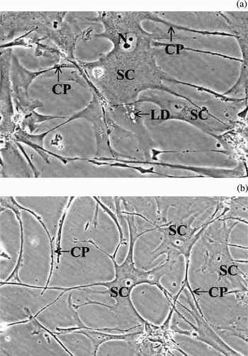
The cells cultured from the maculae flavae were stellate in shape and possessed slender cytoplasmic processes (Figure 1). They were larger than conventional fibroblasts. The nuclei were oval, and the nucleus:cytoplasm ratio was small (Figure 1). In the primary culture, small lipid droplets were present in the cytoplasm (Figure 1a); however, these disappeared in the second culture. The stellate cells proliferated by attaching their cytoplasmic processes to each other (Figure 1b).
Phase-contrast photomicrographs showing cultured stellate cells from the maculae flavae, primary culture: (a) ×75; (b) ×50. SC = stellate cell; LD = lipid droplet; CP = cytoplasmic process; N = nucleus

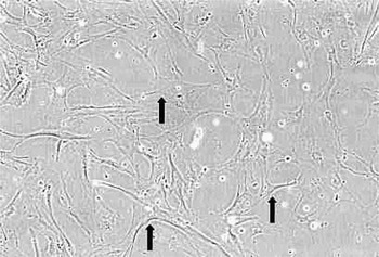
During the seven to 10 month subculture period, 19 to 26 subcultures were made. During the subculture period, cells continued to exhibit the same morphological characteristics (Figure 2).
Phase-contrast photomicrograph showing cultured stellate cells from the maculae flavae, 20th subculture. Arrow = stellate cell (×25).

Reinke's space cultured cells
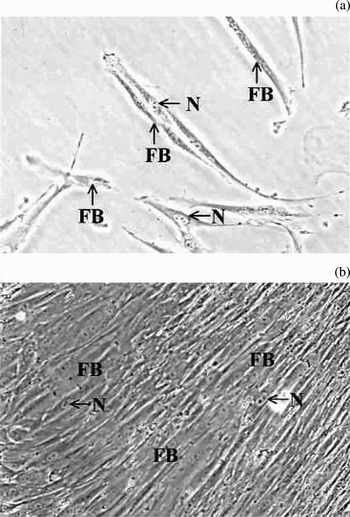
The cells cultured from Reinke's space were spindle-shaped, conventional fibroblasts. They did not have cytoplasmic processes (Figure 3). The nuclei in the cells were oval, and the nucleus:cytoplasm ratio was large (Figure 3). No lipid droplets were present in the cytoplasm (Figure 3). These fibroblasts proliferated by attaching their cytoplasm to each other (Figure 3b).
Phase-contrast photomicrograph showing cultured fibroblasts from Reinke's space, primary culture: (a) ×75; (b) ×50. FB = fibroblast; N = nucleus

During the three to 10 month subculture period, three to 13 subcultures were made. During the subculture period, cells continued to exhibit the same morphological characteristics.
Discussion
Human vocal fold mucosa contains dense masses of cells and extracellular matrix, which have been termed maculae flavae. These are located at the anterior and posterior ends of the membranous portion of the bilateral vocal folds. The extracellular matrix of the maculae flavae is composed of fibrillar protein (such as collagenous fibres, reticular fibres and elastic fibers), glycoprotein and glycosaminoglycan.Reference Sato and Hirano1–Reference Sato, Hirano and Nakashima4 These components are necessary to facilitate the vibratory function of the vocal fold mucosa. The maculae flavae, located at both ends of the human vocal fold mucosa, are thought to be involved in the metabolism of the extracellular matrix of the vocal fold mucosa lamina propria. They are considered to be important in the growth, development and ageing of the vocal fold mucosa.Reference Sato and Hirano1–Reference Sato, Hirano and Nakashima6
• The maculae flavae are located at both ends of the human vocal fold mucosa. They are thought to be involved in the metabolism of vocal fold mucosa extracellular matrix, and to form the characteristic layered structure of the human vocal fold mucosa
• In this study, for the first time, human vocal fold stellate cells in the maculae flavae were isolated and subcultured
• Human vocal fold stellate cells should be considered a new, independent category of cell within the human vocal fold
In our previous studies, we discovered interstitial cells with a star-like appearance within the human maculae flavae, which we termed vocal fold stellate cells.Reference Sato, Hirano and Nakashima7–Reference Sato and Nakashima10 There is a high density of these cells in the human maculae flavae. The cells are stellate in shape and possess slender cytoplasmic processes, intracellular organelles, a small nucleus:cytoplasm ratio and well developed rough endoplasmic reticulum. These cells possess lipid droplets and store vitamin A.Reference Sato, Hirano and Nakashima7, Reference Sato, Hirano and Nakashima8 They synthesise fibrous protein, and collagenous and elastic fibers are constantly formed around the cells.Reference Sato, Hirano and Nakashima7 There are many vesicles at the periphery of the cytoplasm, and newly released amorphous materials are present on the cell surface.Reference Sato, Hirano and Nakashima7 Microfibrils are observed around this amorphous material and collagenous fibrils are detected near the microfibrils.Reference Sato, Hirano and Nakashima7 There are microfibril assemblies on which elastin is deposited.Reference Sato, Hirano and Nakashima7 Vocal fold stellate cells and CD44 (a cell surface glycoprotein which acts as a cell surface receptor for hyaluronic acid) interact to play important roles in maintaining hyaluronic acid within the human vocal fold mucosa.Reference Sato, Sakamoto and Nakashima5 Our previous studies indicated that vocal fold stellate cells are not quiescent but active. From our results, we inferred that these cells form a new, independent cell category involved in the metabolism of extracellular matrix in the human vocal fold mucosa.Reference Sato, Hirano and Nakashima7–Reference Sato and Nakashima10
To the best of our knowledge, there have been no previous studies addressing the isolation and subculture of human vocal fold stellate cells. Only porcine vocal fold stellate cells have been isolated and cultured.Reference Fuja, Probst-Fuja and Titze11 The histological structure of the lamina propria of the vocal fold mucosa significantly varies between animals.Reference Kurita, Nagata, Hirano and Kirchner12, Reference Nagata13 Furthermore, the structure of other mammalian vocal fold mucosa differs from that of humans.Reference Kurita, Nagata, Hirano and Kirchner12, Reference Nagata13 For example, porcine vocal folds have no vocal ligament, the anterior and posterior maculae flavae are not clear, and their cellular component is not dense.Reference Kurita, Nagata, Hirano and Kirchner12, Reference Nagata13 As human vocal folds differ in histology, physiology and pathology from those of other mammals, vocal fold stellate cells should ideally be collected from human samples.
If human vocal fold stellate cells in the maculae flavae form an independent cell category, some morphological differences should be detected between subcultured human vocal fold stellate cells and subcultured conventional fibroblasts. In this study, several morphological differences were detected between isolated, subcultured cells from the maculae flavae and from Reinke's space. Cultured cells from the maculae flavae were stellate in shape and possessed slender cytoplasmic processes. The nucleus:cytoplasm ratio was small, and lipid droplets were present in the cytoplasm. These findings characterised these cells as human vocal fold stellate cells. Small lipid droplets were present in the cytoplasm in the primary culture; however, they disappeared in the second culture. This phenomenon may depend on supplements within the growing medium.
Cultured human vocal fold stellate cells proliferated by attaching their cytoplasmic processes to each other. In many types of cells, the interiors of adjacent cells communicate with each other through cell-to-cell channels.Reference Kanno14 Cell communications are proposed to play an important role in cell growth and differentiation.Reference Kanno14 Subcultured human vocal fold stellate cells may therefore communicate with each other during growth and differentiation.
During the isolation and subculture period, the vocal fold stellate cells continued to exhibit the same morphological characteristics, which differed from those of conventional fibroblasts.
Conclusion
Human vocal fold stellate cells in the maculae flavae have been isolated and subcultured for the first time. This study revealed several morphological differences between subcultured vocal fold stellate cells and conventional, subcultured fibroblasts. This finding corroborates our theory that the vocal fold stellate cells in the human maculae flavae form a new, independent cell category within the human vocal fold.