INTRODUCTION
Water molds is a common name for a group of multinucleated organisms that superficially resemble fungi, although they are now recognized as an independent evolutionary lineage and placed in the new kingdom Stramenopila (Dick, Reference Dick2001). There are 2 important phyla (divisions) of water molds, the chytrids (Chytridiomycota) and the oomycetes (Oomycota). Many oomycetes are aquatic, and most live in water at some point during their life-cycle. They can be saprobes, obtaining their metabolic energy from decaying organic material, or parasites, attacking plants, fungi, and animals. Moreover, they can infect as primary parasites and as opportunists, which can grow wherever the epidermal barrier of a victim is broken (Leclerc et al. Reference Leclerc, Guillot and Deville2000). In general, it seems that parasitism in oomycetes has evolved independently several times, as it occurs in phylogenetically distinct lineages (Riethmuller et al. Reference Riethmuller, Weiss and Oberwinkler1999). Both parasitic and saprobic strains can be found even within a single oomycete species. For example, although Saprolegnia diclina has been frequently reported as a fish pathogen (e.g. Wood and Willoughby, Reference Wood and Willoughby1986), some strains are acting as saprotrophs (e.g. Hatai and Hoshiai, Reference Hatai and Hoshiai1993).
Fish oomycete infections are frequently reported from nature as well as from commercial farms (e.g. Bangyeekhun et al. Reference Bangyeekhun, Quiniou, Bly and Cerenius2001), where they cause substantial economic losses. In contrast to a large body of work investigating fish oomycete diseases, the nature of such epidemics on aquatic invertebrate populations has rarely been studied. Only a specific kind of oomycete infection has drawn the attention of aquatic ecologists, those that produce pathology restricted to host brood clutches, but not the adults themselves (e.g. Burns, Reference Burns1985; Stazi et al. Reference Stazi, Mantovani, Fuglieni and Di Delupis1994). Although the mortality of oomycete-infected eggs was often 100% in copepods or waterfleas (Daphnia), such infections could be cleared after aborting the infected clutch (Burns, Reference Burns1985; Tellenbach et al. Reference Tellenbach, Wolinska and Spaak2007). Among the dozens of parasite species adversely affecting adult Daphnia beyond a single clutch (reviewed by Green, Reference Green1974 and Ebert, Reference Ebert2005), only 2 oomycete species have been reported: Aphanomyces daphniae (Prowse, Reference Prowse1954) and A. petersonii (Scott, Reference Scott1961). These descriptive studies, however, provide only limited knowledge about the interactions between Daphnia hosts and oomycete parasites. For example, the results of controlled laboratory infections were restricted to a short observational note (Prowse, Reference Prowse1954).
Over the last 2 decades, advances in molecular biology have aided in the identification of oomycete diseases. Previously, species identification was often difficult, as it relied on the description of the sexual reproductive organs (Dick, Reference Dick1969a). The majority of reproduction, however, is asexual, by division of a sporangium into individual flagellated spores. The sexual organs are sometimes formed only after several weeks (Dick, Reference Dick1969a); and some strains do not even have sexual reproduction (e.g. Ballesteros et al. Reference Ballesteros, Martin and Dieguez-Uribeondo2006). To identify species, we can now use genetic markers, like the sequences of the internal transcribed spacer regions (ITS1 and ITS2) together with large subunit of ribosomal DNA (Leclerc et al. Reference Leclerc, Guillot and Deville2000).
Here we describe the infectivity, virulence, cultivating conditions and phylogenetic positions of naturally occurring oomycete parasites of Daphnia hosts. We exposed Daphnia pulex clones to sediment collected from several ponds inhabited by this Daphnia species, and found that some individuals died and were then covered by oomycetes. A series of experiments was conducted in order to determine: (1) if the oomycetes are parasitic or saprobic; (2) the effect of infection on host survival, and (3) the conditions under which the oomycetes could be cultivated in vitro. Subsequently, we conducted genetic analyses to determine whether the isolates belonged to different species. The sequence information was also used to examine the phylogenetic relationships with well-known oomycete parasites of phytoplankton, zooplankton and fish.
MATERIALS AND METHODS
Daphnia sampling and general cultivating conditions
Crustacean waterfleas, Daphnia, are small (1–5 mm) planktonic organisms that inhabit various water bodies, from temporary ponds to large permanent lakes. They reproduce by cyclical parthenogenesis. Daphnia are important components of aquatic foodwebs, as they feed on algae and are themselves a main food item of many fish species (Lampert and Sommer, Reference Lampert and Sommer1999). Seven Daphnia pulex clones were isolated from temporary ponds in Michigan and Indiana, and then propagated in the laboratory as clonal lineages for 2–3 years prior to use (each clone originated from a different pond). The following cultivating conditions were applied for 2 generations prior to the experiments (to reduce any maternal effects), and then in all experimental surveys, if not stated otherwise: standardized medium (autoclaved, 0·45 μm filtered lake water) changed every third day; 20±0·5°C; 16:8 h light-dark cycle; daily diet of batch-cultured Scenedesmus obliquus of final concentration 105 algae cells ml−1 (~1 μg C). All experiments were started with 2 to 3-day-old juvenile Daphnia.
Sediment as the parasite source
Exp. 1
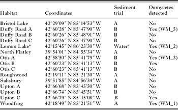
Surface sediment samples (collected from the littoral zone of 13 ponds and 1 lake) were stored prior to use at 4°C in darkness (see Table 1 for sediment identities). In Exp. 1A, 5 D. pulex clones were separately exposed to 5 different sediments. In addition, 2 other clones were tested against 3 sediments only (due to limited sediment sources). The clone×sediment combination was replicated 3 times. A control treatment consisted of a combination of clones exposed to an autoclaved mixture of all 5 sediments. On experiment day 1, six juveniles of a single clone were introduced into each experimental beaker, containing 8 ml of sediment in 100 ml of medium. Twice a day, the beakers were turned to stir up the sediment and increase the encounter rates of the Daphnia with parasite spores. On day 7, only Daphnia were transferred to fresh media. The media were refreshed every 3 days; newborns were removed daily, and dead Daphnia were examined for signs of any infection (at 50×magnification). Dead Daphnia were placed individually in 1 ml of medium and monitored during the next 48 h for symptoms of infection. The trial was terminated on day 17, at which point all Daphnia were observed for external signs of infection. During Exp. 1B, 1 clone and 9 other sediments were used (see Table 1). To increase the likelihood of successful infection, Daphnia were exposed to the sediment for 17 days. The medium was not changed, but any evaporated medium was replaced daily. The number of beakers with at least 1 infected Daphnia and the number of beakers without a given infection was compared among the sediment sources (Fisher exact: 2×5 and 2×9 table in Exp. 1A and 1B, respectively). For Exp. 1A, the corresponding numbers (i.e. beakers with and without any infected Daphnia) were compared among the clones (Fisher exact: 2×7 table), and only the infectious sediments were included.
Sediment as the parasite source
(Sediment trials A and B refer to Exp. 1A and 1B, respectively. With the exception of Bristol Lake and Lemon Lake, the other habitats are small, temporary ponds.)

a In addition to controlled exposures to different sediment sources, oomycete-infected Daphnia were found in laboratory cultures. The WM_2 isolate probably originates from Lemon Lake, as water from this lake was used in the laboratory as Daphnia culture medium.
Oomycetes cultivated in vitro
Isolation on agar
A small part of oomycete hypha was cut from dead, infected Daphnia, obtained from the sediment exposures. The tissue was placed in the centre of a Petri dish (8·7 cm diameter) filled with Sabouraud (SB) agar (Difco Lab., USA), and incubated at 28°C for 48 h. If necessary, the procedure was repeated to obtain 1 isolate per sediment treatment.
Exp. 2: colony growth on agar
A square of SB agar (1 cm2) with a given oomycete isolate was inoculated in the centre of an SB agar Petri dish (8·7 cm diameter). This procedure was replicated 4–6 times for each of 4 obtained isolates: WM_1, WM_2, WM_3 and WM_5 (WM stands for water molds, see Table 1 for isolate identities). After 24 h of incubation at 28°C, half of the plates were left at 28°C, and half were transferred to 4°C. Every 24 h, two measurements of the colony diameter were taken, and the average was calculated. The experiment lasted until all plates were covered by the colony. Colony growth was analysed with a two-way repeated measures ANOVA, across oomycete isolates, with time as a repeated measure (separate tests were conducted per 28°C and 4°C temperatures). The differences across the isolates were analysed with a posthoc Scheffe's test to account for the increased type-I error for multiple comparisons.
Exp. 3: viability of isolates preserved in different media
A square of SB agar (0·5 cm2) with an oomycete isolate was inoculated in a glass culture tube containing 5 ml of one of the following media: SB agar, LB broth (Luria-Bertani, Difco Lab.); PYG broth (Peptone Yeast extract Glucose, Difco Lab.), and HS baits (Hemp Seeds autoclaved in a distilled water). Each treatment was replicated 4 times. After 48 h incubation at 28°C, the presence/absence of growth was assessed. The culture tubes were sealed and stored in darkness: 2 replicates at 22°C; and the other 2 at 4°C. After 6 weeks, a small piece of preserved hypha was plated on 2 SB agar Petri dishes and incubated for 72 h at 28°C. The oomycete isolate was classified as ‘successfully preserved’ on a given medium and at a given temperature if it grew on 1 of the 2 inoculated Petri dishes. The number of successfully preserved oomycete isolates versus the number of non-successful cases was compared among the different media, and separate tests were conducted per 22°C and 4°C preservation temperatures (Fisher exact: 2×4 table).
Infectivity of oomycete isolates
Exp. 4: Daphnia exposed to oomycetes grown on agar
In Exp. 4A, 6 Daphnia (mixture of 3 clones) were exposed in 50 ml of medium to 4 treatments: 1 cm2 of SB agar with WM_1 oomycete isolate, 1 cm2 of SB agar with WM_2 oomycete isolate, 1 cm2 of SB agar (i.e. AGAR control), and nothing (i.e. BLANK control). Each treatment was replicated 6 times. On day 5, half of the medium was replaced, and the experiment was terminated after 8 days. The treatments were fed daily and checked for dead Daphnia, which were then placed individually in 1 ml of water and monitored for 48 h for symptoms of infection. To test for host-to-host transmission, visibly infected dead Daphnia (20 individuals per treatment) were placed individually in 2 ml of medium with a single healthy Daphnia from 1 of 3 available clone sources. If a previously healthy Daphnia died within 48 h, it was monitored during the next 48 h for infection. In Exp. 4B, 4 Daphnia were exposed to 2 treatments: 1 cm2 of SB agar with WM_3 oomycete isolate and 1 cm2 of SB agar (i.e. AGAR control), with 4 replicates per treatment. To reduce the high mortality observed in the AGAR control in Exp. 4A, half of the medium was replaced every other day. Daphnia mortality was analysed with a two-way repeated measures ANOVA, across the exposure treatments, with time as a repeated measure. In Exp. 4A, the number of successful host-to-host transmissions was compared between the two oomycete isolates (Fisher exact: 2×2 table).
Exp. 5: Daphnia exposed to oomycetes grown on liquid media
Due to the high mortality caused by the agar alone (see above), the infectivity of oomycetes was tested using the liquid media. In Exp. 5A, culture tubes with different liquid media (LB broth, PYG broth and HS baits) were inoculated with oomycete isolates (WM_1 or WM_2). This was replicated 6 times. After 72 h of incubation at 28°C, the mycelia were placed into 1 ml of medium at 20°C. The media were refreshed every 12 h. Infection trials were started approximately 30 h after the first zoospores were observed. Sporulating mycelia were placed in the fresh media for 2 h, at which time, the mycelia were removed and 1 Daphnia was placed in this zoospore-containing medium. After 24 h, the Daphnia were transferred to new oomycete-free medium. Transfers were repeated and Daphnia were fed every 24 h, and monitored for mortality and infection every 4 h (with a break of 7 h at night) for 4 days. This procedure was repeated 4 times, each with new Daphnia. In Exp. 5B, the risk of zoospore encounters was increased as Daphnia were exposed to mycelia for 10 days. Three Daphnia were exposed to each mycelium treatment (i.e. WM_2 or WM_5) in 30 ml of water, with 3 replicates. To reduce the probability of Daphnia being tangled or injured by the growing mycelia, the water molds were enclosed in a 1 cm long silicon cage, with 50 μm nylon mesh attached on both ends. This allowed zoospores to pass into the media. To control for any cage effects, Daphnia were placed in 6 controls with empty parasite cages. All media were replaced on day 5, and the experiment was terminated on day 7. The infection rates (Exps 5A and B) and time to sporulation (Exp. 5A) were analysed with two-way ANOVAs, across media types and oomycete isolates. In Exp. 5A, to exclude the possibility of post-mortality infection (i.e. saprobic oomycetes), the time until death was compared between the individuals that eventually became colonized and those that did not (t-test: media and oomycete isolates were pooled). In Exp. 5B, to exclude the possibility that Daphnia died due to the presence of parasite cages, the number of dead Daphnia was compared between exposure treatments and the controls (t-test).
Genetic identification of the oomycete isolates
DNA extraction and sequencing
Oomycete isolates were grown on the HS baits for 72 h at 28°C. DNA extraction was performed using a DNeasy Tissue Kit (Qiagen). DNA fragments containing internal transcribed spacers ITS1 and ITS2 including 5·8 S (hereafter referred to as ITS) were amplified, using a BioMix from Bioline, with a primer pair ITS5/ITS4 (Martin et al. Reference Martin, Raidl and Tellaria2004). The nuclear large subunit ribosomal DNA sequences (hereafter referred to as LSU) were amplified with a primer pair NL1/NL4 (O'Donnell, Reference O'Donnell, Reynolds and Taylor1993). The obtained DNA fragments were cleaned with a QIAquick PCR Purification Kit (Qiagen) on a spin column. The eluted DNA was sequenced using the same set of primers and a BigDye Terminator v3.1 Cycle Sequencing Kit (Applied Biosystems). The reactions were analysed on an ABI Prism 3730 DNA Sequencer.
Phylogenetic analyses
The obtained sequences were checked for inaccurate base calling using the CodonCode's Aligner. Homologies of the sequences were searched in BLAST sequence data; however, only the sequences of isolates identified to the species level were selected. Two outgroup taxa were chosen per alignment, based on the previously published phylogenies. The sequences were aligned in Clustal X, and manually corrected in BioEdit (gaps were treated as missing data). The neighbour-joining method was used to construct the phylogenetic trees, with Kimura two-parameter distances (Kimura, Reference Kimura1980) and the complete deletion option (using MEGA Kumar et al. Reference Kumar, Tamura and Nei2004). The robustness was improved by the bootstrap proportions with 2000 replicates.
RESULTS
Sediment as the parasite source
Exp. 1
Two groups of parasites were detected: epibionts (Vorticella sp.) and oomycetes. The oomycetes were found on dead Daphnia only and the filamentous zoosporangia emerged through the wall of the host. The sediments differed in infectiveness with epibionts (2 of 5 sediments were infectious in Exp. 1A, Fisher exact: P=0·0007, Table 1) and oomycetes (1 of 5 in Exp. 1A: P=0·038; and 4 of 9 in Exp. 1B: P=0·049; in Exp. 1B, only oomycete infections were looked for). There were no clonal differences in epibiont infection (Fisher exact: P=0·800); however, a single clone suffered significantly higher infection by oomycetes (Fisher exact: P=0·025).
Oomycetes cultivated in vitro
Isolation on agar
We attempted to obtain 1 isolate from each of 5 infectious sediments, but only 3 sediments served as a parasite source (WM_1, WM_3, WM_5, Table 1). In addition, an extra isolate was obtained from the accidental infection of Daphnia laboratory cultures (WM_2). The latter might have originated from lake water used as culture media for Daphnia clones. The installation of a UV-passing filter on the water filtration apparatus successfully prevented any future contaminations of media.
Exp. 2: colony growth on agar
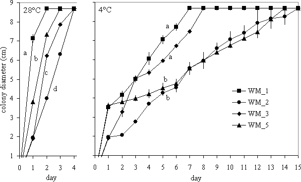
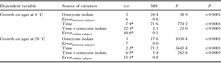
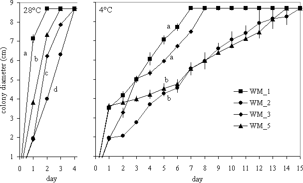
The hyphae of 4 obtained oomycete isolates gradually radiated from the centre of inoculation until the entire Petri dish was covered (see ‘time’ in Table 2). Colony growth was faster at 28°C (<4 days) than at 4°C (up to 15 days, Fig. 1), and there was a significant difference in growth among the isolates (‘oomycete isolate’ in Table 2). For example, WM_1 isolate grew significantly faster than all 3 isolates at 28°C, and faster than 2 other isolates at 4°C (Fig. 1). Each isolate colony had a specific morphology; for example, WM_2 had 8 radiating ‘arms’, whereas other isolate colonies were homogeneous in shape.
Growth of 4 oomycete isolates on agar (Exp. 2) under different temperatures (28°C and 4°C). Mean (±s.e.) colony diameter is shown. In the 28°C treatment, all plates were completely covered by day 4. Groups with different letters were significantly different in pairwise comparisons.
The effects of oomycete isolate and time on oomycete growth on agar (two-way repeated measures ANOVAs)
(All 4 oomycete isolates were tested (Exp. 2, see Fig. 1.)
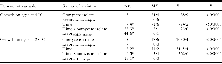
* Due to heterosphericity (Mauchly's test), the degrees of freedom were adjusted by the Huynh-Feldt epsilon.
Exp. 3: viability of isolates preserved on different media
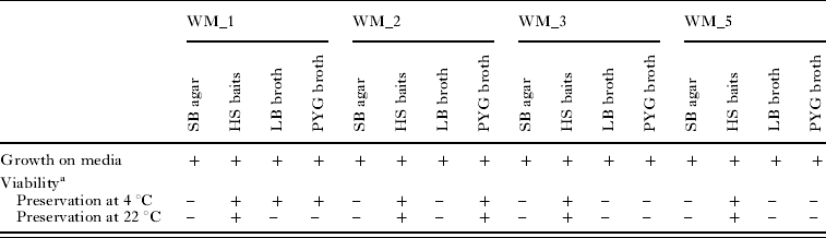
The oomycete isolates grew on all 4 tested media (Table 3). However, the viability of isolates differed among the preservation media; the isolates on HS baits were significantly more viable than others when preserved at 22°C (Fisher exact: P=0·011) and showed a similar, but non-significant, tendency at 4°C (P=0·054). In general, the preservation temperature had no significant influence on the viability of the isolates (Fisher exact: P=0·716).
Growth of 4 oomycete isolates on 4 different media and their viability after a 6-week preservation period (Exp. 3)
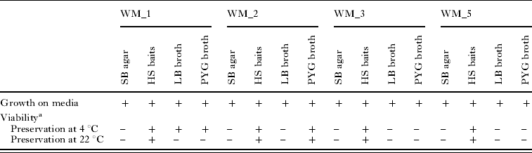
a A small portion of material was plated in the centre of an SB agar Petri dish and incubated at 28°C for 72 h.
+/− positive and negative results, respectively.
Infectivity of oomycete isolates
Exp. 4: Daphnia exposed to oomycetes grown on agar
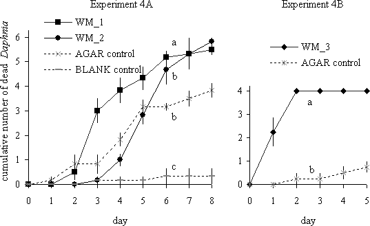
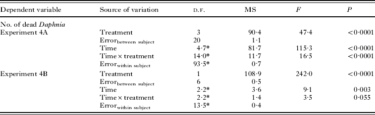
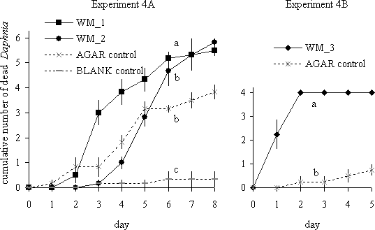
Daphnia died over time, but the mortality rate was treatment-dependent (Table 4). In Exp. 4A, Daphnia exposed to the WM_1 isolate suffered significantly higher mortality than those exposed to WM_2. The lowest mortality was detected in the BLANK control; however, the mortality rate in the AGAR control was comparable to that for the WM_2 isolate (Fig. 2). In Exp. 4B, the medium was changed more frequently, which reduced the mortality caused by agar alone (Fig. 2). None of the Daphnia that died in the controls (AGAR and BLANK) became a substrate for oomycete mycelia. In contrast, 90·9%, 91·4% (Exp. 4A), and 75·0% (Exp. 4B) of dead Daphnia were covered by mycelia in the oomycete treatments (WM_1, WM_2 and WM_3, respectively). Of those infected with WM_1 and WM_2 isolates, 65·0% and 85·0%, respectively, transmitted the parasite to another Daphnia host (i.e. host-to-host transmission in Exp. 4A: Fisher exact: P=0·273).
Daphnia mortality after exposure to oomycetes grown on agar. The mean cumulative number of dead Daphnia (±s.e.) is shown (there were 6 or 4 Daphnia per experimental unit; Exp. 4A, B; respectively). Due to the high mortality observed in the ‘AGAR control’ (Exp. 4A), the medium was changed more frequently (Exp. 4B), which resulted in reduced mortality caused by agar alone. Groups with different letters were significantly different in pairwise comparisons.
The effects of treatment and time on Daphnia mortality after exposure to oomycetes grown on agar (two-way repeated measures ANOVAs)
(The following treatments were tested: WM_1, WM_2, AGAR control, and BLANK control (Exp. 4A); WM_3, AGAR control (Exp. 4B, see Fig. 2.)
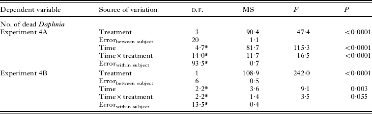
* Due to heterosphericity (Mauchly's test), the degrees of freedom were adjusted by the Huynh-Feldt epsilon.
Exp. 5: Daphnia exposed to oomycetes grown on liquid media
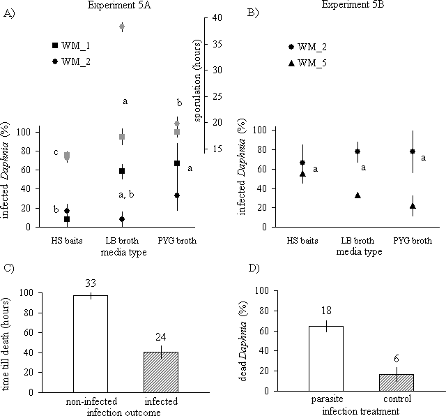
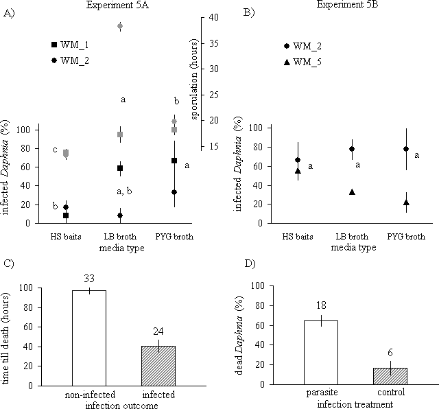
The first spores were observed within 15 to 38 h after mycelia were placed in water. The WM_1 isolate sporulated significantly earlier than WM_2 (Exp. 5A; F 1,12=56·02, P<0·0001, Fig. 3A). The WM_2 isolate sporulated especially late when grown on LB broth (‘isolate×media’ interaction: F 2,12=49·05, P<0·0001). In addition, the WM_1 isolate was more infectious than WM_2 (Exp. 5A; F 1,12=5·4, P=0·039, Fig. 3A), whereas WM_2 was more infectious than WM_5 (Exp. 5B; F 1,12=10·0, P=0·008, Fig. 3B). Furthermore, the oomycetes grown on the PYG broth were significantly more infectious than those obtained from the HS baits (Exp. 5A; F 2,12=4·07, P=0·045, Fig. 3A), although the differences were not significant in Exp. 5B (Fig. 3B). The hypothesis that Daphnia had died prior to infection was rejected as infected Daphnia died significantly faster than those without infection (Exp. 5A; t=7·25, d.f.=31·85, P<0·001, Fig. 3C). Furthermore, the parasite cages had no impact on Daphnia mortality, as the mortality in the parasite treatments was significantly higher than that in the controls with empty parasite cages (Exp. 5B; t=3·55, d.f.=22, P=0·002, Fig. 3D).
Daphnia exposed to oomycetes grown on liquid media. Left and right panels: Experiment 5A, B; respectively: (A) time to sporulation (grey symbols) and the infection rate (black symbols) (mean±s.e.), (B) the infection rate, (C) time till death, (D) mortality rate. Groups with different letters were significantly different in pairwise comparisons; sample size is given at the top of each bar.
Genetic identification of the oomycete isolates
The WM_1, WM_2 and WM_3 isolates were phylogenetically distinct from the WM_5 isolate. The analyses were thus performed in 2 sets.
Phylogenetic analyses of the WM_1, WM_2 and WM_3 isolates
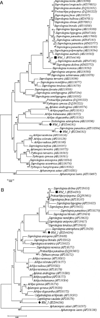
The sequences of 3 isolates (the GenBank Accession numbers on Fig. 4A, B) were aligned with 45 (ITS) and 31 (LSU) published sequences. The obtained alignments were 720 bp and 636 bp long, respectively. There were no large (>10 bp) insertions or deletions. The neighbour-joining analyses showed that WM_1, WM_2 and WM_3 isolates all belong to the Saprolegnia genera, but are distinct species. Specifically, the WM_1 isolate clusters with S. australis and S. diclina on the ITS tree, or with S. diclina, S. hypogyna, S. ferax and Protoachlya polysporus on the LSU tree (the cluster groups are well sustained in terms of bootstrap proportions: 66% and 74%, respectively, Fig. 4A, B). The WM_2 isolate clusters with Scoliolegnia asterophora or Saprolegnia asterophora (ITS and LSU: 100% bootstrap support). The WM_3 isolate clusters with Saprolegnia parasitica or Leptolegnia caudata (ITS and LSU: 100% and 90% bootstrap support).
A neighbour-joining phylogeny of oomycetes including WM_1, WM_2 and WM_3 isolates, based on the (A) ITS and (B) LSU rDNA sequences. Branch lengths are scaled in terms of expected numbers of nucleotide substitutions per site. Numbers within the tree represent the bootstrap values (2000 replications). Numbers beside species names are the GenBank Accession numbers.

Phylogenetic analyses of the WM_5 isolate
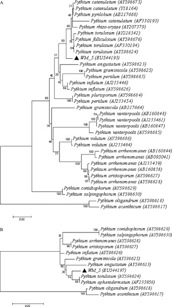
The sequence of the WM_5 isolate was aligned with 32 (ITS) and 11 (LSU) published sequences. The obtained alignments were 818 bp and 679 bp long, respectively. There was one large insertion in the ITS alignment (16 bp, all 4 Pythium vanterpolii isolates). The neighbour-joining analyses showed that the WM_5 isolate belongs to the Pythium genus. The isolate clustered with few independent isolates of P. torulosum, as well as with single isolates of other Pythium species (Fig. 5A, B).
A neighbour-joining phylogeny of oomycetes including WM_5 isolate, based on the (A) ITS and (B) LSU rDNA sequences. Branch lengths are scaled in terms of expected numbers of nucleotide substitutions per site. Numbers within the tree represent the bootstrap values (2000 replications). Numbers beside species names are the GenBank Accession numbers.

DISCUSSION
Oomycete parasites, similar to those studied in the present survey, have rarely been reported from natural Daphnia populations (but see Scott, Reference Scott1961). In previously controlled exposures to sediment, daphnids became infected with bacteria or microsporidia (e.g. Decaestecker et al. Reference Decaestecker, Lefever, De Meester and Ebert2004), but never with oomycetes. Similarly, not all 14 tested sediments were infectious; only 5 led to oomycetes detection. Interestingly, even across sediments collected from ponds in close proximity, some were infectious whereas others were not (e.g. ‘Otis B’ and ‘Otis C’, see Table 1). It could also be, however, that exposure time (i.e., time to activate parasite sporulation) plays an important role as well. In the 7-day exposure, only 1 of 5 sediments was infectious, whereas in the 17-day exposure, 4 of 9 sediments produced infections. Indeed, most infections occurred towards the end of both experiments (data not shown).
Since in the sediment exposures, all Daphnia covered by the oomycete mycelium were dead, we needed to determine whether the isolates tested here were indeed parasitic, meaning that we could rule out post-mortality colonization. Looking only at the data from the ‘Daphnia exposed to oomycetes grown on agar’ survey; we could not be certain if the high mortality in the oomycete treatment was not caused by the presence of agar alone. However, when mortality in the AGAR control was substantially reduced by the frequent media exchange, the mortality in the oomycete treatment remained high. Moreover, when exposed to pure oomycetes (isolated from liquid cultures), daphnids died and were again covered by oomycete mycelium. An additional test indicated that these Daphnia were not dead prior to infection. Moreover, when oomycetes were enclosed in the cages to reduce injury to the Daphnia, mortality in the exposed treatments was significantly higher than that observed in the controls (with empty cages). These different lines of evidence all suggest that the tested oomycetes were parasitic and not saprobic. The fact that oomycetes are more visible when their Daphnia host were already dead suggests that their prevalence may be underestimated in the field. First, researchers often look for ‘external signs’ of Daphnia infection (see review by Ebert, Reference Ebert2005) and thus the early stages of oomycete infections might be overlooked. Second, only live members of sampled Daphnia populations are screened. Several hours (e.g. Little and Ebert, Reference Little and Ebert1999; Wolinska et al. Reference Wolinska, Keller, Manca and Spaak2007) or even a few days (e.g. Ebert et al. Reference Ebert, Hottinger and Pajunen2001) can pass between collecting and examining the zooplankton sample, whereas oomycetes kill Daphnia very rapidly (see also Prowse, Reference Prowse1954). Discarding the dead animals, therefore, might substantially lower the likelihood of detecting oomycete infections.
Oomycete parasites are cosmopolitan in distribution, occurring in most freshwater habitats. Yet for some oomycetes, there is evidence of strong host specificity for infection. For example, Saprolegnia ferax infected only one of the many coexisting frog species (Kiesecker and Blaustein, Reference Kiesecker and Blaustein1999). Aphanomyces daphniae, the oomycete parasite of Daphnia hyalina, was host-specific on a higher taxonomic level, as it was unable to infect other crustaceans (Euryercus, Bosmina, Diaptomus and Cyclops, in Prowse, Reference Prowse1954). In our experiment, a single D. pulex clone was infected significantly more often than the other 6 clones tested. In contrast, the epibiont Vorticella sp. infected all exposed Daphnia clones to the same extent. We cannot exclude that the detected host specificity was caused by indirect traits. For example, it has been shown that the D. magna clones, which dwell on the bottom of the pond, are more exposed to parasite spores hidden in the sediment than clones which prefer to stay in the upper water column (Decaestecker et al. Reference Decaestecker, De Meester and Ebert2002). Although further work is needed to confirm the results on the host specificity of oomycetes, the present study clearly shows that the tested oomycetes differed in their virulence on Daphnia. The WM_1 isolate was significantly more virulent than WM_2, independent of whether isolates grew on agar or liquid media, whereas the WM_2 isolate was consistently more virulent than WM_5 (only liquid media were tested here).
It is clear that all 4 oomycete isolates that we obtained were distinct species. First, they showed different characteristics in the experimental assays: differences in colony growth rate on agar plates, virulence levels and time to sporulation. Second, the ITS and LSU phylogenetic analyses showed that 1 of the analysed isolates belong to the genus Pythium, and the other 3 to the genus Saprolegnia. Importantly, the 3 later isolates were distinct within the Saprolegnia group. Surprisingly, none of the tested isolates belong to the genus Aphanomyces, although the only 2 oomycete species previously reported to affect the entire Daphnia body belong to that genus (Prowse, Reference Prowse1954; Scott, Reference Scott1961).
There are close relationships among the isolates studied here to the oomycete parasites of other organisms. For example, the WM_1 isolate was closely related (on the ITS and LSU trees) to Saprolegnia diclina, a parasitic strain isolated from salmon (Hatai and Hoshiai, Reference Hatai and Hoshiai1993). Other relatives to the WM_1 isolate, including S. australis, S. ferax, and S. hypogyna, also infect fish (e.g. Kiesecker et al. Reference Kiesecker, Blaustein and Miller2001). The WM_2 isolate clustered with Scoliolegnia asterophora (ITS) and Saprolegnia asterophora (LSU). Those two are actually the same species, but with interchangeable names (Dick, Reference Dick1969b). Both isolates were of unknown origin, but S. asterophora is a common parasite of green algae (Beck and Erb, Reference Beck and Erb1984). The WM_3 isolate clustered with S. parasitica (ITS), a known parasite of fish (Kitancharoen et al. Reference Kitancharoen, Yuasa and Hatai1996). For the LSU, the WM_3 clusters with Leptolegnia caudata, this parasite was briefly reported from a large cladoceran Leptodora kindti (Green, Reference Green1974), a common Daphnia predator. Moreover, Leptolegnia sp. has been shown to cause mass mortality in copepods (Vallin, Reference Vallin1951). The WM_5 isolate belonged to the Pythium genera, and clustered with WM_5 isolates originated from various grasses or, alternatively, their origin was unknown. Interestingly, Green (Reference Green1974) briefly mentioned a record of Daphnia being infected by Pythium daphnidarum.
The small phylogenetic distance of the studied isolates to oomycetes infecting other species is not so surprising. It is well documented that oomycete parasites easily cross host species boundaries. For instance, laboratory experiments have shown that hatchery-reared rainbow trout spread Saprolegnia ferax to amphibians (Kiesecker et al. Reference Kiesecker, Blaustein and Miller2001). However, so far, no Daphnia parasites are known to be shared with Daphnia predator or prey communities. The obtained phylogenies, however, question this belief. The molecular similarities of Daphnia oomycete parasites with those attacking species on other trophic levels are an intriguing finding. This is in contrast to a large body of empirical and theoretical work on shared parasite communities which generally refers to hosts being members of the same food web level (see references in Tompkins et al. Reference Tompkins, Greenman, Robertson and Hudson2000). The notion of a single oomycete parasite attacking various members of the trophic cascade needs further testing and inquiry. For example, one should compare the sequences of oomycetes isolated from host species coexisting in the same water body. One can also expose Daphnia to isolates obtained from a fish host as there are many successfully cultivated fish oomycetes in various laboratories and tissue bank collections.
In recent years, Daphnia and their parasites have been commonly used as a model system for the ecological and evolutionary interactions between host and parasites (reviewed by Ebert, Reference Ebert2005, Reference Ebert2008). However, none of these studied Daphnia parasites can currently be cultivated outside its original host, whereas our survey showed that oomycete parasites can be maintained and grown on artificial media (see also Prowse, Reference Prowse1954). Importantly, we demonstrated in a series of experiments that oomycetes cultivated in vitro are still able to infect Daphnia, and those Daphnia infected with the artificially grown oomycetes were infectious to others. The tested artificial media were variously useful in different aspects of experimental assays. For example, the HS baits seemed to be best for preservation of oomycetes, but, at the same time, oomycetes grown on such media were relatively less infectious than those cultivated on the PYG broth. The SB agar was sufficient for isolating and growing the oomycetes, but desiccation prevented a colony from surviving well on such medium. Using liquid media might be generally a better choice, because this procedure decreases the mortality caused by the presence of agar alone, as it is not possible to separate the agar-grown mycelia from their substrate.
In summary, oomycetes parasites are expected to induce significant selective pressure on Daphnia populations in nature, due to their extraordinary virulence and possible high abundance as indicated in this study. If oomycetes additionally act in a genotype-specific way, as our survey suggests, they could cause strong negative frequency-dependent selection on host genotypes (Hamilton, Reference Hamilton1980). In contrast to the frequently studied bacterial parasite, Pasteuria ramosa (Ebert, Reference Ebert2008), which requires up to seven weeks to kill its host (Ebert et al. Reference Ebert, Lipsitch and Mangin2000), the oomycetes studied here killed Daphnia in a few days. Thus, the oomycete parasites might have the capacity to remarkably influence Daphnia host population and gene-frequency dynamics. These suggestions, however, remain to be tested in the field. Both their extreme virulence and amenability to in vitro cultivation, make oomycete parasites, along with Daphnia hosts, a powerful model system in ecological and evolutionary host-parasite research.
We thank Spencer Hall who collected sediment samples, Piotr Madej and Golda Collamat who helped conduct the experiments, and Emily Williams, who prepared the Daphnia food. John Colbourne advised us on the phylogenetic analyses. Hadas Hawlena, and anonymous referees provided valuable comments on the manuscript. Our work was supported by a National Science Foundation FIBR grant (DEB-0328516, P.I.: Michael Lynch), and by the Indiana METACyt Initiative of Indiana University, funded in part through a major grant from the Lilly Endowment, Inc.