Significant outcomes
• Use of a simple and complex oral picture description task can reliably discriminate the performance of healthy elderly controls and mild–moderate Alzheimer's disease (AD) patients.
• The findings indicate the need for a screening task that assesses multiple components of language.
• The inclusion of lexico-semantic language measures in screening assessments would increase their initial sensitivity, whereas visual and phonological measures would be useful in documenting the progression of the disease.
Limitations
• The longitudinal findings are limited by subject attrition; as a result, the analyses and conclusions drawn are based on a small sample, thus limiting the wider application of the findings.
• Although the sample included minimal, mild and moderate AD patients, they were not separated in terms of their disease severity because of the small sample size.
• Therefore, it is extremely difficult to identify the impairments characteristic of those in the earliest stages of the disease.
Introduction
Numerous prospective cross-sectional studies have reported observations of subtle speech deficits even in the preclinical [mild cognitive impairment (MCI)] or minimal stage of Alzheimer's disease (AD) (Reference Vuorinen, Laine and Rinne1–Reference Salmon9).
These studies found ineffective communication of information, failure to identify pictorial themes, to judge semantic features and loss of vocabulary acquired late in life, indicating that impairments at the level of the semantic system were present very early in the course of the disease. There is no evidence of a similar decline in normal elderly controls. On the contrary, those aspects of language, which show deterioration as a consequence of AD, appear to be unaffected by the process of normal ageing-related decline (Reference Forbes-McKay, Ellis, Shanks and Venneri5,Reference Verhaeghen10–Reference Zec, Markwell, Burkett and Larsen12).
Therefore, the presence of subtle changes in the semantic aspects of spontaneous speech might represent a cognitive marker of pathological cognitive decline and help the early and differential diagnosis between AD and normal ageing-related cognitive decline.
Because AD symptomatology is extremely heterogeneous in the early stages, cross-sectional studies may provide inaccurate data regarding the gradual degenerative course of the disease. In addition, cross-sectional studies must also deal with the confounding influence of cohort differences such as education level and age changes. Given the debate as to the influence of age at onset, education level and cognitive reserve on the decline experienced by AD patients (Reference Faber-Langendoen, Morris, Knesevich, Labarge and Begg13–Reference Staff, Murray, Deary and Whalley16), longitudinal studies may provide a richer understanding of the decline.
A longitudinal study, which followed the language deterioration of 94 mildly demented patients and 60 healthy controls over 5 years, found that deterioration was confounded by task difficulty (Reference Bayles, Tomoeda and Trosset17). Simple tasks such as oral reading failed to track deterioration among early-stage AD patients, because patients performed at ceiling on the task. Similarly, complex tasks such as superordinate naming did not reveal deterioration among late-stage patients, because performance was at floor (Reference Bayles, Tomoeda and Trosset17).
Using a comprehensive aphasia battery, which comprised the Boston Diagnostic Aphasia Examination (Reference Goodglass and Kaplan18) and the Aphasia Language Performance Scales (Reference Keenan and Brassell19), Faber-Langendoen et al. (Reference Faber-Langendoen, Morris, Knesevich, Labarge and Begg13) found that the prevalence of aphasia ranged from 36% in mild AD to 100% in severe AD. Furthermore, in a comparison study of early-stage and late-stage AD patients, Hier et al. (Reference Hier, Hagenlocker and Shindler20) found that increasing severity was linked to an increase in the use of empty words, increased anomia, increased errors in the selection of prepositions and a decrease in relevant observations made.
According to a description by Obler and Albert (Reference Obler and Albert21), mild AD patients produce speech, which is characterised by naming difficulties, word-finding pauses and word substitutions. Although patients may show an awareness of such deficits, they will not necessarily correct them. Discourse remains informative at this stage, but patients may dwell on irrelevant topics and indicate an unawareness of their intentions. However, intact grammar, fluency and production of familiar greetings may mask speech impairments (Reference Lyons, Kemper, Labarge, Ferraro, Balota and Storandt22). In the moderate to severe stages of AD, discourse begins to resemble that of Wernicke's aphasics, whereby speech becomes empty and meaningless. Finally, in the late stages of AD, patients show poverty of speech and often become mute. Indeed, Appell et al. (Reference Appell, Kertesz and Fisman23) reported that it was only in the most advanced cases of AD that echolalia, non-linguistic utterances and mutism became apparent. Thus, disease progression clearly has a detrimental influence on speech production.
Numerous longitudinal studies have documented a decline in language performance among AD patients, including a deterioration in idea density and grammatical complexity of written language (Reference Snowdon, Greiner and Markesbery24,Reference Kemper, Marquis and Thompson25), verbal fluency (Reference Papagno26) semantic skills and sophistication of vocabulary in writing (Reference Garrard, Maloney, Hodges and Patterson27,Reference Lee, Lancashire, Hirst and Jokel28).
However, very few studies have specifically focused on documenting the progressive decline of spontaneous speech in AD. One such study was a 12-month follow-up study conducted by Romero and Kurz (Reference Romero and Kurz29) to assess the performance of 30 mild and 33 moderate AD patients on the spontaneous speech section of the German Aphasia Battery (Aachener Aphasie Test). With the exception of phonemic structure, results indicated that performance on all six language scales was significantly more impaired at the follow-up examination. These authors found a pattern of impairment characterised by a disruption in communicative and semantic processing. Interestingly, they also noted that patients with a less severe initial assessment tended to show a more rapid decline at the 1-year follow-up.
Aims of the study
The main aim of this study was to determine the nature of spontaneous speech impairments among mild to moderate AD patients compared with healthy elderly controls and to document the progressive decline of speech over time. In particular, this study aims to build up a profile of the deterioration in performance, on a simple and complex oral description task, over a period of 12 months.
If the speech impairment experienced by AD patients is because of an initial breakdown in semantic processing, their deficits should be most evident on those aspects of spontaneous speech that deal with lexical semantic processing. Furthermore, if the initial breakdown in lexical processing is followed by a subsequent breakdown in articulatory, syntactic, phonological and visuo-spatial skills, the sensitivity of the latter four measures should increase over time.
Materials and methods
Participants
Baseline cross-sectional study
The participants comprised 30 healthy elderly individuals and 31 probable AD patients. All patients underwent structural and/or dynamic imaging, clinical assessment and neuropsychological testing. All probable AD patients spoke English as their first language, and met the National Institute of Neurological and Communicative Disorders and Stroke–Alzheimer's Disease and Related Disorders Association (NINCDS–ADRDA) clinical criteria for a diagnosis of probable dementia of the Alzheimer type (Reference McKahnn, Drachman and Folstein30). Those patients who were in the minimal dementia severity range were re-examined after at least 6 months and clinical diagnosis was confirmed. Informed consent was obtained from all patients, as specified by the Grampian Health Board and the University of Aberdeen Joint Ethics Committee. Before administering the oral and written picture description task, a brief interview was carried out with each patient and their relative/guardian. Only those patients who were able to give consent were tested.
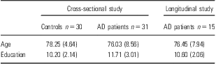
The 30 healthy controls (18 men and 12 women) were recruited from the University of Aberdeen Participation Panel. All controls spoke English as their first language, were literate, met the exclusion criteria of Wechsler Memory Scale–Revised (WMS III) and Wechsler Adult Intelligence Scale–Revised (WAIS III) standardisation samples (Reference Wechsler31) and had visual acuity sufficient to read newsprint. As shown in Table 1, they were aged between 71 and 89 years (mean 78.25, SD 4.64), with a mean of 10.20 (SD 2.14) years of education. All controls over the age of 80 were assessed on the mini-mental state examination (MMSE) to ensure normal cognitive functioning. None of these participants scored below 27/30.
Mean (SD) age, education and MMSE for AD patients and healthy controls
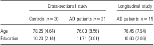
AD, Alzheimer's disease; MMSE, mini-mental state examination.
As shown in Table 1, the 31 patients (19 men and 12 women) were aged between 53 and 90 years (mean 76.03, SD 8.56), with a mean of 11.71 (SD 3.01) years of education. The patient group had a mean MMSE of 22.29 (SD 4.11), with a minimum of 16 and a maximum of 29. According to the cut-off scores established by Folstein et al. (Reference Folstein, Folstein and McHugh32), 16 patients fell within the minimal range (24–30), nine within the mild range (19–24) and six within the moderate range (12–18) of dementia severity.
To ensure that the patients and controls were matched in terms of age and education, a one-way ANOVA was carried out with age and education as the dependent factors. Results indicated that there was no significant difference for age (F (3,60) = 2.32, NS) or education (F (3,60) = 2.45, NS). As there was no significant difference between any of the groups in terms of age or education, all analyses were carried out using the raw scores.
Neuropsychological assessment
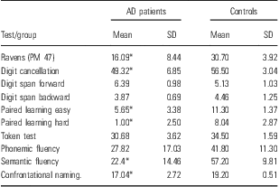
All patients and controls were assessed at the time of entering the study using a comprehensive battery of neuropsychological tests including the paired associate learning test (Reference Wechsler31), a forward and backward digit span task (Reference Wechsler31), a semantic and phonemic word fluency task (Reference Forbes-McKay and Venneri4), a 20-item naming task, the shortened version of the Token Test (Reference De Renzi and Faglioni33), a digit cancellation task (Reference Spinnler and Tognoni34) and the coloured progressive matrices (Reference Raven35) (Table 2).
As is indicated in Table 2, the AD patients performed within the normal range on the Token task, the digit span and the measure of phonemic fluency; however, deficits were evident on confrontation naming, semantic fluency, paired associate learning, digit cancellation and coloured progressive matrices.
Mean and SD scores on the neuropsychological battery for AD patients and healthy controls
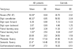
AD, Alzheimer's disease.
*Performance fell two standard deviations below the mean of controls.
Longitudinal study
For various reasons, such as illness, departure from the area, loss of contact or inability to complete the task, the full 12-month assessment of spontaneous speech was not possible for 16 (52%) of the original 31 AD samples. Of the 15 participants who were reassessed, five patients fell within the minimal range (24–30), six within the mild range (19–24) and four within the moderate range (12–18) of dementia severity. The AD patients had a mean age of 76.45 (SD 7.94) and a mean education of 10.60 years (SD 2.06). To determine whether those patients who completed the longitudinal assessment differed from those who dropped out in terms of initial MMSE, age or education, a one-way ANOVA was carried out with age, education and initial MMSE as the dependent factors. Results indicated that there was no significant effect for age (F (1,29) = 2.46, NS), but there was a significant effect for education (F (1,29) = 4.38, p < 0.05) and MMSE (F (1,29) = 4.95, p < 0.05). The subgroup of ‘survivors’ had significantly less years of education (mean 10.60, SD 2.06) and lower MMSE scores (mean 21.13, SD 2.79) than those who dropped out (years of education – mean 12.75, SD 2.06; MMSE – mean 24.12, SD 4.44). However, the two groups did not differ in terms of their performance on any of the psychometric or neuropsychological measures carried out at baseline.
Materials
Spontaneous speech assessment
Spontaneous speech was assessed using an oral description task comprising two simple line drawings that depict a domestic scene, namely, ‘The Cookie Theft Picture’ and ‘The Tripping Woman Picture’, and two complex line drawings that depict a traffic scene, ‘The Traffic Chaos Picture’ and the ‘The Bus Stop Picture’ (see Forbes-McKay and Venneri (Reference Forbes-McKay, Ellis, Shanks and Venneri5), for further details on complexity, scoring, validity and reliability).
Procedure
Baseline cross-sectional study
Each individual was instructed to describe orally everything that they could see happening in one simple and one complex picture. The descriptions were tape recorded and later analysed on a speech analysis scale, which measured 12 different aspects of speech, namely, melodic line, articulation, grammatical form, phrase length, phonological paraphasias, semantic paraphasias, visual paraphasias, word-finding delays, response to word-finding delays, error monitoring, information content and number of pictorial themes. Each measure was scored on a seven-point Likert-type scale ranging from 1 (severely impaired functioning) to 7 (no abnormality) [see Forbes-McKay and Venneri (Reference Forbes-McKay, Ellis, Shanks and Venneri5)]. Ratings were carried out by KFM, who devised the rating scales and has extensive experience of using them [see Forbes-McKay et al. (Reference Forbes-McKay, Venneri and Shanks2,Reference Forbes-McKay, Venneri and Shanks3)]. As demonstrated in Forbes-McKay and Venneri (Reference Forbes-McKay, Ellis, Shanks and Venneri5), the scales have high validity, inter-rater, parallel forms and test–retest reliability among healthy individuals and AD patients.
Longitudinal assessment
The procedure for study 2 was the same as study 1; however, to document the language deterioration suffered by AD patients over time, spontaneous speech was reassessed after a period of 6 months and 12 months. To control for practice effects, the stimuli were rotated every 6 months; for example, if presented with the ‘Bus Stop’ and ‘Cookie Theft’ pictures at baseline, patients were presented with the ‘Traffic Chaos’ and the ‘Tripping Woman’ pictures at 6 months.
Statistical analysis
Baseline cross-sectional assessment
To examine the effects of group (Healthy controls and AD patients), on the performance score of each speech variable, a one-way ANOVA was carried out for each speech measure.
Longitudinal assessment
To examine the effects of time (baseline, 6 months and 12 months) on the performance score of each speech variable, a one-way repeated ANOVA was carried out for each speech measure across all patients.
Results
Baseline cross-sectional analysis
Simple task
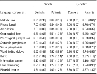
Results from a series of one-way ANOVAs indicated that the group had a significant effect on the measure of grammatical form (F (1,60) = 25.65, p = 0.00; η2 = 0.30), word-finding delays (WFDs; F (1,60) = 89.33, p = 0.00; η2 = 0.60), response to WFDs (F (1,60) = 34.98, p = 0.00; η2 = 0.37), error monitoring (F (1,60) = 161.63, p = 0.00; η2 = 0.81) and information content (F (1,60) = 47.80, p = 0.00; η2 = 0.45). The group had no significant effect on melodic line (F (1,60) = 4.95, p = 0.03), phrase length (F (1,60) = 3.77, p = 0.06), articulation (F (1,60) = 5.58, p = 0.02), phonological paraphasias (F (1,60) = 0.15, p = 0.70), semantic paraphasias (F (1,60) = 8.44, p = 0.005), visual paraphasias (F (1,60) = 7.29, p = 0.09) or number of pictorial themes (F (1,60) = 4.47, p = 0.04). Tests were conducted using Bonferroni-adjusted α levels of 0.004 per test (0.05/12). Table 3 shows a summary of mean scores of control and patient groups on this task.
Mean (SD) scores on the simple and complex task for AD patients and healthy controls
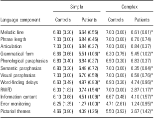
*Significant main effect of group, p < 0.004 tests were conducted using Bonferroni-adjusted α levels of .004 per test (0.05/12).
Complex task
Results from a series of one-way ANOVAs indicated that the group had a significant effect on the measure of melodic line (F (1,60) = 11.87, p = 0.01; η2 = 0.16), grammatical form (F (1,60) = 12.95, p = 0.001; η2 = 0.18), semantic paraphasias (F (1,60) = 17.74, p = 0.00; η2 = 0.23), visual paraphasias (F (1,60) = 9.01, p = 0.004; η2 = 0.13), WFDs (F (1,60) = 136.74, p = 0.00; η2 = 0.69), response to WFDs (F (1,60) = 369.67, p = 0.00; η2 = 0.86), error monitoring (F (1,60) = 43.63, p = 0.00; η2 = 0.47), information content (F (1,60) = 73.00, p = 0.00; η2 = 0.55) and number of pictorial themes (F (1,60) = 34.63, p = 0.00; η2 = 0.37). The group had no significant effect on phrase length (F (1,60) = 4.62, p = 0.04), articulation (F (1,60) = 5.58, p = 0.02) or phonological paraphasias (F (1,60) = 0.49, p = 0.48).
Tests were conducted using Bonferroni-adjusted α levels of 0.004 per test (0.05/12). Table 3 shows a summary of mean scores of control and patient groups on this task.
Longitudinal analysis
Simple task
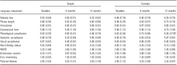
Results from the within subjects ANOVA demonstrated no significant effect of time on the measures of melodic line (F (2,28) = 1.91, p = 0.19), grammatical form (F (2,28) = 1.93, p = 0.16), information content (F (2,28) = 1.81, p = 0.18), phrase length (F (2,28) = 0.41, p = 0.66), pictorial themes (F (2,28) = 1.61, p = 0.22), semantic paraphasias (F (2,28) = 2.63, p = 0.09), visual paraphasias (F (2,28) = 0.63, p = 0.01), phonological paraphasias (F (2,28) = 3.55, p = 0.04), WFDs (F (2,28) = 2.93, p = 0.07) or response given to WFDs (F (2,28)=4.89, p = 0.02).
As the patients performed at floor on the measure of error monitoring, and scores remained constant over time for articulation, ANOVA could not be calculated on these measures. Tests were conducted using Bonferroni-adjusted α levels of 0.005 per test (0.05/10). Table 4 summarises the mean patient group score for each variable.
Mean (SD) scores on the simple and complex task at baseline, 6 and 12 months in the patient group

*Significant main effect of time, p < 0.004. Tests were conducted using Bonferroni-adjusted α levels of .004 per test (0.05/12).
Complex task
Results demonstrated a significant main effect of time on phonological paraphasias (F (2,28) = 7.57, p = 0.002; η2 = 0.4). Further analysis using Bonferroni pairwise comparisons indicated that the patients produced significantly more phonological paraphasias at 12 months than at baseline (p < 0.05).
There was no significant effect of time on the measures of articulation (F (2,28) = 0.48, p = 0.62), error monitoring (F (2,28) = 1.68, p = 0.21), melodic line (F (2,28) = 0.45, p = 0.64), grammatical form (F (2,28) = 2.82, p = 0.08), information content (F (2,28) = 0.18, p = 0.98), phrase length (F (2,28) = 1.76, p = 0.19), response to WFDs (F (2,28) = 1.81, p = 0.18), visual paraphasias (F (2,28) = 1.97, p = 0.16), semantic paraphasias (F (2,28) = 5.01, p = 0.01), number of pictorial themes described (F (2,28) = 3.70, p = 0.04) or WFDs (F (2,28) = 1.76, p = 0.19). Tests were conducted using Bonferroni-adjusted α levels of 0.004 per test (0.05/12).
Discussion
The cross-sectional study found that impairments at the level of the semantic system were present early in the course of the disease. In addition to a minor breakdown in articulation and melodic line, the mild–moderate AD patients produced speech, which contained significantly more WFDs, more semantic paraphasias, more empty and indefinite phrases and fewer repaired errors than the healthy elderly controls. These findings are in line with previous research (Reference Vuorinen, Laine and Rinne1–Reference Forbes-McKay, Ellis, Shanks and Venneri5,Reference Biundo, Gardini and Caffarra8,Reference Salmon9,Reference Lyons, Kemper, Labarge, Ferraro, Balota and Storandt22).
However, as the disease progressed over time, the number of indefinite and empty phrases, semantic paraphasias, WFDs and unresolved errors did not increase with time. Performance levelled off and these measures were no longer sensitive in monitoring decline. Moreover, the motoric and syntactic aspects of speech remain relatively well preserved over a period of 12 months. The measure of phonological paraphasias did, however, demonstrate a significant decline over 12 months.
Therefore, consistent with the description given by Obler and Albert (Reference Obler and Albert21), the early stage AD patients produced fluent speech, which was characterised by word-finding pauses and semantic substitutions. Although their narratives remained informative at this stage, patients did introduce irrelevant topics and produce indefinite and empty phrases. In contrast to Obler and Albert (Reference Obler and Albert21), however, these patients also showed a slight reduction in grammatical form. Although their descriptions were grammatically intact, sentences were less complex than those produced by the controls. As the disease progressed, discourse became increasingly empty and meaningless, and there was an increase in the number of indefinite and irrelevant terms. Finally, in the late stages of AD, a few patients showed a reduction in fluency and articulation, and two patients were no longer testable on the language task.
With the exception of minor word retrieval problems and a simplification of grammatical form, language abilities tend to be well maintained in later life (Reference Heine, Ober and Shenaut36). Impairments evident in the healthy elderly appear linked to a more general decline in other cognitive functions such as memory, attention and executive function. Although the current control sample showed a slight decline in grammatical form and error monitoring, they experienced very few word-finding delays and produced very few semantic paraphasias. Even when controls did fail to produce the target word, errors were typically phonological in nature. Considering the qualitative and quantitative differences shown between the two groups, the speech impairment in AD cannot be explained by exaggerated ageing.
In line with the findings of Hodges et al. (Reference Hodges, Salmon and Butters37), the proportion of phonologically related errors produced by the AD patients increased as the disease progressed.
Although the group was not separated in terms of their disease severity, the results of the current study indicate that the majority of mild–moderate AD patients progress from an initial impairment in semantic knowledge, which is later joined by a secondary impairment in visuo-perceptual and phonological processing. Therefore, despite the heterogeneity of AD, there is a relatively common pattern of impairments shown across the current sample. Although the conclusions drawn are limited by a small sample, which limits the wider application of the findings, the findings are consistent with those shown in previous studies. Further, analysis of the patients’ biographical characteristics and neuropsychological performance indicates that there was very little difference between those who survived and those who dropped out. Although the speed of deterioration may vary, the pattern of impairment appears relatively consistent.
Consistent with the findings of Ehlrich et al. (Reference Ehrlich, Obler and Clark38), the need to integrate more information and identify more pictorial themes in the complex task increases the sensitivity of the narrative description task in both the cross-sectional and longitudinal study, whereby patients produce more paraphasias, less complex grammar and less pictorial themes.
Conclusion
In conclusion, as AD progresses in severity, patients show a qualitative and quantitative deterioration in language production. Despite the heterogeneity of patients, the current results show a pattern of impairment dominated by semantic errors, which are later joined by a disruption to the phonological, visual and motoric aspects of speech. By the final stages of the assessment, the patients show a global language problem characterised by indefinite and empty phrases, short grammatically simple sentences, word-finding difficulties, visual, phonological and semantic errors and an inability to repair such errors. Although a number of patients did show impairments in articulation and melodic line, results indicate that the motoric aspects of speech remain relatively preserved.
The language deterioration shown by the current AD sample is consistent with findings from neuropathological and neuroimaging studies, which have demonstrated a pattern of cortical degeneration that is initially centred in the temporal and parietal lobes and later spreads to the frontal lobes of the brain. These temporoparietal areas are associated with naming, word retrieval and semantic processing and undergo the earliest and most severe destruction in AD (Reference Harasty, Halliday and Code39,Reference McGeown, Shanks, Forbes-McKay and Venneri40) and pre-clinical AD (Reference Vuorinen, Laine and Rinne1,Reference Biundo, Gardini and Caffarra8,Reference Kaye, Swithart and Howieson41). The frontal areas responsible for motor output and phonological processing, on the other hand, remain relatively spared until the later stages of the disease (Reference Hirono, Mori and Ishii42–Reference Braak, Braak, Yilmazer, Vos, Jansen and Bohl44). This pattern of cortical degeneration may contribute to an inability to access specific semantic attributes about concepts (Reference Forbes-McKay, Venneri and Shanks45) determining production of indefinite terms and semantic paraphasias. However, as the disease progresses into the frontal regions of the brain, phonological processing and motoric output become impaired, and moderately demented patients produce phonological errors in keeping with the progression of regional neuropathological and functional changes.
Acknowledgements
This study was supported by an educational studentship to KFM at The University of Aberdeen. These data were collected while KFM and AV were employed by the University of Aberdeen and MFS was employed by Grampian Healthcare NHS Trust.
Authors contributions
Katrina Forbes-McKay, Michael Shanks, Annalena Venneri contributed to the conception, design of and interpretation of data and to the drafting and revising of the article. Katrina Forbes-McKay was additionally responsible for the acquisition and analysis of data.
Conflict of interest
None.