Introduction
The Merodon aureus Fabricius, 1805 (Diptera: Syrphidae: Eristalinae: Merodontini) species group is within the most speciose hoverfly genus in Europe. The species group has been morphologically defined by Radenković et al. (Reference Radenković, Vujić, Ståhls, Pérez-Bañón, Rojo, Petanidou and Šimić2011). Molecular analyses using cytochrome c oxidase I (COI) and 28S rRNA gene sequences have supported it as a monophyletic lineage (Mengual et al. Reference Mengual, Ståhls, Vujić and Marcos-García2006; Radenković et al. Reference Radenković, Vujić, Ståhls, Pérez-Bañón, Rojo, Petanidou and Šimić2011; Vujić et al. Reference Vujić, Radenković, Ståhls, Ačanski, Stefanović and Veselić2012). The M. aureus species group comprises small-sized species possessing a short, rounded abdomen; metatrochanter with an inner calcar with two pointed angles; and a characteristic structure of the male genitalia. The anterior lobe of surstylus is undeveloped, while the posterior surstyle lobe has parallel margins and a rounded apex and a narrow, elongate, sickle-shaped hypandrium without a lateral sclerite of the aedeagus (Radenković et al. Reference Radenković, Vujić, Ståhls, Pérez-Bañón, Rojo, Petanidou and Šimić2011). Previous studies have indicated that the group may have an especially large number of cryptic taxa (Šašić et al. Reference Šašić, Ačanski, Vujić, Ståhls, Radenković and Milić2016, Reference Šašić, Ačanski, Đan, Kočiš Tubić, Veličković, Radenković and Vujić2018; Veselić et al. Reference Veselić, Vujić and Radenković2017; Radenković et al. Reference Radenković, Šašić Zorić, Djan, Obreht Vidaković, Ačanski and Ståhls2018).
Šašić et al. (Reference Šašić, Ačanski, Vujić, Ståhls, Radenković and Milić2016) defined five species subgroups within the Merodon aureus species group (M. aureus, M. bessarabicus Paramonov, 1924, M. cinereus (Fabricius, 1794), M. chalybeus Wiedemann in Meigen, 1822, and M. dobrogensis Bradescu, 1982 species subgroups, respectively) based upon the morphological similarity of the species. Each of these species subgroups contains at least one complex of cryptic species, although they may contain multiple complexes of species such as the M. bessarabicus species subgroup (see Veselić et al. Reference Veselić, Vujić and Radenković2017).
Šašić et al. (Reference Šašić, Ačanski, Vujić, Ståhls, Radenković and Milić2016) defined species complexes as morphologically inseparable species, which can only be resolved by employing an integrative taxonomy approach including different data types such as molecular, geometric morphometry, and ecology. Applying this approach for the Merodon aureus species group has led to the discovery of previously unknown species complexes. For the M. cinereus species subgroup, Šašić et al. (Reference Šašić, Ačanski, Vujić, Ståhls, Radenković and Milić2016) resolved the M. atratus (Oldenberg, 1919) complex of species, and found that it consisted of three species, two of which were new. Veselić et al. (Reference Veselić, Vujić and Radenković2017) provided evidence for the presence of four species complexes in the M. bessarabicus species subgroup. Radenković et al. (Reference Radenković, Šašić Zorić, Djan, Obreht Vidaković, Ačanski and Ståhls2018) resolved M. luteomaculatus Vujić, Ačanski, and Šašić, 2018 as a complex of six cryptic species. Additionally, the same approach was used to resolve the M. caerulescens Loew, 1869 species complex (Šašić et al. Reference Šašić, Ačanski, Đan, Kočiš Tubić, Veličković, Radenković and Vujić2018).
The taxonomy of the Merodon dobrogensis species subgroup has not been evaluated using an integrative taxonomy approach. This species subgroup is monotypic, containing only the species M. dobrogensis, which was first discovered in Romania and is easily recognised by its red abdomen and reddish-orange tibiae. However, the description of M. puniceus Vujić, Radenković, and Pérez-Bañón, 2011 (Radenković et al. Reference Radenković, Vujić, Ståhls, Pérez-Bañón, Rojo, Petanidou and Šimić2011), a morphologically similar species from the Aegean island of Lesvos, raises the question of whether or not M. dobrogensis is a species complex and provides good reason to conduct an in-depth taxonomic investigation, and to check for additional undiscovered species within the M. dobrogensis species subgroup.
Material and methods
We explored species borders within the M. dobrogensis subgroup by applying an integrative taxonomy approach using molecular data and morphological data, specifically wing and surstylus geometric morphometry.
Taxonomy
For the present study, material from continental Greece, Turkey, and Romania (the type locality of Merodon dobrogensis) was collected to explore overall diversity of the M. dobrogensis species subgroup. Material examined includes specimens collected during the last 30 years (1982–2014) as well as any available specimens, including types, previously cited in the literature. Collections from which specimens were obtained are listed below.
The following acronyms of museums and entomological collections are used in the text: Natural History Museum, London, United Kingdom (BMNH); Colección Entomológica de la Universidad de Alicante, Alicante, Spain (CEUA); Faculty of Sciences, Department of Biology and Ecology, University of Novi Sad, Novi Sad, Serbia (FSUNS); Melissotheque of the Aegean, University of the Aegean, Mytilene, Greece (MAUA); Michael de Courcy Williams collection, Alexandroupoli, Greece (MCWC); Muzeul de Istorie Naturala “Grigore Antipa”, Bucharest, Romania (MGAB); Naturhistorisches Museum, Vienna, Austria (NMW); Naturalis Biodiversity Centre, Leiden, The Netherlands (RMNH); and World Museum Liverpool, Liverpool, United Kingdom (WML).
A short diagnosis is provided for the Merodon aureus species group, the M. dobrogensis species subgroup, and the newly defined M. dobrogensis species complex, including the description of a new species, with drawings and photographs of adult morphology. Terminology follows Thompson (Reference Thompson1999) for non-genitalic morphology and Marcos-García et al. (Reference Marcos-García, Vujić and Mengual2007) for the morphology of male terminalia. Methodology from Vujić et al. (Reference Vujić, Radenković, Likov, Trifunov and Nikolić2013) was followed for studying the male genitalia. Digital photographs were taken with Leica (Wetzlar, Germany) DFC 320 digital camera, attached to a Leica MZ16 binocular microscope. Measurements were taken with an eyepiece graticule or micrometer. Morphological characters were observed using a Nikon (Tokyo, Japan) SMZ 745T stereomicroscope.
Data on the natural history of species from the Merodon dobrogensis species subgroup, presented in the database for European Syrphidae by Speight (Reference Speight2018), have been updated.
Geometric morphometric analysis
As part of an integrative taxonomic approach, two different geometric morphometric analyses were employed to identify species of the Merodon dobrogensis species subgroup: landmark-based analysis of the wing shape, and outline contours of the posterior surstyle lobe of male genitalia (hereinafter referred to as the surstylus).
High-resolution photographs of the wings and surstylus were made using a Leica DFC320 video camera attached to a Leica MZ16 stereomicroscope. Landmarks and semi-landmarks were drawn on every picture using the TpsDig version 2.30 (Rohlf Reference Rohlf2017a).
A principal component analysis was carried out on the Procrustes shape variables of surstylus to reduce the dimensionality of the dataset. A stepwise discriminant function analysis and canonical variate analysis were used to explore wing and surstylus shape variation among the taxa. Additionally, a Gaussian naïve Bayes classifier was used to delimit species boundaries based on wing and surstylus shape variation without a priori defined groups. Phenetic relationships among the taxa were characterised using an unweighted pair group method with arithmetic mean cluster analysis based on squared Mahalanobis distances computed from the discriminant function analysis applied to shape variables. Superimposed outline drawings were produced using MorphoJ version 2.0 (Klingenberg Reference Klingenberg2011) to visualise differences in mean wing and surstylus shape among species pairs. All statistical analyses were done using Statistica for Windows (TIBCO Software 2017).
Wing morphometry
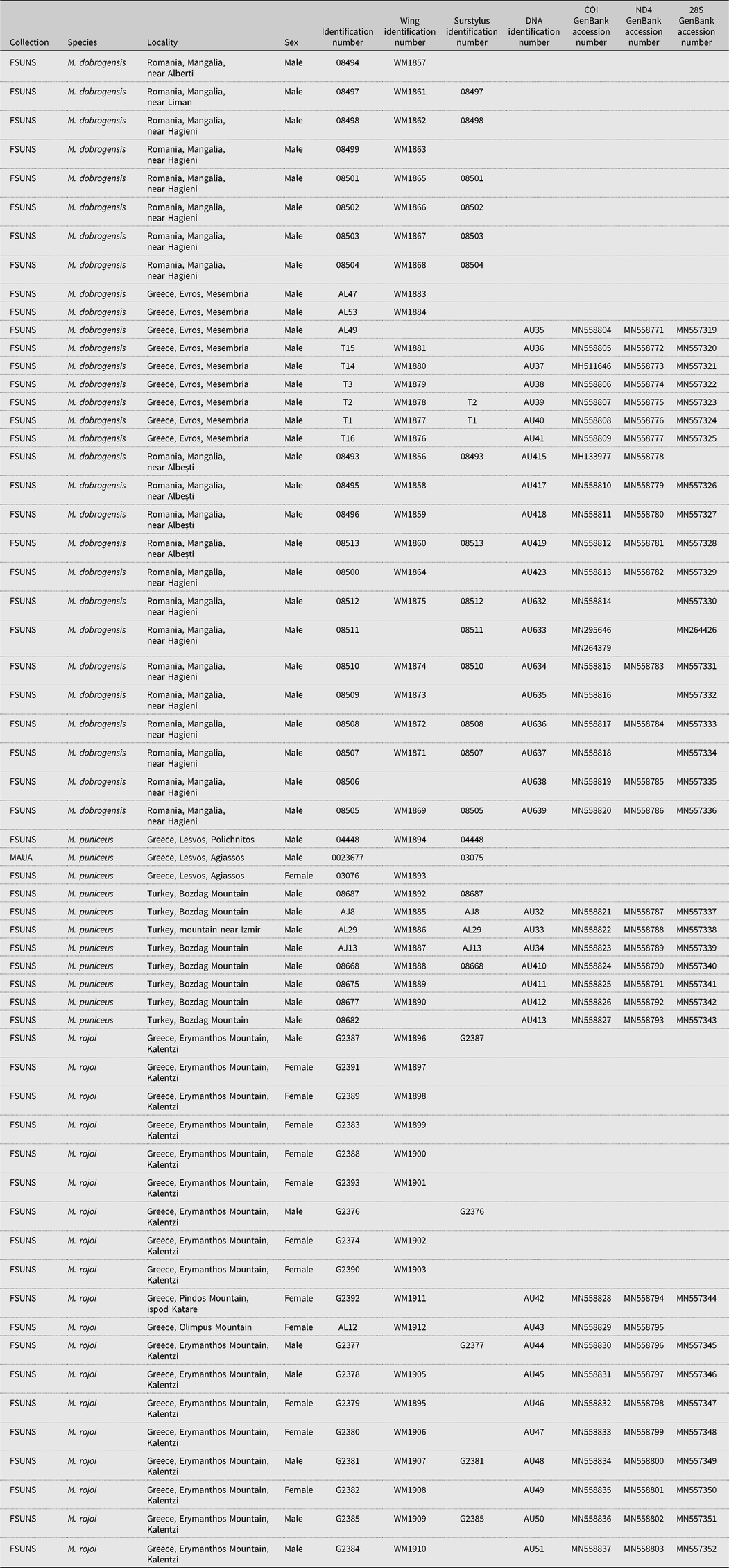
Landmark-based geometric morphometric analysis of wing shape was conducted on 53 specimens of the Merodon dobrogensis species subgroup (Table 1). The right wing of each specimen was taken off using micro-scissors and mounted in Hoyer’s medium on a microscopic slide. Wings were archived and labelled with a unique code in the FSUNS, together with other data relevant to the specimens. Eleven homologous landmarks at vein intersections or terminations (that could be reliably identified) were selected. Due to an uneven distribution of sexes within taxa, analyses were conducted on a dataset in which both sexes are pooled and for the male dataset separately. Wing shape components were size-corrected to remove the allometric component of the wing shape.
List of the Merodon dobrogensis species subgroup specimens used in molecular and geometric morphometric analyses.

Generalised least squares Procrustes superimposition on the raw coordinates was done using TpsRelw version 1.68 (Rohlf Reference Rohlf2017b) to minimise nonshape variations in location, scale, and orientation of wings, and to superimpose the wings in a common coordinate system (Rohlf and Slice Reference Rohlf and Slice1990; Zelditch et al. Reference Zelditch, Swiderski, Sheets and Fink2004).
Surstylus morphometry
Shape analysis of the right posterior surstylus lobe of male genitalia was carried out on 28 specimens of the Merodon dobrogensis species subgroup (Table 1). The right posterior surstylus lobe was removed using a scalpel and placed on its side in glycerol on a microscopic slide, with a cover slip placed on top of the surstylus to immobilise it.
Fifteen semi-landmarks were digitised from the membranous part of the epandrium to the end of the surstylus. CoordGen version 7.14 (Sheets Reference Sheets2012) with an integrated Semiland module was used for semi-landmark superimposition using a distance-minimising protocol, minimising the shape differences due to the arbitrary nature of semi-landmark positions along the curve (Bookstein Reference Bookstein1997; Zelditch et al. Reference Zelditch, Swiderski, Sheets and Fink2004).
Molecular study
Total genomic DNA was extracted from mesothorcic and metathoracic legs of 37 hoverfly specimens using the sodium dodecyl sulphate extraction protocol (Chen et al. Reference Chen, Rangasamy, Tan, Wang and Siegfried2010). Detailed information on the specimens included in molecular analyses and GenBank (www.ncbi.nlm.nih.gov/genbank) accession numbers are presented in Table 1. Genomic DNA vouchers have been conserved at the Faculty of Sciences, Department of Biology and Ecology, University of Novi Sad (FSUNS), Serbia.
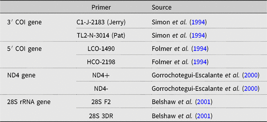
The primers used for amplification of 3′ and 5′ ends of the mitochondrial COI gene, fragment of NADH dehydrogenase subunit 4 (ND4) gene, as well as the D2–3 expansion segment of the 28S rRNA gene are provided in Table 2. Polymerase chain reactions were carried out in 25-μL reaction volumes. The reaction mixture for 3′ and 5′ COI and 28S rRNA gene amplification contained 1× reaction buffer (ThermoScientific, Vilnius, Lithuania), 2.5 mM MgCl2, 0.1 mM of each nucleotide, 1.25 U Taq polymerase (ThermoScientific), 5 pmol of each primer, and approximately 50–100 ng of template DNA. For ND4 gene amplification, we used 1.25 U Dream Taq polymerase (ThermoScientific) and corresponding 1× Dream Taq reaction buffer (ThermoScientific) with included MgCl2. We used the following polymerase chain reaction conditions for COI and 28S rRNA gene amplification: 95 °C for two minutes; 29 cycles of 94 °C for 30 seconds, 49 °C (for 3′ COI) and 50 °C (for 5′ COI and 28S rRNA gene) for 30 seconds, 72 °C for two minutes; with a final extension at 72 °C for eight minutes. The applied polymerase chain reaction conditions for ND4 gene were: 95 °C for five minutes; 35 cycles of 94 °C for one minute, 45 °C for 30 seconds, 72 °C for two minutes; with a final extension at 72 °C for eight minutes. The purification of polymerase chain reaction products was done using Exonuclease I and FastAP Thermosensitive Alkaline Phosphatase (ThermoScientific) according to the manufacturer’s instructions. Sequencing was performed in a forward direction for each amplified fragment using the BigDye Terminator version 3.1 cycle sequencing kit (ThermoScientific) on an ABI3730xl Genetic Analyzer (Applied Biosystems, Foster City, California, United States of America) in the Sequencing Service Laboratory of the Finnish Institute for Molecular Medicine (FIMM), University of Helsinki, Finland, and the Macrogen EZ-Seq service (Macrogen Europe, Amsterdam, The Netherlands).
Primers used for the amplification of COI, ND4, and 28S rRNA gene fragments.

Molecular genetic data analyses
COI and 28S rRNA gene sequences were edited for base-calling errors using BioEdit version 7.0.9.0 (Hall Reference Hall1999), aligned manually, and trimmed to a final length by eye.
The 5′ COI sequence matrix with included Merodon puniceus sequence (Ståhls et al. Reference Ståhls, Vujić, Pérez-Bañón, Radenković, Rojo and Petanidou2009; Radenković et al. Reference Radenković, Vujić, Ståhls, Pérez-Bañón, Rojo, Petanidou and Šimić2011; GenBank accession number FR717827) was used for uncorrected pairwise (p) distance calculation to assign Turkish specimens to the particular species. Uncorrected p distances were calculated using MEGA 7 (Kumar et al. Reference Kumar, Stecher and Tamura2016). In all further analyses, sequences were assigned to a particular species. Because of low variability, 28S rRNA gene sequences were only used for genotypes determination. ND4 and COI gene sequences were analysed more thoroughly. The 3′ and 5′ COI gene sequences were concatenated and analysed together.
Maximum parsimony and maximum likelihood analyses were applied on COI and combined COI+ND4 sequence matrices. The maximum parsimony analysis was performed in NONA (Goloboff Reference Goloboff1999) spawned with the aid of ASADO (Nixon Reference Nixon2008) using the heuristic search algorithm with 1000 random addition replicates (mult*1000), holding 100 trees per round (hold/100), maxtrees set to 100 000, and applying tree-bisection-reconnection branch swapping. The maximum likelihood tree was constructed using RAxML 8.2.8 (Stamatakis Reference Stamatakis2014) using the CIPRES Science Gateway web portal (Miller et al. Reference Miller, Pfeiffer and Schwartz2010) under the general-time-reversible evolutionary model with a gamma distribution (GTRGAMMA) (Rodríguez et al. Reference Rodríguez, Oliver, Marín and Medina1990). Clade supports were calculated using 1000 bootstrap replicates for both maximum parsimony and maximum likelihood trees. The trees were rooted on Eristalinus aeneus (GenBank accession number NC_042911). The detection of COI and ND4 haplotypes, as well as 28S rRNA gene genotypes, was done using DnaSP version 5 (Librado and Rozas Reference Librado and Rozas2009). The average uncorrected pairwise distances (p) between taxa were calculated using MEGA7 (Kumar et al. Reference Kumar, Stecher and Tamura2016).
Correlation among wing shape, surstylus shape, genetic, spatial, and climatic differentiation
To test pairwise correlations between morphometric (wing and surstylus), genetic (COI and ND4), and geographic and climatic distances among the species, two-tailed Mantel tests were performed (Mantel Reference Mantel1967) with 9999 permutations in PaSSaGe (Rosenberg and Anderson Reference Rosenberg and Anderson2011). Additionally, partial Mantel tests were used to explore the relationships between: morphometric and geographic differentiation while accounting for the effect of climatic distances; and morphometric and climatic differentiation while accounting for the effect of geographic distances. Morphometric distances were represented as a matrix of pairwise squared Mahalanobis distances, and genetic distances as a matrix of uncorrected p distances. Geographic distances were calculated as the minimum distance between two species using QGIS (Quantum GIS Development Team 2012). Climatic distances were represented as Euclidean distances of the factor scores calculated based on 19 bioclimatic variables generated for each locality from the current climate WorldClim dataset (2.5 arc-minute resolution) (Hijmans et al. Reference Hijmans, Cameron, Parra, Jones and Jarvis2005).
Results
Geometric morphometric evidence
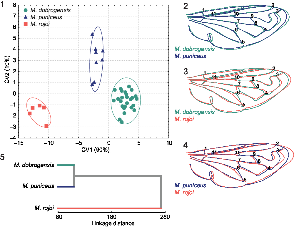
Wing shape. Geometric morphometric analyses of wing shape show the same pattern for pooled sexes and for the males separately; thus, only results based on the male dataset are presented. Stepwise discriminant analysis provides evidence for highly significant wing shape differences among all species pairs (P < 0.01; Merodon dobrogensis–M. rojoi new species F 13, 25 = 45.233; M. dobrogensis–M. puniceus F 13, 25 = 11.875; M. punicues–M. rojoi F 13, 25 = 18.162). Additionally, a discriminant function analysis with cross-validation based on wing shape showed correct species assignment for all male specimens (100%). A congruent classification was obtained by the Gaussian naive Bayes classifier through which all male specimens of M. puniceus and M. rojoi were correctly classified, and only 1 out of 27 specimens of M. dobrogensis was misclassified as M. puniceus.
The succeeding canonical variate analysis conducted on shape variables of males produced two highly significant canonical axes linked to shape variation among the species (CV1: Wilks’ lambda = 0.0098; χ 2 = 138.768; P < 0.01; CV2: Wilks lambda = 0.2598; χ 2 = 40.440; P < 0.01). The first canonical axis represents the majority of wing shape variation (90%) and clearly differentiates all three species, with the main focus on differences between Merodon rojoi and the M. dobrogensis species complex (Fig. 1). The second canonical axis with 10% of wing shape variation clearly separates M. dobrogensis and M. puniceus (Fig. 1). This pattern of shape variation among the species continues in superimposed outline drawings of mean wing shapes (Figs. 2–4). The more subtle differences in mean shape were between M. dobrogensis and M. puniceus (Fig. 2), while the most obvious differences were between M. rojoi and M. puniceus (Fig. 4). Wing shape differentiation among the species was most apparent in the central and distal parts of the wing (Figs. 2–4).
Congruent with the previous results, Merodon dobrogensis and M. puniceus were most similar to each other, while M. rojoi had the most distinct wing shape, based on the unweighted pair group method with arithmetic mean dendrogram (Fig. 5).
Geometric morphometric analysis of the wing shape of the Merodon dobrogensis species subgroup. 1, Scatter plot of individual scores of CV1 and CV2; 2–4, superimposed outline drawings of average wing shape for each species. Differences between the species were exaggerated threefold to make them more visible; 5, unweighted pair group method with arithmetic mean phenogram constructed using Mahalanobis square distances of wing shape.

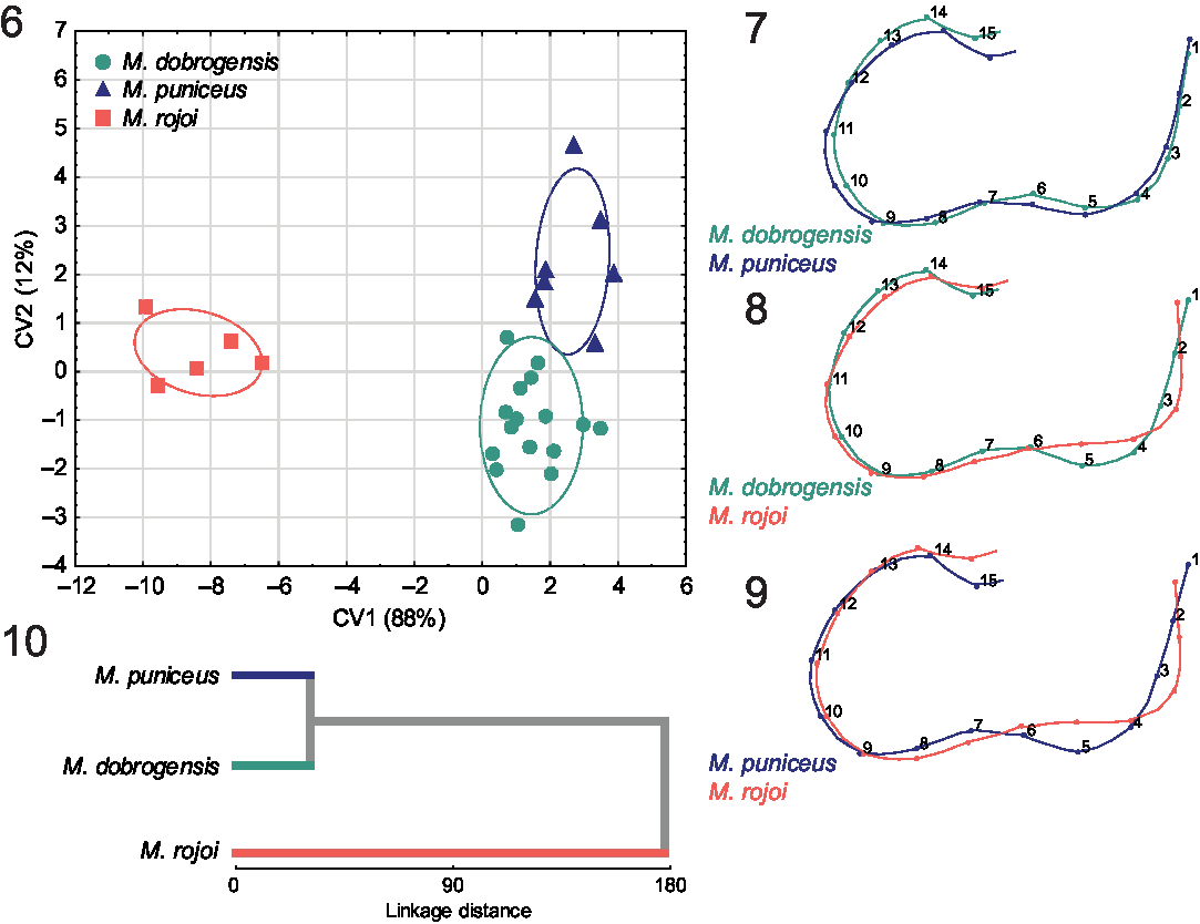
Surstylus shape. A principle components analysis carried out on the Procrustes shape variables produced 26 principal components. A stepwise discriminant analysis revealed that the 12 principal components represent the highest overall classification percentage of investigated taxa. A discriminant function analysis showed that species within the Merodon dobrogensis species subgroup differed significantly in surstylus shape (M. dobrogensis–M. rojoi F 12, 15 = 17.539, P < 0.01; M. dobrogensis–M. puniceus F 12, 15 = 2.925, P < 0.05; M. puniceus–M. rojoi F 12, 15 = 17.032, P < 0.01). Importantly, all specimens were correctly classified (100%) to a priori defined groups. Moreover, a congruent classification was obtained by the Gaussian naive Bayes classifier through which all specimens were correctly classified to the belonging species, demonstrating that surstylus shape is a reliable character for interspecific discrimination. A canonical variates analysis produced two significant axes (CV1: Wilks lambda = 0.0167; χ 2 = 79.757; P < 0.01; CV2: Wilks lambda = 0.3068; χ 2 = 23.039; P < 0.05). As in wing shape, the first canonical axis represented the majority of surstylus shape variation (88%) and clearly differentiated M. rojoi from the M. dobrogensis species complex (Fig. 6). With 12% of total shape variation, CV2 clearly separated M. dobrogensis and M. puniceus (Fig. 6).
Differences in surstylus shape between Merodon dobrogensis and M. puniceus are mostly related to the posterior part of the posterior surstyle lobe (Fig. 7), while the greatest differences between M. rojoi and species from the M. dobrogensis species complex are in the anterior part of the posterior surstyle lobe (Figs. 8–9). Phenetic relationships constructed on the basis of surstylus shape followed the pattern obtained for wing shape; M. dobrogensis and M. puniceus were closest to each other, while M. rojoi had the most distinct surstylus shape (Fig. 10).
Geometric morphometric analysis of the surstylus shape of the Merodon dobrogensis species subgroup. 6, Scatter plot of individual scores of CV1 and CV2; 7–9, superimposed outline drawings of average surstylus shape for each species. Differences between the species were exaggerated twofold to make them more visible; 10, unweighted pair group method with arithmetic mean phenogram constructed using Mahalanobis square distances of surstylus shape.

Molecular data
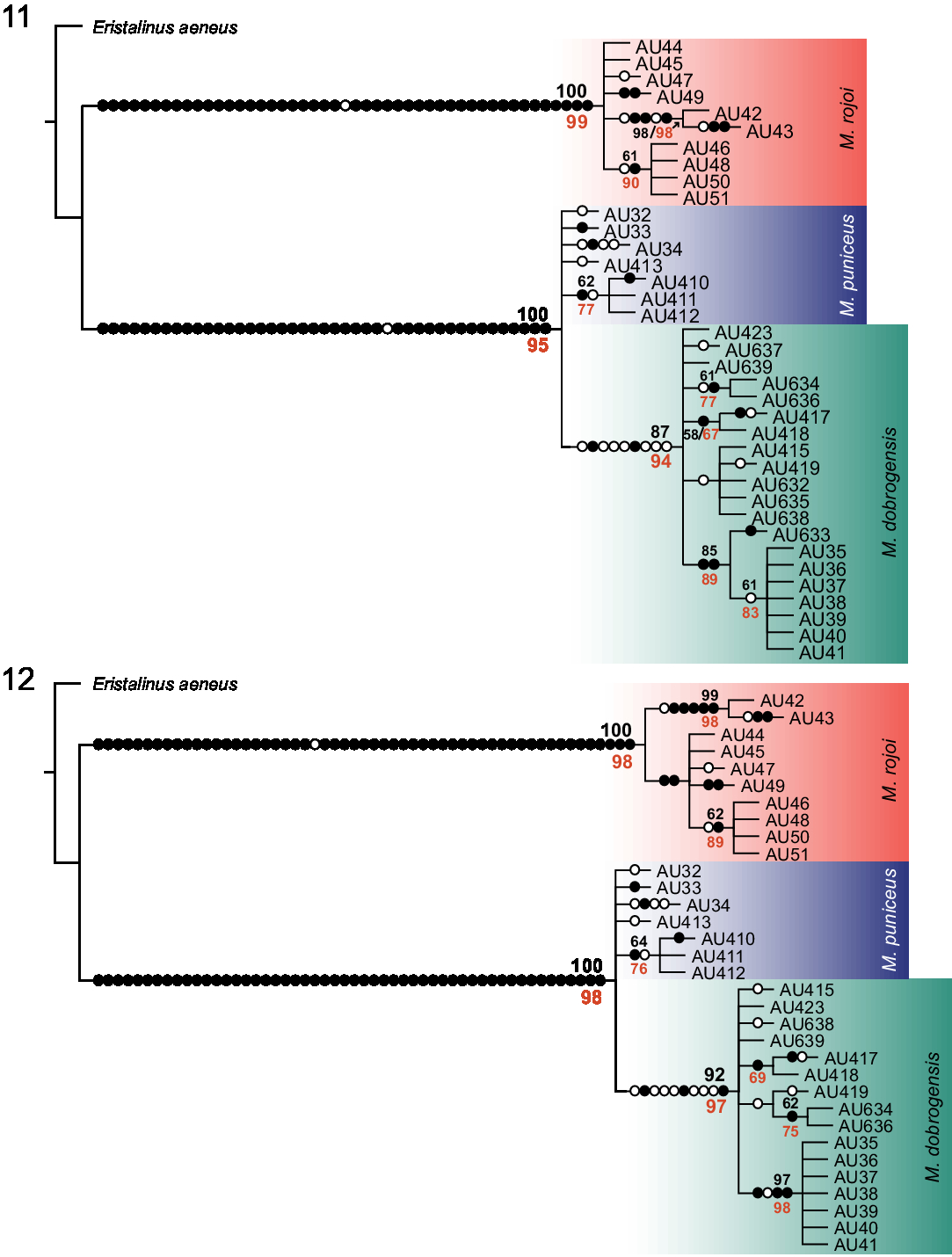
COI sequences were generated for 37 specimens belonging to the Мerodon dobrogensis species subgroup. Based on the uncorrected pairwise (p) distances between 5′ COI sequences, Turkish specimens were assigned to M. puniceus as the p distances of specimen from Lesvos were the lowest (0–0.2%) compared with Turkish specimens (see Supplementary Table S1). The combined 3′ and 5′ COI sequence alignment comprised 1400 base pairs. There were 144 variable positions, while 133 were parsimony informative. The resulting maximum parsimony tree (Fig. 11) was a strict consensus of 46 equally parsimonious trees (length = 299, consistency index = 95%, retention index = 98%). It had almost identical topology to the maximum likelihood tree. The M. rojoi clade and the cluster of M. dobrogensis complex taxa (M. puniceus + M. dobrogensis) were clearly separated on both trees (Fig. 11). Merodon rojoi was resolved as a monophyletic clade with a high bootstrap support (maximum parsimony/maximum likelihood = 100/99), while M. dobrogensis was resolved as nested within the M. puniceus clade. The M. dobrogensis clade was also well supported with bootstrap values of 87/94 (maximum parsimony/maximum likelihood).
ND4 sequences were generated for 33 specimens, and the alignment was 360 base pairs long (only one sequence was shorter; 329 base pairs). The number of variable positions when excluding the missing data was 30, and all were parsimony-informative. The combined COI+ND4 alignment used for maximum parsimony and maximum likelihood tree constructions was 1760 base pairs long. The combined COI+ND4 trees were similar in topology with COI gene trees, and the main clades were supported with similarly high bootstrap values (Fig. 12).
Maximum parsimony trees of the Merodon dobrogensis species subgroup (filled circles stand for unique changes; open circles stand for non-unique changes; bootstrap values ≥ 50 are presented near nodes; bootstrap values in red correspond to those from maximum likelihood trees). 11, COI strict consensus tree of 46 equally parsimonious trees; 12, COI+ND4 strict consensus tree of 23 equally parsimonious trees.

The average COI-uncorrected pairwise distances (p COI) between species pairs showed variations: 0.8% between Merodon puniceus and M. dobrogensis, 8.7% between M. rojoi and M. puniceus, and 8.8% between M. rojoi and M. dobrogensis. The average ND4-uncorrected pairwise distances (p ND4) between species pairs were lower than p COI values (0.4% M. puniceus–M. dobrogensis; 7.8% M. rojoi–M. puniceus; 8.2% M. rojoi–M. dobrogensis). Additionally, ND4 gene sequences were less variable compared to COI gene sequences. We detected only five ND4 haplotypes and 19 COI haplotypes (Supplementary Table S2). The haplotypes were not shared among subgroup taxa.
The 28S rRNA gene sequence matrix contained 35 sequences with length varying from 578 to 579 base pairs, while the length of alignment was 579 base pairs. The total number of variable and parsimony-informative sites was three. The sequences formed two genotypes from which one (I) was shared between Merodon dobrogensis and M. puniceus, and the other (II) corresponded to M. rojoi. The genotypes differed in three mutational steps.
Correlation among wing shape, surstylus shape, and genetic, spatial, and climatic differentiation
Simple two-tailed Mantel tests revealed that geographic and climatic distances exhibited no association with wing and surstylus shape distances or genetic distances among Merodon dobrogensis, M. puniceus, and M. rojoi (P > 0.05). Additionally, the partial Mantel test showed that geographic distance had no impact on wing and surstylus shape differentiation while accounting for climatic distance, nor did climatic distance while accounting for geographic distance (P > 0.05).
Merodon aureus species group sensu Radenković et al. (Reference Radenković, Vujić, Ståhls, Pérez-Bañón, Rojo, Petanidou and Šimić2011)
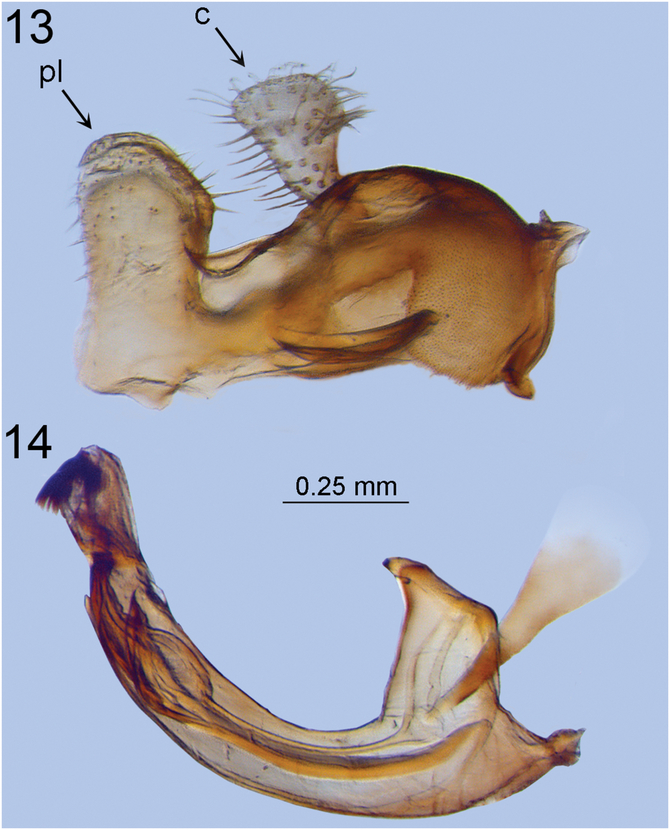
Diagnosis. The following combination of characteristics distinguishes this group from other Merodon Meigen, 1803 groups: mesocoxa pilose posteriorly, anterior anepisternum with pilose area ventral to postpronotum, in male genitalia anterior lobe of surstylus undeveloped. Small-sized (8–13 mm) species with a short, rounded abdomen; a distinct calcar on the metatrochanter in males; and a characteristic structure of the male genitalia: posterior surstylus lobe with parallel margins and rounded apex (Fig. 13: pl) and a narrow, elongate, sickle-shaped hypandrium without lateral sclerite of aedeagus (Fig. 14).
Merodon rojoi, male genitalia, lateral view. 13, Epandrium; 14, hypandrium. pl, posterior surstyle lobe; c, cercus.

Merodon dobrogensis species subgroup sensu Šašić et al. (Reference Šašić, Ačanski, Vujić, Ståhls, Radenković and Milić2016)
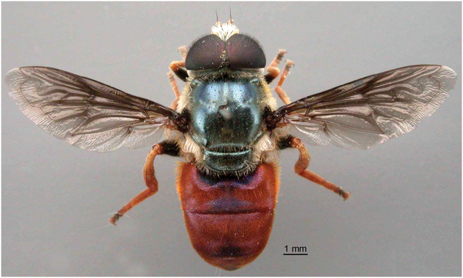
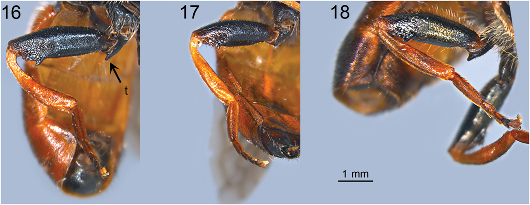
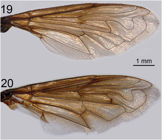
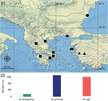
Diagnosis. Very distinctive species, clearly recognisable in the field by its mostly red terga (Fig. 15) and reddish-orange tip of femora, tibiae, and tarsi (except two or three apical segments dark) (Figs. 16–18). Medium-sized species with metallic blue-green mesonotum and predominantly reddish-brown, oval abdomen with purple lustre. Wings brown, darkened especially basally, anteriorly, and along the veins (Figs. 19–20). Species subgroup restricted to southeastern Europe (Romania, Greece) and the western part of Turkey (Fig. 21). The M. dobrogensis species subgroup comprises the M. dobrogensis species complex and the species M. rojoi.
Habitus of Merodon puniceus, male, dorsal view (from Radenković et al. Reference Radenković, Vujić, Ståhls, Pérez-Bañón, Rojo, Petanidou and Šimić2011: 55, fig. 66).

Metathoracic leg, lateral view. 16, Merodon rojoi male; 17, M. rojoi female; 18, M. dobrogensis male (genitalia dissected). t, calcar on metatrochanter.

Right wing, dorsal view, male. 19, Merodon dobrogensis; 20, M. puniceus.

Distribution range of the Merodon dobrogensis species subgroup. 21, Species distribution: •, M. dobrogensis (n = 31); ▴, M. puniceus (n = 16); ▪, M. rojoi (n = 25); 22, variability plot of species elevational gradients (n = 31).

Merodon dobrogensis species complex
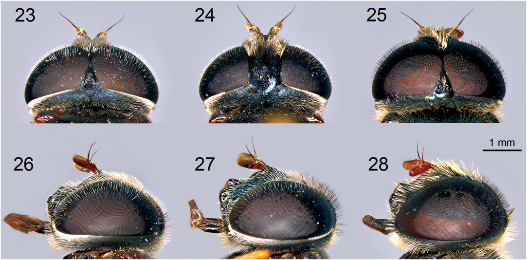
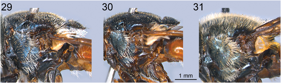
Diagnosis. Eyes completely covered with black pile (Figs. 25, 28), as long as pedicel (grey and short in M. rojoi). Pile on mesoscutum longer than pedicel, white, except for a patch of black pile near wing basis (Figs. 15, 31). Posterior quarter of sternum IV in the male covered with pale pile.
Species distributed in Eastern Mediterranean and Balkan Peninsula. The complex comprises two species: Merodon dobrogensis and M. puniceus. Morphological differences between the species are small.
Radenković et al. (Reference Radenković, Vujić, Ståhls, Pérez-Bañón, Rojo, Petanidou and Šimić2011) stated that Merodon puniceus is very similar to M. dobrogensis except for the denser and longer pile on its mesonotum, more darkened wing especially along the veins, and in the female with more dense pile on the eyes. However, these differences actually apply to M. puniceus and M. rojoi, because Radenković et al. (Reference Radenković, Vujić, Ståhls, Pérez-Bañón, Rojo, Petanidou and Šimić2011) had misidentified specimens from the continental part of Greece as M. dobrogensis because they did not have the type material of the real M. dobrogensis from Romania for comparison. Based on the analysis of two available populations of both species, the only morphological difference is the infuscation of the wings: much darker in M. puniceus than in M. dobrogensis (Figs. 19–20).
Merodon dobrogensis Bradescu, Reference Bradescu1982
Merodon dobrogensis Bradescu, Reference Bradescu1982: 13.
Type locality. Romania: Plateau of Dobrudja, Hagieni (100 m).
Type specimens examined. Holotype: male, 27.viii.1977, (MGAB); Paratype: female 30.viii.1977, V. Bradescu (FSUNS).
Material examined: Greece. Evros, Mesembria: 6♂♂, 2.x.2011, 127 m, Michael de Coursy (MCWC T1-T3, T14-T16); 3♂♂, 8.ix.2013, 127 m (FSUNS AL47, AL49, AL53); Romania: Mangalia: 15♂♂, near Hagieni, 28.ix.2014, 58 m, A. Vujić, J. Ačanski, L. Šašić Zorić (FSUNS 08498-08512); 1♂, near Liman, 28.ix.2014, 30 m, A. Vujić, J. Ačanski, L. Šašić Zorić (FSUNS 08497); 5♂♂, near Albes,ti, 29.ix.2014, 50 m, A. Vujić, J. Ačanski, L. Šašić Zorić (FSUNS 08493-08496, 08513).
Diagnosis. Differs from Merodon puniceus by the infuscation of the wings (lighter in M. dobrogensis, and much darker in M. puniceus; see Figs. 19–20), molecular data (COI and ND4 gene sequence variability; see Figs. 11–12), by morphometric characters of wing and male genitalia (Figs. 2, 7), and distribution. It is geographically separated from M. puniceus, being distributed in southeastern Europe near the Black Sea and Aegean Sea (Fig. 21).
Natural history. Preferred environment: forest; small, open areas maintained by goat grazing, in evergreen Quercus coccinea Münchhausen (Fagaceae) matorral on limestone. Flowers visited: Prospero autumnale (Linnaeus) Speta (Asparagaceae). Flight period: August to October. Larvae unknown (Speight Reference Speight2018).
Merodon puniceus Vujić, Radenković, and Pérez-Bañón, 2011
Merodon sp. nova 4: Ståhls et al. Reference Ståhls, Vujić, Pérez-Bañón, Radenković, Rojo and Petanidou2009: 1436–1437.
Merodon puniceus Radenković et al. Reference Radenković, Vujić, Ståhls, Pérez-Bañón, Rojo, Petanidou and Šimić2011: 48.
Type locality. Greece: Lesvos.
Type specimens examined. Holotype: ♂, Greece: Lesvos, Agiassos, 14.x.2005, 760 m, C. Perez-Bañón (MAUA 0023677). Paratypes: Greece: Lesvos: 1♀, Agiassos, 8.x.2005, 760 m, C. Perez-Bañón (MAUA 0023663); 1♂, Polichnitos, 24.x.2008, 37 m, A. Vujić (FSUNS 04448); 1♂, River Almiropotamos, 1.7 km WWN Vatera, from fennel, 8.x.2008, 55 m, M. Hull (WML 1771).
Additional material. Greece: Lesvos, Agiassos: 1♀, vi.2005, C. Perez-Bañón, S. Rojo, G. Ståhls (FSUNS); 2♀♀, 10.iv.2007, T. Petanidou (CEUA); 1♂, 23.iv.2007, C. Perez-Bañón (CEUA); 1♀, 27.iv.2007, T. Petanidou (CEUA). Turkey: 1♂, Mountain near Izmir, 16.ix.2013, A. Vujić (FSUNS AL29); Bozdag Mountain: 2♂♂, Sart to Bozdag 1, 16.ix.2013, 1320 m, A. Vujić (FSUNS AJ8, AJ13); 4♂♂, Bozdag 2, after Allahdiyen, 12.x.2014, 1070 m, A. Vujić, S. Šimić, S. Radenković (FSUNS 08675, 08677, 08682, 08687); 1♂, Bozdag 3, 12.x.2014, 1229 m, A. Vujić, S. Šimić, S. Radenković (FSUNS 08668).
Diagnosis. Differs from Merodon dobrogensis by the infuscation of the wings (much darker in M. puniceus and lighter in M. dobrogensis; see Figs. 19–20), by morphometric characters of wing and male genitalia (Figs. 2, 7), distribution in western Anatolia and Lesvos Island (Figs. 21–22). The two species differ also based on COI and COI+ND4 tree topologies (Figs. 11–12).
Natural history. Preferred environment: forest; small, open areas in Castanea Miller (Fagaceae) and evergreen Quercus Linnaeus and Pinus Linnaeus (Pinaceae) forests. Flowers visited: Prospero autumnale, Foeniculum vulgare Miller (Apiaceae). Flight period: spring (April–June) and autumn (September–October). Larva unknown (Speight Reference Speight2018).
Merodon rojoi Radenković and Vujić, new species
http://zoobank.org/urn:lsid:zoobank.org:act:F2208118-FA92-4F29-834D-2307F7FA0665
Type specimens. Holotype: ♂, Greece: Mountain Erymanthos, Kalentzi, 37.95039ºN, 37.95039ºE, 2.ix.2012, 1212 m, A. Vujić (NMW). Paratypes: Greece: 1♂, Euboea, Nea Artaki, 18.ix.1982, Bette (BMNH) (misidentified as M. dobrogensis in Radenković et al. Reference Radenković, Vujić, Ståhls, Pérez-Bañón, Rojo, Petanidou and Šimić2011); 2♀♀, Attiki, Dafni, 10 km W of Athena, 28.ix.1985, 199 m (RMNH 03074) (misidentified as M. dobrogensis in Petanidou et al. Reference Petanidou, Vujić and Ellis2011 and Radenković et al. Reference Radenković, Vujić, Ståhls, Pérez-Bañón, Rojo, Petanidou and Šimić2011); 1♂, Argolida, Korfos-Epidavros, 15–27.ix.1986, 136 m, Wolf (RMNH 04157) (misidentified as M. dobrogensis in Radenković et al. Reference Radenković, Vujić, Ståhls, Pérez-Bañón, Rojo, Petanidou and Šimić2011); 8♂♂, 10♀♀, Mountain Erymanthos, Kalentzi, 2.ix.2012, 1212 m, A. Vujić (FSUNS G2374-G2384, G2386-G2391, G2393); 1♀, Mountain Pindos, above Katara, 3.ix.2012, 1200 m, A Vujić (FSUNS G2392); 1♀, Mountain Olympus (on Prospero autumnale), 22.ix.2013, 632 m, A. Vujić (FSUNS AL12).
Diagnosis. Eyes covered with sparse, grey pile, shorter than pedicel (Figs. 23, 26), mostly black in the Merodon dobrogensis species complex (as on Figs. 25, 28). In the male, pile on scutum is as long as pedicel; mostly pale, with two maculae of black pile around wing basis and transverse suture; in the female, short black pile (as long as half of pedicel) covers most of the scutum (Figs. 29–30). Wing infuscation similar to M. dobrogensis. Posterior quarter of sternum IV in the male with sparse, black pile. Additionally, it differs from taxa belonging to the M. dobrogensis species complex by morphometric characters of wing and male genitalia (Figs. 3–4, 8–9), distribution (Fig. 21), COI and COI+ND4 tree topologies (Figs. 11–12), COI and ND4 unique haplotypes, as well as unique 28S rRNA gene genotype. Differs from M. dobrogensis in its elevational range (Fig. 22).
Head, the Merodon dobrogensis species subgroup. 23, Merodon rojoi, male dorsal view; 24, M. rojoi, female dorsal view; 25, M. dobrogensis, male dorsal view; 26, M. rojoi, male lateral view; 27, M. rojoi, female lateral view; 28, M. dobrogensis, male lateral view.

Mesoscutum, lateral view. 29, Merodon rojoi, male; 30, M. rojoi, female; 31, M. dobrogensis, male.

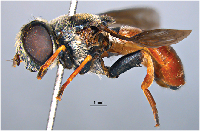
Description. Male (Fig. 32): Head (Figs. 23, 26): Antenna (Fig. 26) reddish brown, basoflagellomere 2.5 times as long as wide, 1.6–1.7 times longer than pedicel, concave, and darkened dorsally, apex rectangular; arista dark brown and thickened basally, slightly shorter than the basoflagellomere. Face and frons black with blue lustre, covered with long whitish-yellow pile. Oral margin bare with black lustre. Vertical triangle isosceles, shiny black, predominantly covered with black pile, and posteriorly with long, white pile. Ocellar triangle equilateral. Eye contiguity about 11 ommatidia long. Vertical triangle: eye contiguity: ocellar triangle = 1.5:1.5:1. Eye pile sparse, grey. Occiput with whitish-yellow pile, along the eye margin with dense white microtrichia and posteriorly with metallic bluish-green lustre.
Habitus of Merodon rojoi, male, lateral view.

Thorax: Postpronotum with conspicuous tooth-like process posteriorly. Scutum and scutellum black with metallic blue lustre, covered with short, erect white to yellow pile, except black pile around wing basis and transverse suture. Posterior anepisternum, anepimeron, and posterodorsal part of katepisternum with long whitish-yellow pilosity and metallic blue lustre. Wing with dense microtrichia, brown, infuscate especially basally, anteriorly and along veins; veins dark brown except light brown C, Sc, and basal part of R1. Calypter grey yellow. Halter with light brown pedicel and dark brown capitulum. Femora dark brown with pale apex; profemur and mesofemur with a stripe of light yellow pile posteriorly and very short, black pile anteriorly and dorsally; metafemur with very short, black pile. Tibiae and tarsi orange, except 2 (3) apical tarsomeres darkened (fourth tarsomere distinctly brown), covered with yellow pilosity and some black pile. Metatrochanter with an inner calcar with two pointed angles.
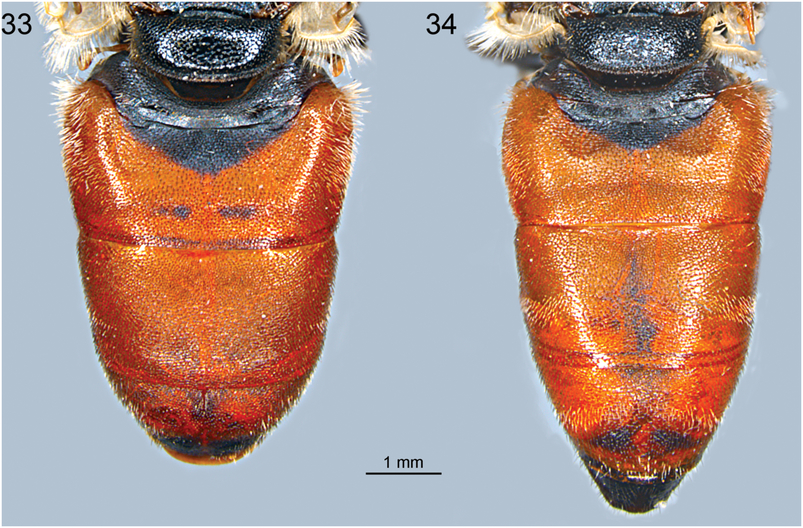
Abdomen (Fig. 33): Oval, slightly longer than mesonotum; reddish brown; tergum I black with golden lustre; tergum II reddish-brown with small black marking medially, and two lateral indistinct white microtrichose spots near the black marking; tergum III reddish brown; tergum IV reddish brown, black posteriorly; pile on terga predominantly short golden, except black pile on black markings and posterior margins of terga II and III. Sterna translucent, from reddish yellow to reddish brown towards the end of abdomen, black lined, covered with long whitish-yellow pile, except few black pile on posterior quarter of sternum IV.
Male genitalia: Similar to all other species of the Merodon aureus species group (Figs. 13–14): posterior surstylar lobe with parallel margins and rounded apex (Fig. 13) and a narrow, elongate, sickle-shaped hypandrium without lateral sclerite of aedeagus (Fig. 14).
Female. (Figs. 17, 24, 27, 34): Similar to the male except for the normal sexual dimorphism and for the following characteristics: metatrochanter (Fig. 17) without thorn; black markings on terga (Fig. 34) narrower or absent; tergum II with more conspicuous two lateral white microtrichose spots and terga III and IV with distinct stripes of white pilosity; tergum V and sternum V black. Length: body 11–12 mm, wing 8–9 mm.
Merodon rojoi, abdomen, dorsal view. 33, Male; 34, female.

Etymology. The new species is named after the Spanish entomologist Santos Rojo and is dedicated to his comprehensive work on syrphids. Additionally, “rojo” in Spanish language means red, indicating the red abdomen in this species.
Natural history. Preferred environment: forest; small, open areas in evergreen Quercus and Pinus forests. Flowers visited: Prospero autumnale. Flight period: Autumn (September). Larva unknown.
Discussion
With their red abdomen, species from the Merodon dobrogensis species subgroup have clearly different morphological features compared to all other species within the M. aureus species group. Merodon dobrogensis was described from Romania (Bradescu Reference Bradescu1982), and for a long time, there were no additional records of this species. Radenković et al. (Reference Radenković, Vujić, Ståhls, Pérez-Bañón, Rojo, Petanidou and Šimić2011) described the morphologically very similar species M. puniceus from the island of Lesvos, but since there were no genetic data on M. dobrogensis, it was impossible to unambiguously resolve the taxonomy of this species. Continental material from central, south, and west of Greece, referred to as M. dobrogensis in Petanidou et al. (Reference Petanidou, Vujić and Ellis2011) as well as Radenković et al. (Reference Radenković, Vujić, Ståhls, Pérez-Bañón, Rojo, Petanidou and Šimić2011), are the here-described M. rojoi. During 2013 and 2014, we collected new specimens of M. dobrogensis in Romania and eastern Greece and here we confirm that they belong to one nominal species based on molecular data, morphology, wing geometric morphometry, and distribution. Additionally, we validate M. puniceus as a separate species based on genetic, wing, and surstylus morphometry differentiations. These two species form the M. dobrogensis species complex.
New distributional data of Merodon puniceus have been recorded in the western part of the Anatolian Peninsula, representing the first known mainland sites for this species. Previously, this species has been listed as endemic for Lesvos Island, Greece (Radenković et al. Reference Radenković, Vujić, Ståhls, Pérez-Bañón, Rojo, Petanidou and Šimić2011). The specimens from Turkey are firstly assigned to M. puniceus based on the similarity of DNA barcode sequences to one from Lesvos Island (Supplementary Table S1), which is then supported based on the integrative taxonomy approach applied here. The distributional pattern of M. puniceus is in agreement with the earlier study of Merodon fauna in the Aegean region (Vujić et al. Reference Vujić, Petanidou, Tscheulin, Cardoso, Radenković and Ståhls2016). Namely, the fauna of large islands not very remote from the nearest mainland, such as Lesvos or Samos, is closely related to the adjacent mainland fauna. These two islands host approximately half of the Merodon species previously classified as Anatolian (Ståhls et al. Reference Ståhls, Vujić, Petanidou, Cardoso, Radenković and Ačanski2016; Vujić et al. Reference Vujić, Petanidou, Tscheulin, Cardoso, Radenković and Ståhls2016).
Merodon rojoi, which is morphologically similar to both abovementioned taxa, has been discovered in the central, south, and west parts of Greece. This species has some unique morphological features that separate it from the M. dobrogensis species complex, and it is resolved as a separate monophyletic clade, supported with high bootstrap values on COI and COI+ND4 trees. Additionally, it has unique COI and ND4 haplotypes as well as 28S rRNA genotype. Moreover, M. rojoi differs significantly in its wing and surstylus shape from species from the M. dobrogensis species complex, with an excellent percentage of correct species classification by both discriminant analyses (both wing and surstylus 100%) and the Gaussian-naïve Bayes classifier analyses (wing 97%; surstylus 100%). Apart from the significant differences in wing and surstylus shapes among all the three species, the position of specimens in both canonical variate analysis scatter plots clearly illustrates the division of the M. dobrogensis species subgroup into M. rojoi and M. dobrogensis species complex. This segregation is also noticeable when comparing species mean wing and surstylus shapes, with the lowest differences being within the M. dobrogensis species complex.
In general, molecular findings support wing and surstylus shape differentiation among species from the Merodon dobrogensis species subgroup. These results are not surprising, given that congruence between molecular and geometric morphometrics results in the recent taxonomic studies of syrphid species becomes standard (Nedeljković et al. Reference Nedeljković, Ačanski, Vujić, Obreht, Djan, Ståhls and Radenković2013, Reference Nedeljković, Ačanski, Djan, Obreht-Vidaković, Ricarte and Vujić2015; Vujić et al. Reference Vujić, Radenković, Likov, Trifunov and Nikolić2013; Ačanski et al. Reference Ačanski, Vujić, Djan, Obreht Vidaković, Ståhls and Radenković2016; Šašić et al. Reference Šašić, Ačanski, Vujić, Ståhls, Radenković and Milić2016, Reference Šašić, Ačanski, Đan, Kočiš Tubić, Veličković, Radenković and Vujić2018; Chroni et al. Reference Chroni, Grković, Ačanski, Vujić, Radenković and Veličković2018; Kočiš Tubić et al. Reference Kočiš Tubić, Ståhls, Ačanski, Djan, Obreht Vidaković and Hayat2018; Radenković et al. Reference Radenković, Šašić Zorić, Djan, Obreht Vidaković, Ačanski and Ståhls2018). Moreover, based on both simple and partial Mantel tests, there is no significant correlation of wing and surstylus shape and genetic differentiation with climate and geographic proximity among the investigated species.
The low average uncorrected p distances (p COI, p ND4) between the two species from the Merodon dobrogensis species complex suggest that they diverged recently and probably during the late Pleistocene. However, a high COI sequence divergence between the M. rojoi and M. dobrogensis complex species indicates a much earlier divergence of the M. rojoi lineage from the M. dobrogensis complex ancestor lineage. The speciation process is probably related to the complex geological history of the Aegean region and severe climatic oscillation during the Pleistocene epoch, as has been suggested for many other Merodon taxa inhabiting the Mediterranean and sub-Mediterranean regions (e.g., Petanidou et al. Reference Petanidou, Vujić and Ellis2011; Ačanski et al. Reference Ačanski, Vujić, Djan, Obreht Vidaković, Ståhls and Radenković2016; Šašić et al. Reference Šašić, Ačanski, Vujić, Ståhls, Radenković and Milić2016; Vujić et al. Reference Vujić, Petanidou, Tscheulin, Cardoso, Radenković and Ståhls2016; Radenković et al. Reference Radenković, Šašić Zorić, Djan, Obreht Vidaković, Ačanski and Ståhls2018). Also, it may be connected to their larval natural history/host plant relationships, which remain unknown.
The applied integrative approach once again proved useful in resolving the taxonomy of Merodon species. The M. dobrogensis species subgroup was successfully resolved using molecular data, geometric morphometry, and distributional data in addition to morphological character states analyses.
Acknowledgements
This work was funded by the Ministry of Education, Science, and Technological Development of the Republic of Serbia (grant numbers: OI173002 and III43002), the Provincial Secretariat for Science and Technological Development (0601-504/3) and H2020 Project “ANTARES” (664387).
Supplementary material
To view supplementary material for this article, please visit https://doi.org/10.4039/tce.2019.72.