Introduction
The cobia Rachycentron canadum (Linnaeus, Reference Linnaeus1766) is an extant carangiform fish, the sole representative of the monotypic family Rachycentridae (Nelson, Reference Nelson2006; Sajeevan and Madhusoodana Kurup, Reference Sajeevan and Madhusoodana Kurup2014). Cobias are marine teleosts that occur nearly worldwide in a variety of tropical, subtropical, and warm temperate habitats and occasionally enter estuaries and other brackish-water environments feeding on crustaceans, cephalopods, and other fishes (Shaffer and Nakamura, Reference Shaffer and Nakamura1989). Other than the review by Carnevale and Godfrey (Reference Carnevale and Godfrey2018) of a single partially complete specimen from the Calvert Formation referred to Rachycentron sp., the genus apparently has no fossil record based on skeletal remains. Neither is it known on the basis of fossil otoliths (Nolf, Reference Nolf2013). Here, we describe a new fossil species of the genus Rachycentron from the Tortonian of the St. Marys Formation, Calvert Cliffs, Calvert County, Maryland, USA, on the basis of a single nearly complete neurocranium. This fossil therefore represents the second known record of the family Rachycentridae, thereby providing relevant information about the evolutionary history of these fishes.
Geology
The specimen derives from the Little Cove Point Member of the Miocene St. Marys Formation from north of Driftwood Beach along Calvert Cliffs, Maryland, USA. The Miocene deposits of the Atlantic Coastal Plain of North America include the siliciclastic sediments of the St. Marys Formation. These fossiliferous shallow marine deposits constitute the uppermost portion of the Neogene Chesapeake Group as exposed in the Calvert Cliffs along the Chesapeake Bay shoreline in Calvert County (Shattuck, Reference Shattuck, Clark, Shattuck and Dall1904; Kidwell, Reference Kidwell and Schlee1984; Kidwell et al., Reference Kidwell, Powars, Edwards and Vogt2015). The deposits of the St. Marys Formation are located along the western margin of the Salisbury Embayment, a paleogeographic domain that was occupied by a shallow arm of the Atlantic Ocean during the Miocene (Ward and Andrews, Reference Ward and Andrews2008), structurally representing a westward extension of the Baltimore Canyon Trough (see Poag, Reference Poag1979; Kidwell, Reference Kidwell and Schlee1984; Kidwell et al., Reference Kidwell, Powars, Edwards and Vogt2015). Overall, the Miocene succession of the Chesapeake Group records a gradual shallowing within the Salisbury Embayment (e.g., Kidwell, Reference Kidwell and Schlee1984, Reference Kidwell1988, Reference Kidwell1989, Reference Kidwell1997; Ward and Strickland, Reference Ward, Strickland and Poag1985; Ward, Reference Ward1992; Ward and Andrews, Reference Ward and Andrews2008). In this context, the deposits of the St. Marys Formation (specifically, the Little Cove Point Member) are quite different from those of the Calvert and Choptank formations, which are the other two Miocene formations making up Calvert Cliffs. Sediments of this St. Marys Formation member show more rapid lateral (and vertical) facies changes and include facies from shallower water and more tide-dominated environments, reflecting a transition from open marine to tidally influenced low salinity muddy embayments (Kidwell, Reference Kidwell1988, Reference Kidwell1997; Kidwell et al., Reference Kidwell, Powars, Edwards and Vogt2015). The age of the Little Cove Point Member has been estimated at 9–10 Ma (Perez et al., Reference Perez, Godfrey, Kent, Weems and Nance2019, fig. 1), which is entirely within the Tortonian stage.
The St. Marys Formation has yielded a rich record of marine and terrestrial organisms (Vogt and Eshelman, Reference Vogt, Eshelman and Roy1987; Carnevale and Godfrey, Reference Carnevale and Godfrey2018; Godfrey, Reference Godfrey2018). A large number of predominantly marine species have been described from these deposits, with representatives from several groups of microfossils and invertebrates as well as an array of vertebrate taxa, including sharks and rays, actinopterygian fishes, turtles, crocodiles, pelagic birds, seals, sea cows, odontocete and mysticete cetaceans, and rare isolated remains of large terrestrial mammals (e.g., Clark et al., Reference Clark, Shattuck and Dall1904; Ward and Andrews, Reference Ward and Andrews2008; Carnevale et al., Reference Carnevale, Godfrey and Pietsch2011; Godfrey, Reference Godfrey2018).
Remains of actinopterygian fishes are relatively common in the St. Marys Formation, consisting of isolated bones, partially articulated skeletons, teeth, and otoliths (Carnevale and Godfrey, Reference Carnevale and Godfrey2018). However, until recently, our knowledge of the ichthyofaunal assemblage was limited to short descriptions of isolated remains incidentally collected from several localities in Maryland and Virginia (Cope, Reference Cope1867, Reference Cope1869; Leidy, Reference Leidy1873; Eastman, Reference Eastman, Clark, Shattuck and Hall1904; Smith, Reference Smith1909; Lynn and McLelland, Reference Lynn and Melland1939; Blake, Reference Blake1940; Leriche, Reference Leriche1942; Dante, Reference Dante1953; Kimmel and Purdy, Reference Kimmel, Purdy, Ward and Krafft1984; Weems, Reference Weems1985). More recently, Müller (Reference Müller1999) provided a comprehensive overview of the otolith assemblage. Finally, on the basis of the analysis of the skeletal remains in the collections of the Calvert Marine Museum, Solomons, Maryland, and the Department of Paleobiology, National Museum of Natural History, Smithsonian Institution, Washington, DC, Carnevale and Godfrey (Reference Carnevale and Godfrey2018) presented a broad overview of the fish diversity of the successions of the Chesapeake Group, recognizing on the basis of skeletal elements 19 genera from the St. Marys Formation. Thirty-six genera were reported from the St. Marys Formation on the basis of the presence of otoliths (Müller, Reference Müller1999; Carnevale and Godfrey, Reference Carnevale and Godfrey2018). Carnevale and Godfrey (Reference Godfrey2018, fig. 3.8) also described a set of associated bones (CMM-V-3694), (sclerotic ring, right premaxilla, left maxilla, left suspensorium and preopercle, right second pharyngobranchial, right posttemporal, vertebrae) from the Calvert Formation that were referred to an indeterminate species of the genus Rachycentron.
Materials and methods
The nearly complete neurocranial remains were collected on 27 and 28 April 2019 from the St. Marys Formation outcropping along Calvert Cliffs, Maryland. Detailed osteological examinations were made possible following the removal of the surrounding matrix. This was achieved using needles working under a stereomicroscope. Measurements were taken with a caliper to the nearest 0.1 mm. To improve contrast in the figures, the specimens were lightly dusted with sublimed ammonium chloride. After the specimens were photographed under fluorescent lighting, the ammonium chloride was removed by rinsing them under running water.
Repositories and institutional abbreviations
Type, figured, and one other specimen examined in this study are deposited in the Calvert Marine Museum comparative osteology collection (CMM-O) and vertebrate paleontology collection (CMM-V), Solomons, Maryland, USA.
Systematic paleontology
Division Percomorphacea Wiley and Johnson, Reference Wiley, Johnson, Nelson, Schultze and Wilson2010
Order Carangiformes Jordan, Reference Jordan1923
Family Rachycentridae Gill, Reference Gill1895
Genus Rachycentron Kaup, Reference Kaup1826
Type species
Rachycentron canadum (Linnaeus, Reference Linnaeus1766).
Rachycentron stremphaencus new species
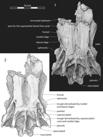
Rachycentron stremphaencus n. sp., CMM-V-10,000, neurocranium: (1) dorsal view; (2) interpretive drawing. Specimen dusted with sublimed ammonium chloride to improve contrast.

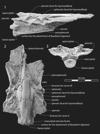
Rachycentron stremphaencus n. sp., CMM-V-10,000, neurocranium: (1) right lateral view; (2) ventral view; (3) posterior view. Specimen dusted with sublimed ammonium chloride to improve contrast.

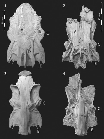
(1, 3) Neurocranium of Rachycentron canadum, CMM-O-0063: (1) dorsal view; (3) ventral view. (2, 4) Neurocranium of Rachycentron stremphaencus n. sp., CMM-V-10,000: (2) dorsal view; (4) ventral view. The letters A–E enumerate the major differences between the neurocranium of Rachycentron canadum and that of Rachycentron stremphaencus n. sp. Specimens dusted with sublimed ammonium chloride to improve contrast.

Holotype
CMM-V-10,000, relatively complete neurocranium lacking the ethmoid portion and the anterior half of the basicranium (Figs. 1, 2, 3.2, 3.4).
Diagnosis
A Rachycentron species with outer surface of the bones of the skull roof smooth or scarcely ornamented; skull roof with two longitudinal bony ridges, a lateral ridge formed by the frontal and pterotic and a flat medial ridge formed by the frontal, parietal, and epioccipital bordering a shallow medial frontal depression; sphenotic located in the posterior half of the neurocranium and not extending laterally at the level of the pterotic; epioccipital not extending posteriorly to the level of the articular facet of the exoccipital; trough formed by the medial ridges on either side of the supraoccipital; intercalar prong apparently absent.
Occurrence
Calvert Cliffs north of Driftwood Beach, Chesapeake Ranch Estates, Calvert County, Maryland, USA. Shattuck Zones 22–23, Little Cove Point Member of the St. Marys Formation, Tortonian Stage, late Miocene. GPS coordinates are approximately 38.3538591°N, 76.3929062°W.
Description
Measurements (in millimeters): neurocranial length = 119.0; minimum neurocranial width across the frontals at the supraorbital margin of the orbit = 46.5; maximum neurocranial width across the pterotics = 67.0; neurocranial width across the epioccipitals = 39.0; neurocranial width across the exoccipitals = 22.5.
CMM-V-10,000 consists of a dorsoventrally compressed neurocranium (Figs. 1, 2). Overall, the lateral profile of the neurocranium is wedge shaped, deepest at the back of the skull. The width of the skull across the pterotics is approximately equal to 1.9 times the maximum height of the back of the neurocranium (which is 35 mm deep). There are two paired continuous bony ridges and a median supraoccipital crest (damaged in CMM-V-10,000) on the dorsal face of the neurocranium. The lateral ridge is formed by the frontal, sphenotic, and pterotic; the medial ridge is formed by the frontal, parietal, and epioccipital. The lateral ridge arises at the level of the anterior third of the orbit and continues posteriorly along the central margin of the pterotic, ending as the posterolateral corner of the neurocranium. The flattened medial ridge begins slightly anteriorly, runs longitudinally through the whole length of the frontal, and continues posteriorly on the parietal and epioccipital. In the posterior third of the neurocranium, a deep and oblong trough is bounded by the lateral and medial ridges. Another shorter trough is demarcated by the medial ridge and the supraoccipital crest.
The right lateral ethmoid is partially preserved in the specimen along the ventral surface of the neurocranium (Fig. 2); it is a wing-like flattened bone, almost quadrangular in shape, with a smooth ventral surface. The lateral ethmoid articulates posteriorly with the frontal.
The frontals are the largest bones of the skull roof. They comprise more than half of the length of the preserved portion of the neurocranium and the full width of the neurocranium anteriorly. There is a shallow medial depression that reaches its maximum depth at the level of the suture of the two contralateral frontals. The contralateral pores of the supraorbital lateral-line canal can be observed in the anterior portion of this longitudinal depression. Lateral to the medial ridge, the lateral ridge stands above the wide supraorbital margin of the frontal, and a deep furrow separates the lateral ridge from the supraorbital margin of the frontal. In ventral view (Fig. 2.2), the frontal exhibits a relatively smooth and wide supraorbital surface. Each frontal articulates anteriorly with the lateral ethmoid, posterolaterally with the sphenotic and pterotic, posteriorly with the parietal and supraoccipital, and ventromedially with the pterosphenoid.
The parietal occurs on the lateral side of the supraoccipital and articulates anteriorly with the frontal, medially with the supraoccipital, posteriorly with the epioccipital, and posterolaterally with the pterotic. The supraoccipital occupies the median posterodorsal portion of the skull roof and supports a shallow and relatively thin supraoccipital crest (Fig. 1); the supraoccipital crest is incomplete posteriorly, but in life, it would have risen above the exoccipitals.
From the posterolateral margin of the frontal, the tongue-shaped sphenotic projects laterally to form the dorsolateral margin of the orbit. In dorsal view, a ridge crosses the bone diagonally (in an anteromedial to posterolateral direction) to join with one on the pterotic. Medial to this ridge, the sphenotic preserves the anterior end of a wedge-shaped depression that broadens posteriorly onto the pterotic and is bounded medially by the bold ridge of the epioccipital. In lateral and ventral views, the sphenotic displays a posteroventrally directed deep cavity that represents the articular surface for the anterior dorsal head of the hyomandibula (Fig. 2.1, 2.2).
The sphenotic articulates posteriorly with the pterotic and ventrally with the prootic.
The pterotic forms the posterolateral corner of the neurocranium. In dorsal view, it bears a posterolaterally projecting tubular ridge; medial to this ridge, the surface of the bone is depressed into the longitudinal furrow that started on the sphenotic; ventrally, the pterotic has a deep articular surface for the posterior dorsal head of the hyomandibula. The pterotic articulates medially with the epioccipital and posteriorly with the exoccipital.
The two contralateral epioccipitals are separated from each other by the supraoccipital. Each epioccipital is robust, is expanded posteriorly, and forms the posterior portion of the medial ridge of the dorsal face of the neurocranium (Fig. 1); the epioccipital portion of the medial ridge splits the difference in distance between the supraoccipital crest and the lateral ridge on the pterotic.
The exoccipitals are visible in dorsal, ventral, and posterior views (Figs. 1, 2). Posteriorly, each exoccipital bears a shallow and medially oriented concave facet that overhangs the basioccipital for the articulation with the fovea of the first vertebra. Ventrally, each exoccipital shows a large foramen for the nervus vagus. The basioccipital occupies the posteroventral portion of the neurocranium. In posterior view, the basioccipital is concave and subcircular in outline. The attaching surface for Baudelot's ligament is well recognizable ventrolaterally in the posterior sector of this bone. There is a 2 mm wide trough that separates the basioccipital from the exoccipitals (Fig. 2.3).
In a ventral view of CMM-V-10,000, the parasphenoid is deeply sutured to the basioccipital, showing no clear physical separation between these two bones. The elongate parasphenoid is unremarkable in its smooth ventral surface except for a medially situated shallow longitudinal trough in the midsection of its preserved length. The intercalar is difficult to recognize. The bilaterally swollen prootics are the largest bones of the lateral wall of the neurocranium; they are preserved dorsolateral to the anterior end of the preserved length of the parasphenoid (Fig. 2.2). The structure of the trigemino-facialis chamber is difficult to determine although a large facial foramen can be easily recognized. Each prootic contacts the sphenotic dorsolaterally and the pterotic posterodorsally, the exoccipital and basioccipital posteriorly, and the parasphenoid ventrally. The pterosphenoid articulates dorsally with the frontal and sphenotic, and posteriorly with the prootic.
Etymology
The specific epithet honors Stephen Groff and Marcus Jones, who co-discovered the specimen. The name ‘stremphaencus’ is derived from a combination of the letters of ‘Stephen’ and ‘Marcus’ with some rearrangement to make the specific name mellifluous.
Remarks
Despite being represented solely by a single partially complete neurocranium, the attribution of CMM-V-10,000 to the family Rachycentridae and, more particularly to the genus Rachycentron, is clearly justified by its overall morphology and configuration, as well as by the mutual relationships of the recognizable skull bones (see O'Toole, Reference O'Toole2002). In particular, the remarkable posterior elongation of the epioccipitals and the medially oriented articular facets of the exoccipitals are considered as diagnostic of the genus Rachycentron (O'Toole, Reference O'Toole2002; Friedman et al., Reference Friedman, Johanson, Harrington, Near and Graham2013). Compared with the extant cobia Rachycentrum canadum, the neurocranium of R. stremphaencus n. sp. exhibits a number of differences that unquestionably support its assignment to a new previously undescribed species. These differences mostly regard the relative proportions and development of the neurocranial portions, which are well exemplified in Figure 3. First, the external bony texture of the skulls is broadly different (Fig. 3, feature A). While that of the extant cobia consists of fine to bold anastomosing sharp ridges, the outer surface of the neurocranial bones of Rachycentron stremphaencus n. sp. is nearly smooth or scarcely ornamented.
In CMM-V-10,000, the lateral ridge is pronounced, resulting in a considerable gradient from the dorsal-medial exposure of the frontal to its lateralmost supraorbital margin (Fig. 3, feature B). In the extant cobia, the typical carangoid lateral ridge is weakly developed, and the frontal presents a gently curving outer surface.
The size, shape, and relative position of the sphenotic are conspicuously different between these two taxa (Fig. 3, feature C). In Rachycentron stremphaencus n. sp., the sphenotic is located in the posterior half of the neurocranium, and its lateralmost edge is adjacent to the anteriormost extent of the wedge-shaped trough in the dorsal surface of the skull formed by the lateral and medial ridges. In Rachycentron canadum, the sphenotic extends laterally at the same level of the pterotic and located further forward on the skull, approximately at the midpoint of its maximum length.
In Rachycentron stremphaencus n. sp., the two contralateral medial ridges form a proportionately much wider trough on either side of the supraoccipital than in Rachycentron canadum, in which the epioccipitals are much elongated posteriorly, reaching the rear edge of the neurocranium (Fig. 3, feature D). Finally, in Rachycentron canadum, there is a small but conspicuous posterolateral prong of the intercalar, which is apparently not present in Rachycentron stremphaencus n. sp. (Fig. 3, feature E).
Although the partial remains of one individual of Rachycentron have been described from the Calvert Formation (Carnevale and Godfrey, Reference Carnevale and Godfrey2018), because there was no neurocranium preserved in that specimen, there is no way of knowing whether the Calvert cobia is conspecific with the one described herein from the St. Marys Formation. In comparing the fossil Rachycentron remains from the Calvert Formation with those of extant cobia, significant differences were not found.
As documented in the preceding, a set of associated bones (sclerotic ring, right premaxilla, left maxilla, left suspensorium and preopercle, right second pharyngobranchial, right posttemporal, vertebrae) from the Calvert Formation has been assigned by Carnevale and Godfrey (Reference Carnevale and Godfrey2018) to an indeterminate species of Rachycentron. Unfortunately, it is not possible to provide a reliable scrutiny of the potential affinities between Rachycentron stremphaencus n. sp. and the fossil cobia from the Calvert Formation due to the absence of comparable structures. As a consequence, any possible attribution of the fossil cobia from the Calvert Formation to the new species described herein must be excluded until additional comparable material would be available.
In summary, on the basis of the ecological requirements of its living congenerics (e.g., Shaffer and Nakamura, Reference Shaffer and Nakamura1989) and the paleoenvironmental reconstructions of the depositional context of the St. Marys Formation (Kidwell, Reference Kidwell1988, Reference Kidwell1997; Carnevale et al., Reference Carnevale, Godfrey and Pietsch2011), it is reasonable to conclude that Rachycentron stremphaencus n. sp. was a predatory fish that inhabited the subtropical to warm temperate Atlantic coast of North America during the late Miocene, where it occurred in shallow marine and paralic waters, in a way similar to the other fossil and extant cobias.
Acknowledgments
We are grateful to S. Groff, M. Jones, and J.R. Nance for having quarried the remains of the fossil cobia neurocranium described herein. We are further obliged to J.R. Nance (Paleontology Collections Manager, Calvert Marine Museum, Solomons, MD) for access to fossil and extant material in his care. We are delighted to acknowledge A.F. Bannikov and one anonymous reviewer for offering constructive comments on our manuscript. Furthermore, we wholeheartedly thank associate editor A. Murray and journal editor H.-D. Sues for shepherding this work through the review and publication process. Research by S.J.G. was made possible by funding from the County Board of Calvert County Commissioners, the citizens of Calvert County, and the Clarissa and Lincoln Dryden Endowment for Paleontology at the Calvert Marine Museum. The research of G.C. was supported by grants (ex-60% 2020) from the Università degli Studi di Torino.