Introduction
Lichen planus is a common mucocutaneous disorder affecting approximately 0.5–1 per cent of the world's population.Reference Mollaoglu1 It can affect the skin or mucous membranes of any part of the body. Most cutaneous lesions are seen on the flexor surface of the wrists, lower back, ankles and scalp. Common mucosal sites include the oral cavity (especially the buccal mucosa, hard palate and lips), oropharynx, oesophagus, the vulvovaginal region in females and the penis in males.
The larynx has been reported as a rare site of mucosal lesions. A Medline and Pubmed search (using the key words ‘larynx’ and ‘lichen planus’) identified only a single published case of lichen planus involving the laryngeal mucosa, reported in Russian.Reference Kunel'skaia and Arievich2 We could find no previous publications in the English language literature.
Case
A 57-year-old Caucasian man presented with a two-month history of hoarseness of voice. He was noted to be a heavy drinker and smoker, drinking 80 units of alcohol per week and smoking 30 cigarettes a day. He reported no pain or abnormal swallowing. There was no history of weight loss, fever, anorexia, dysphagia, odynophagia, blood-tinged sputum, chronic cough or gastric reflux. His past medical history was otherwise unremarkable, and there was no relevant family or medication history.
Examination of the oral cavity, oropharynx, nose and neck was unremarkable.
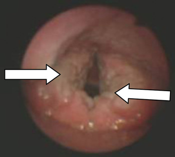
Fibre-optic laryngoscopy showed bilaterally oedematous false vocal folds and arytenoids. The laryngeal mucosa was non-homogeneous both in colour and texture. There was marked cicatrisation and hypertrophy of both false vocal folds and arytenoids; cicatrisation was symmetrical and more noticeable in the mid-portion of both false folds. The true vocal folds were normal in appearance and mobility.
Systemic examination was unremarkable, and there were no other mucocutaneous lesions elsewhere in the body.
Routine blood investigations and chest radiography were within normal limits.
The patient underwent direct laryngoscopic examination, which confirmed the above findings (Figure 1).
Direct laryngoscopic view showing marked cicatrisation of both false vocal folds and arytenoids (arrows).

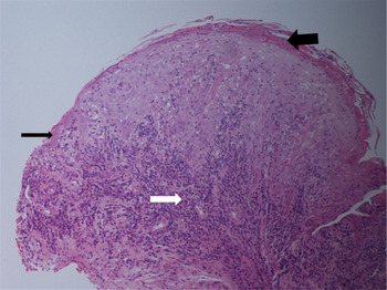
Histopathological examination of biopsy specimens revealed degeneration and atrophy of the basal epithelial layer, with marked hyperkeratosis and parakeratosis. Abundant lymphocytic infiltration and fibrinous exudates were found, indicating an underlying chronic inflammatory process. Lymphocytes were found to be infiltrating into the subepithelial tissue (Figure 2). Cellular architecture was normal and fungal staining was negative, ruling out dysplasia and fungal infection, respectively.
Photomicrograph showing degeneration and atrophy of basal layers (thin black arrow), as well as hyperkeratosis and hyperparakeratosis with fibrinous exudates (thick black arrow) rich in lymphocytes infiltrating the subepithelial tissue (white arrow), indicating an underlying chronic inflammatory process. (H&E; ×100)

Based on the above findings, a diagnosis of lichen planus was made.
Since the patient’s only symptom was a hoarse voice, he was advised to commence and maintain good oral hygiene, and to cease his tobacco and alcohol intake. He was also given a two-week course of oral steroids at a dose of 40 mg/day.
After two weeks, the patient reported some improvement of his voice, although there were no visible changes in his larynx. Considering this partial response, oral steroids were continued for another four weeks (30 mg/day for two weeks then 20 mg/day for two weeks), before being gradually tapered off over the next two weeks. At this stage, there was a noticeable change in the patient's voice and laryngeal appearance.
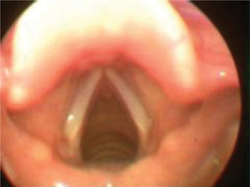
At subsequent follow-up visits, the patient's voice was much improved, as was the appearance of his larynx (Figure 3). The patient was kept under regular follow up, and at the time of writing had remained disease-free for two years.
Direct laryngoscopic appearance 12 months post-treatment, showing a near-normal larynx.

Discussion
Lichen planus is characterized by cutaneous and mucosal lesions that have a tendency to heal with scarring. Stricture formation has been described in sites such as the oesophagus, lacrimal ducts and pharynx.Reference Setterfield, Black and Challacombe3 About one-third of cases present with cutaneous disease only, one-third involve both the skin and the mucous membranes, and the remaining one-third have mucous membrane lesions as the only manifestation. In the natural course of the disease, cutaneous lesions resolve spontaneously in six to 18 months in 85 per cent of cases, but lesions involving the oral cavity, genitals and other mucosal sites often persist longer. Oral lichen planus is a relatively common inflammatory disorder of stratified epithelia, with a prevalence of 0.5–2.2 per cent in the adult population.Reference Setterfield, Black and Challacombe3 The condition classically appears in the fifth to sixth decade of life, and is twice as common in women than men. However, no such epidemiological evidence is available for laryngeal lichen planus. Our understanding is that both oral and laryngeal lichen planus affect mucous membranes and have similar clinical behaviour. Therefore, we believe that both should be grouped together as mucosal lichen planus.
The exact aetiology of lichen planus is still unknown. An autoimmune aetiology triggered by viral infection or drugs has been suggested. The pathogenesis is complex and involves cluster of differentiation 8+ glycoprotein T lymphocytes, mast cells, intercellular adhesion molecule-1 and major histocompatibility complex class II antigens. The immune process results in apoptosis of basal cells, which ultimately leads to liquefaction of the whole basal cell layer.Reference Kim, Chae, Cho, Kim, Kim and Kim4 Triggering factors implicated in the development of lichen planus include systemic drugs (including non-steroidal anti-inflammatory drugs, sulphonylureas and some angiotensin-converting enzyme inhibitors),Reference McCartan and McCreary5 contact-sensitivity reactions to dental amalgam,Reference Koch and Bahmer6 mechanical trauma (Koebner phenomenon) from sharp teeth,Reference Scully, Beyli, Ferreiro, Ficarra, Gill and Griffiths7 candida infection, chronic hepatic disease (e.g. hepatitis C virus infection, autoimmune chronic active hepatitis and primary biliary cirrhosis)Reference Korkij, Chuang and Soltani8 and smoking.Reference Scully, Beyli, Ferreiro, Ficarra, Gill and Griffiths7
Mucosal lichen planus may present in a wide variety of ways and can cause pain, burning, soreness and change in the function of the affected site. Alternatively, it may be completely asymptomatic. In the majority of cases, it is detected as an incidental finding on routine examination.
If symptomatic, the clinical presentation of mucosal disease depends on the anatomical region of involvement. Oral lesions present with soreness, pain and burning, while anogenital lesions present with pruritis. Oesophageal lesions are known to cause dysphagia, while laryngeal lesions may result in hoarseness of voice, as seen in the present case.
On physical examination, oral lesions are most commonly found on the buccal mucosa,Reference Silverman and Bahl9 and are characterized by white or grey streaks forming a linear or reticular pattern on a red-purple background (Wickham striae). These lesions frequently affect multiple sites within the same anatomical region and usually have a symmetrical distribution.
No such definite pattern of lesions could be found in the present case. However, our patient's lesions were symmetrical in distribution with marked cicatrisation and hypertrophy, representing a chronic destructive disease process with partial healing.
The differential diagnosis of mucosal lichen planus includes leukoplakia, fungal infection, carcinoma in situ and frank carcinoma. Although unusual in the western world, laryngeal tuberculosis should also be borne in mind, especially in developing countries.
Chronic mucosal lesions have a tendency to malignant transformation. Reported rates of malignant transformation of oral lichen planus vary from 1.2 to 5.3 per cent. However, no such data are available for laryngeal lichen planus. It is therefore suggested that such patients be followed closely.Reference Silverman and Bahl9 Risk factors implicated for malignant change include smoking, excessive alcohol consumption, and atrophic, ulcerative or erosive disease.Reference Lo Muzio, Mignogna, Favia, Procaccinic, Testaa and Bucci10
The definitive diagnosis of lichen planus is made based on the typical clinical appearance together with the characteristic histopathological appearance of the lesion biopsy specimen. Characteristic histopathological findings comprise a band-like infiltrate of lymphocytes at the dermo-epidermal junction, and damage to the basal cell layer. Characteristic colloid or civatte bodies (degenerated keratinocytes) also appear at the dermo-epidermal junction, especially in cases of cutaneous lichen planus. In mucosal lichen planus, lymphocyte and plasma cell infiltration is more prominent.
• Although rare, laryngeal lichen planus should be considered in the differential diagnosis of chronic hoarseness, especially in the presence of triggering factors or a family history of lichen planus
• Pre-malignant change and fungal infection should be excluded
• A short course of oral steroids may be curative
• Regular follow up is required to detect and manage any malignant change at an early stage
Management of mucosal lichen planus is two-fold, consisting of general measures and specific medical management. General measures include avoidance of potential triggering factors, hygiene maintenance and treatment of coexisting fungal infection. Corticosteroids remain the mainstay of medical management of lichen planus. Asymptomatic patients with non-erosive mucosal disease require no treatment, while steroids are the mainstay of treatment for erosive mucosal lesions. In accessible oral or genital lesions, topical corticosteroids are effective in most patients. However, a few patients may develop candidiasis. Therefore, some clinicians also prescribe prophylactic antifungalsReference Carbone, Conrotto, Carrozzo, Broccoletti, Gandolfo and Scully11 along with topical steroids. A comparative study of topical versus oral steroid treatment showed no difference in response.Reference Carbone, Goss, Carrozzo, Castellano, Conrotto and Broccoletti12
Systemic corticosteroids are used when topical treatment fails, or in cases with erosive or widespread lesions. Patients with inaccessible lesions (e.g. oesophageal or laryngeal lichen planus) or refractory genital lesionsReference Scully and Carrozzo13 are also managed with systemic corticosteroids. The usual dose is 0.5–1.0 mg/kg/day for two to 12 weeks, depending on treatment response.Reference Lozada-Nur and Miranda14, Reference Boyd and Neldner15 In steroid-dependent patients, and those in whom steroids are poorly tolerated or contraindicated, azathioprine may be prescribed. In refractory cases, tacrolimus, dapsone, antimalarials and thalidomide may be considered,Reference Kaliakatsou, Hodgson, Lewsey, Hegarty, Murphy and Porter16, Reference Camisa and Popovsky17 but there has been little evaluation of their efficacy.Reference Scully and Carrozzo13
Acknowledgement
The authors would like to thank Dr Hilary Birch from the Department of Histopathology for her assistance.