Introduction
Toxocara canis (dog roundworm) and Toxocara cati (feline roundworm) are parasites of wide geographic distribution. Human infection (toxocariasis) occurs through accidental ingestion of embryonated eggs found in contaminated soil (geophagia) or by eating contaminated raw vegetables. It is well known that eosinophilia is associated with parasitic diseases, including toxocariasis (Garcia, Reference Garcia2001). Data on T. canis seroprevalence differ between countries. Studies in Europe have shown prevalence rates from 2 to 5% in apparently healthy adults in urban areas compared with 14 to 37% in rural areas (Magnaval et al., Reference Magnaval, Glickman, Dorchies and Morassin2001). Children are more frequently affected than adults (Magnaval et al., Reference Magnaval, Glickman, Dorchies and Morassin2001).
We analysed the seroprevalence of T. canis infection in healthy Croatian children with peripheral blood eosinophilia.
Material and methods
A total of 142 serum samples collected from asymptomatic children with peripheral blood eosinophilia (more than 500 eosinophils/μl blood) in the Zagreb area between 2004 and 2007 were included in the study.
Serum samples were tested using a commercial semiquantitative enzyme-linked immunosorbent assay (ELISA) for the detection of IgG antibodies to T. canis (NovaTec Immundiagnostica GmbH, Germany) according to the manufacturer's recommendations. Initially reactive samples were confirmed using a T. canis Western blot (WB) test (LDBIO diagnostics, Lyon, France) according to the manufacturer's recommendations.
Data analysis was performed using a chi-square test, and Fisher's exact test when more than 20% of expected values were < 5. A P value less than 0.05 was considered to be significant.
Results
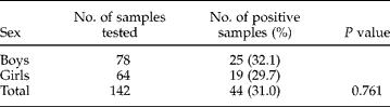
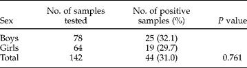
All 142 patients were Caucasians between 3 and 18 years of age. There were 78 (54.2%) boys and 64 (45.8%) girls. Overall 31% (44/142) serum samples were positive by WB for T. canis IgG antibodies. Two serum samples positive by ELISA were negative by WB. Both showed low positive results in the ELISA test. The T. canis seroprevalence rate was 32.1% (25/78) among boys and 29.7% (19/64) among girls, which was not statistically different (χ2 = 0.092, P = 0.761) (table 1).
Seroprevalence of T. canis in children with eosinophilia.
P = χ2 test.
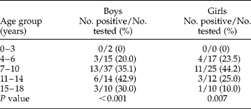
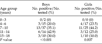
The seroprevalence rate according to age groups is shown in table 2. There was a statistically significant difference in T. canis seropositivity between age groups, both in boys (Fisher's exact test P < 0.001) and girls (Fisher's exact test P = 0.007). In boys, the highest seroprevalence (42.9%) was reported in the 11- to 14-year age group. The majority of the seropositive girls (44.2%) were in the 7- to 10-year age group.
Seroprevalence of T. canis according to age.
P = Fisher's exact test.
Discussion
Although it is known that eosinophilia is associated with T. canis infection (Garcia, Reference Garcia2001), there have been few reports on the seroprevalence of toxocariasis in asymptomatic and symptomatic people with eosinophilia.
In Croatia, no serological studies have been conducted on the relationship between toxocariasis and eosinophilia in children. Our study was carried out on 142 healthy children with peripheral blood eosinophilia. The overall seroprevalence rate of T. canis was 31%. Although Toxocara antigens have been reported to cross-react with other helminthic antigens, cross-reaction was ruled out using a WB test as a confirmatory test (Magnaval et al., Reference Magnaval, Fabre, Maurieres, Charlet and de Larrad1991; Garcia, Reference Garcia2001).
Similar studies on the seroprevalence of toxocariasis have reported rates of 0–4.2% (Fenoy et al., Reference Fenoy, Cuellar and Guillen1996) and 3.1–57.1% (Cilla et al., Reference Cilla, Pérez-Trallero, Gutiérrez, Part and Gomáriz1996) in Spain, 1.9% in Italy (Di Fiore et al., Reference Di Fiore, Virga, Usticano, Di Rosa and Rini1989), 2.1% in Germany (Kimmig et al., Reference Kimmig, Naser and Frank1991), 11% in The Netherlands (Van Gemund et al., Reference Van Gemund, Buijs, Van Dongen and Van den Bergh1989), 13.9% in the United States (Won et al., Reference Won, Kruszon-Moran, Schantz and Jones2008), and up to 24.6% in the Czech Republic (Uhlíková & Hübner, Reference Uhlíková and Hübner1998). The seropositivity in 343 tested Polish children (Zarnowska et al., Reference Zarnowska, Borecka, Gawor, Marczyńska, Dobosz and Basiak2008) was much higher, reaching 73% (249/343 children).
Like most of the similar studies worldwide (Herrmann et al., Reference Herrmann, Glickman, Schantz, Weston and Domanski1985; Buijs et al., Reference Buijs, Borsboom, Van Gemund, Hazebroek, Van Dongen, Van Knapen and Neijens1994; Dogan et al., Reference Dogan, Dinleyici, Bor, Özensoy Töz and Özbel2007), our results showed no difference in T. canis seropositivity between boys (32.1%) and girls (29.7%) (P = 0.761). However, Theodoridis et al. (Reference Theodoridis, Frydas, Papazahariadou, Hatzistilianou, Adamama-Moraitou, Di Gioacchino and Felaco2001) reported that females were significantly more frequently infected than males. In contrast, studies conducted in Ireland (Holland et al., Reference Holland, O'Lorcain, Taylor and Kelly1995) and the Czech Republic (Uhlíková & Hübner, Reference Uhlíková and Hübner1998) showed higher Toxocara seropositivity in male children.
The Toxocara seroprevalence in our study varied significantly between age groups both in boys (P < 0.001) and girls (P = 0.007). Similar results were reported in Ireland (Holland et al., Reference Holland, O'Lorcain, Taylor and Kelly1995), although some studies did not find that age was an important factor related to T. canis seroprevalence (Herrmann et al., Reference Herrmann, Glickman, Schantz, Weston and Domanski1985; Ljungström & Van Knapen, Reference Ljungström and Van Knapen1989). Alonso et al. (Reference Alonso, Bojanich, Chamorro and Gorodner2000) found the highest seroprevalence of toxocariasis in 11- to 12-year-old children (57.1%), while Cilla et al. (Reference Cilla, Pérez-Trallero, Gutiérrez, Part and Gomáriz1996) showed that 6- to 10-year-olds were at the highest risk. We found the highest seroprevalence in boys aged 11–14 (42.9%) and girls aged 7–10 (44.2%).
The main causes of eosinophilia are infectious diseases, especially parasitic infections, allergic diseases, malignancies, drug hypersensitivity reactions and idiopathic hypereosinophilic syndrome. Helminths, which include nematodes, cestodes and trematodes, are typically associated with eosinophilia. Marked eosinophilia is usually seen when helminths invade and migrate through tissues. A wide clinical examination and laboratory analysis in patients with eosinophilia is needed to exclude or confirm other possible causes. This would prevent misdiagnosis that might result in inappropriate clinical interventions.
Humans are considered an incidental host of T. canis and become involved in its life cycle by consuming infective eggs from the environment. Toxocara eggs can survive in the soil for months to years, depending on environmental conditions. An important risk factor for toxocariasis is pica, specifically geophagia (Garcia, Reference Garcia2001). Children are most commonly infected, likely due to unintentional consumption of soil. Contact with puppies is another known risk factor for infection.
A great majority of T. canis infections have no manifest clinical symptoms. None of our patients had any symptoms, either. An accidental lab report for eosinophilia was the reason why they were tested for T. canis infection.
Due to an unexcluded possibility of infection by another parasite, as well as other potential cause of eosinophilia, it was impossible to confirm with certainty that T. canis was, in fact, the cause of eosinophilia in our positive patients. However, it may be concluded that this study has substantiated the hypothesis that toxocariasis is an infection occurring in Croatia. Furthermore, due to a high seroprevalence rate in children and its potential complications, toxocariasis represents an important public health problem. Consequently, it was deemed crucial to either exclude or confirm T. canis as the causative agent of eosinophilia.
Education of veterinarian and medical professionals, as well as the general public, is crucial for the prevention of this disease. Fortunately, T. canis infection can be prevented by simple measures. Pet owners need to be educated about the hazards of Toxocara, be proactive in taking their pets to veterinarians for regulary deworming and treatment, and dispose of pet litter appropriately. Children should not have contact with infected dogs, and should avoid contaminated environments, especially sandboxes.