Case description
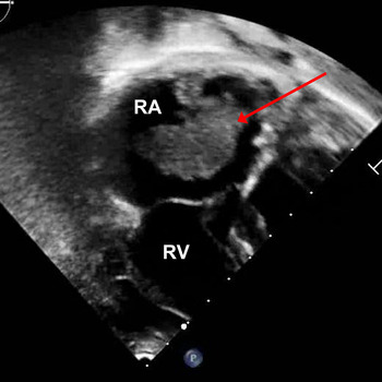
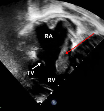
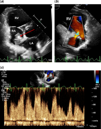
A 15-year-old male recently diagnosed with scimitar syndrome underwent infradiaphragmatic baffling of the scimitar vein to left atrium through an intra-atrial tunnel and patch augmentation of the inferior vena cava using PhotoFix® bovine pericardium. Routine 3-month follow-up echocardiogram revealed the appearance of a large, mobile right atrial thrombus measuring 3 × 1.7 cm adherent to the patch and prolapsing through the tricuspid valve annulus in diastole (Fig 1; Supplementary Clip S1). Surveillance echocardiogram 1 month prior (Supplementary Clip S2) was unremarkable while on prophylactic warfarin, which was started immediately post-operatively and continued to the 3-month follow-up given the extent of the baffle repair. Trials of tissue plasminogen activator and heparin infusions failed to disintegrate the intracardiac thrombus (Fig 2), and he ultimately underwent surgical thrombectomy while leaving the bovine pericardium in place. Two months after thrombectomy, the patient again presented with extensive thrombus burden in both atria (measuring 2 × 1.2 cm on the right atrial side, Fig 3a), and with a new development of systemic and pulmonary venous obstruction (Fig 3b and c; Supplementary Clips S3, S4). He returned to the operating room based on echocardiographic imaging and underwent explantation of the bovine pericardium and reconstruction of the scimitar baffle, augmentation of the right lower pulmonary vein, inferior vena cava, and hepatic veins with Gore-Tex patch. The patient underwent a hypercoagulable evaluation, which resulted negative (including evaluation of protein S and C, Factor V Leiden, Factor VIII, prothrombin 20210A, antiphospholipid antibody panel, and antithrombin III). One-year follow-up demonstrates a normal appearance of the patch by both echocardiogram and cardiac MRI while on therapeutic warfarin and aspirin. Post-operative venous obstruction is common after scimitar vein surgery regardless of surgical technique and frequent imaging surveillance is required, an important consideration that should guide management decisions surrounding corrective surgery. Reference Dusenbery, Geva and Seale1 Given the post-operative complications in this case after using bovine pericardium, our surgical preference is to use a Gore-Tex patch when extensive baffle repair is expected.
Echocardiogram (apical view) at 3 months post-operative with large right atrial thrombus (red arrow) attached to the atrial septum. RA=right atrium; RV=right ventricle.

Echocardiogram (apical view) after tissue plasminogen activator depicting pedunculated thrombus in right atrium (red arrow) prolapsing through the tricuspid valve (TV) in diastole. RA=right atrium; RV=right ventricle.

( a ) Echocardiogram (parasternal short-axis view) depicting a large broad-based thrombus (red arrow) adherent to the right atrial (RA) side of the venous baffle with mural thrombus on the left atrial (LA) side 5 months from the initial surgery. RV=right ventricle. ( b ) Echocardiogram (parasternal long-axis view) demonstrating colour flow acceleration in the systemic venous pathway in the right atrium. RV=right ventricle. ( c ) Pulsed-wave spectral Doppler profile of the systemic baffle obstruction in the right atrium.

Supplementary material
To view supplementary material for this article, please visit https://doi.org/10.1017/S1047951121000822.
Acknowledgements
None.
Financial support
This research received no specific grant from any funding agency, commercial or not-for-profit sectors.
Conflicts of interest
None.
Ethical standards
The authors assert that all procedures contributing to this work comply with the ethical standards of the relevant national guides. This work does not include any experimentation on human or animal subjects.