Introduction
Chicken primordial germ cells (PGCs) are a type of primary undifferentiated pluripotent stem cells that will differentiate towards gametes: spermatozoa or oocytes. PGCs are formed very early in avian development. These cells can be isolated from the embryonic circulatory system and gonads and successfully cryopreserved as genetic resources for future use (Naito et al., Reference Naito, Tajima, Tagami, Yasuda and Kuwana1994; Song et al., Reference Song, D’Costa, Pardue and Petitte2005; Nakamura et al., Reference Nakamura, Usui, Ono, Takeda, Nirasawa, Kagami and Tagami2010, Reference Nakamura, Usui, Miyahara, Mori, Ono, Kagami, Takeda, Nirasawa and Tagami2012). Several histochemical markers are used for PGC identification such as periodic acid–Schiff (PAS) stain (Ji et al., Reference Ji, Guan, Gao, Li, Bai, Ma and Li2016). This reagent stains glycogen or immunohistochemical cell surface antigens such as embryonic mouse antigen‐1 (EMA‐1; Wang et al., Reference Wang, Chen, Chen, Peng, Zhou, Liao and Lu2016) or stage‐specific embryonic antigen‐1 (SSEA-1; Mozdziak et al., Reference Mozdziak, Angerman-Stewart, Rushton, Pardue and Petitte2005). Recently, aldehyde dehydrogenase (ALDH) has received considerable attention as a reliable stem cell marker (Armstrong et al., Reference Armstrong, Stojkovic, Dimmick, Ahmad, Stojkovic, Hole and Lako2004; Cai et al., Reference Cai, Weiss and Rao2004; Corti et al., Reference Corti, Locatelli, Papadimitriou, Donadoni, Salani, Del Bo, Strazzer, Bresolin and Comi2006; Douville et al., Reference Douville, Beaulieu and Balicki2009; Gentry et al., Reference Gentry, Foster, Winstead, Deibert, Fiordalisi and Balber2007; Ginestier et al., Reference Ginestier, Hur, Charafe-Jauffret, Monville, Dutcher, Brown and Schott2007). ALDH family members are strongly active in normal tissue stem cells, therefore they may be considered a marker for stem cells and play a functional role in terms of self-protection, differentiation or expansion of stem cell populations (Tomita et al., Reference Tomita, Tanaka, Tanaka and Hara2016). The ALDEFLUOR assay detects intracellular ALDH expression in viable cells using the green fluorescence channel of a standard flow cytometer. Because ALDEFLUOR-positive cells are viable and sortable using flow-cytometric cell sorting based on their fluorescence, they are readily available for both in vitro and in vivo stem cell studies (Tomita et al., Reference Tomita, Tanaka, Tanaka and Hara2016). The objective of our study was to determine the ‘stemness’ of chicken PGCs using the ALDEFLUOR kit and flow cytometry.
Materials and methods
Gonadal PGC isolation
The experiment using ALDH to detect PGCs was replicated four times (from May to June). Freshly laid Oravka chicken (n=42) eggs (n=200) were obtained from a breeding farm (Lužianky, Nitra, Slovak Republic) and used as the PGC donors. The eggs were incubated at 37.8°C and 80% relative humidity with tilting at a 90° four times per day in a forced air incubator (MG100/150 B.Family) for 5.5 days. Gonads were separated from the mesonephros using microinstruments (needles, tweezer and scissors) under a stereomicroscope and placed in 200 µl minimum essential medium (MEME; Sigma Aldrich, Germany). The PGCs were isolated from the gonads by disaggregation in 0.25% trypsin–0.02% EDTA in Hank’s balanced salt (HBS) solution (Sigma Aldrich, Germany) at 37°C for 1 min and the reaction was stopped by the addition of MEME. Subsequently, cell viability using the 0.4% trypan blue (Sigma Aldrich, Germany) exclusion method and activity of ALDH using flow cytometry were assessed.
Flow cytometry assessment using ALDEFLUORTM kit
ALDH activity of fresh PGCs was evaluated using flow cytometry. PGCs were incubated with ALDEFLUOR substrate for 15 min at 37°C, with or without the ALDH inhibitor diethylaminobenzaldehyde (DEAB), according to the manufacturer’s instructions. The stained samples were analysed by flow cytometer (FACSCalibur, BD Biosciences, San Jose, CA, USA). At least 25,000 events (PGCs) were analysed in each sample.
Results
Flow cytometry assessment using the ALDEFLUOR kit
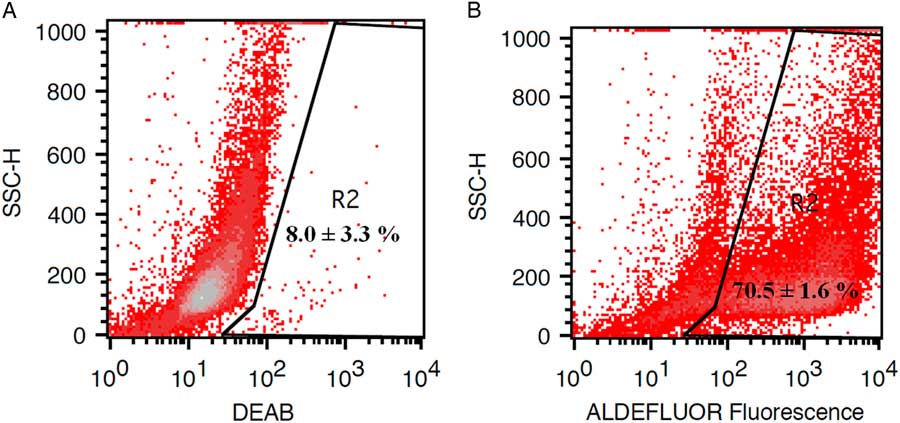
We show, for the first time, that chicken PGCs display high levels of ALDH activity. In our study flow-cytometric analysis the ALDEFLUOR kit was used to determine chicken PGCs stem properties. ALDH activity was analyzed in the cell suspensions dissociated from chicken gonads. A small population was observed upon preincubation of the cells with the specific inhibitor DEAB (Fig. 1A), which deactivated ALDH activity (negative control). However, staining with ALDH (positive cells) caused a fluorescence shift and an increase in the positive cell population (Fig. 1B). The trypan blue exclusion method showed cells with 85.86 ± 1.25% viability.
Representative flow-cytometric analysis of ALDH activity. Baseline fluorescence established in the presence of ALDEFLUOR substrate and the inhibitor DEAB. Gate R2 was defined to DEAB positive (A) and ALDH positive (B) PGCs. Based on the ALDEFLUOR R2 dot plot, cells presented a shift in fluorescence defining the ALDH-positive cell population.

Discussion
ALDEFLUOR is optimized for the detection of ALDH haematopoietic cells in human blood and bone marrow, but it can also be used with non-haematopoietic cells. ALDEFLUOR is a reagent kit that is used to identify stem properties in cells that express high levels of the ALDH enzyme. High levels of ALDH activity in neural cells (Obermair et al., Reference Obermair, Fiorelli, Schroeter, Beyeler, Blatti, Zoerner and Thallmair2010), myogenic cells (Jean et al., Reference Jean, Laoudj‐Chenivesse, Notarnicola, Rouger, Serratrice, Bonnieu, Gay, Bacou, Duret and Carnac2011; Vauchez et al., Reference Vauchez, Marolleau, Schmid, Khattar, Chapel, Catelain, Lecourt, Larghero, Fiszman and Vilquin2009), mammary cells (Starvaggi Cucuzza et al., Reference Starvaggi Cucuzza, Motta, Miretti, Macchi, Martignani, Accornero and Baratta2010), prostate cells (Burger et al., Reference Burger, Gupta, Xiong, Ontiveros, Salm, Moscatelli and Wilson2009) and intestinal crypt cells (Huang et al., Reference Huang, Hynes, Zhang, Ginestier, Dontu, Appelman, Fields, Wicha and Boman2009) have been recorded. Our data indicate that the chicken PGCs express high levels of ALDH activity and that these cells express molecules previously identified in stem cells of different origins. ALDH activity can therefore be used as a new functional marker for the detection of cell ‘stemness’ in chicken PGCs. Further research is necessary to elucidate the role of this functional marker in these cells.
In conclusion, recent evidence suggests that enhanced ALDH activity is a hallmark of stem cells measurable by the ALDEFLUOR assay. While the use of cell surface markers for stem cells identification has met with varying degrees of success, intracellular ALDH activity is emerging as an important and reliable universal stem cell hallmark applicable for most stem cell types, as well as PGCs.
Financial support
The study was financially supported by the Slovak Research and Development Agency (nos. APVV-17-0124), VEGA 1/0611/15, VEGA 1/0160/18 and KEGA 026 SPU-4/2018.
Conflicts of interest
None.
Ethical standards
The authors assert that all procedures contributing to this work comply with the ethical standards of the relevant national and institutional guidelines on the care and use of laboratory animals. Treatment of the animals was approved by the Ministry of Agriculture and Rural Development of the Slovak Republic, no. SK P 28004 and Ro. 1488/06-221/3a.