Introduction
It was suggested long ago that blastozoans (eoccrinoids, blastoids, diploporites, and rhombiferans) and crinoids comprise a monophyletic assemblage, the so-called Pelmatozoa (Leuckart, Reference Leukart1848), largely on the basis of common possession of a superficially similar attachment stalk. Despite doubts about this interpretation that arose during the mid-20th century (Ubaghs, Reference Ubaghs1953, Reference Ubaghs and Moore1968; Sprinkle, Reference Sprinkle1973), this canon was perpetuated in major reference works such as the Treatise on Invertebrate Paleontology and persists in many publications to the present day. Works seeking to separate blastozoans and crinoids as only distantly related pentaradiate forms or to reject the pelmatozoan hypotheses that crinoids and blastozoans share exclusive common ancestry have met with considerable resistance (e.g., Clausen et al., Reference Clausen, Jell, Legrain and Smith2009; Zamora and Smith, Reference Zamora and Smith2012; Kammer et al., Reference Kammer, Sumrall, Zamora, Ausich and Deline2013; Sumrall, Reference Sumrall2017; Sheffield and Sumrall, Reference Sheffield and Sumrall2019a, Reference Sheffield and Sumrallb).
However, even detractors of the idea that crinoids and blastozoans are only superficially similar and do not form an exclusive clade seem to have accepted the importance of the crucial suite of feeding appendage features cited by those who question the validity of Pelmatozoa, or at least argue that any resemblances are superficial (e.g., Mooi and David, Reference Mooi and David1998, Reference Mooi and David2000; David and Mooi, Reference David and Mooi1999; Guensburg and Sprinkle, Reference Guensburg and Sprinkle2007; Guensburg et al., Reference Guensburg, Sprinkle, Mooi, Lefebvre, David, Roux and Derstler2020). For example, proponents for blastozoan ancestry of crinoids posited that an unnamed middle Cambrian blastozoan represented only by disarticulated plates provided evidence that somatocoelar extensions from the main body cavity continued into the arms (Clausen et al., Reference Clausen, Jell, Legrain and Smith2009). However, it is also clear that these supposed blastozoan arms lacked any extraxial elements. The purported coelomic canals pass through floor plates toward the peristome, not through the thecal shoulder, which is an anatomy unlike crinoids. The diminutive nature of these blastozoan canals is also problematic. Other workers cited this and other evidence to reject the idea that any canals within the appendages of blastozoans represented spaces for somatocoelar extensions (Guensburg et al., Reference Guensburg, Mooi, Sprinkle, David and Lefebvre2010). Despite attempts to clarify the issue through precise anatomical descriptions, the debate continues. Here, we shed new light on another purported ‘missing link’ between crinoids and blastozoans.
Recently, a proposal has been published that Eumorphocystis multiporata Branson and Peck, Reference Branson and Peck1940 (hereafter Eumorphocystis Branson and Peck, Reference Branson and Peck1940, because all specimens are conspecific topotypes), a diplopore-bearing blastozoan, represents the nearest-known sister group to crinoids (Sheffield and Sumrall, Reference Sheffield and Sumrall2019b). Evidence central to this suggestion, largely derived from a single specimen, is the description of a longitudinal canal within each of this taxon's feeding appendages. These canals are stated to pierce the theca and connect with the thecal coelomic cavity in a manner similar to that known for crinoids (Sheffield and Sumrall, Reference Sheffield and Sumrall2019b).
Two ancillary putative homologies follow: ‘arm’ construction consisting of triserial axial and extraxial elements, and the presence of ‘radial’ plates from which an aboral uniserial set of ‘brachial’ extraxial elements extends distally out these appendages (here tripartite is used because an aboral series is not in strict one-to-one sequence with the other two). We reconsider and test this proposal using new and existing observations from 12 Eumorphocystis specimens. These data are then compared with an expanded update of early crinoid arm morphology recently made available through examination of the earliest crinoids. These data have only recently been more fully explained in the context of crinoid origins (Guensburg et al., Reference Guensburg, Sprinkle, Mooi, Lefebvre, David, Roux and Derstler2020).
A full understanding of the feeding appendages of Eumorphocystis has been a long-term process, and even now some details, e.g., their full length, remain unknown (Fig. 1). The original description of Eumorphocystis was based on the holotype alone, in which the appendages are broken off close to the theca. This led to the initial conclusion that exothecal feeding appendages were lacking in this taxon. More complete Eumorphocystis specimens provided the first evidence of ‘arms’ (Parsley, Reference Parsley and Sprinkle1982). Initially, these were considered of blastozoan origin and only homoplastic on crinoid arms (Parsley, Reference Parsley and Sprinkle1982). Recently, the discovery that early crinoids express what could also be considered a tripartite pattern (Guensburg and Sprinkle, Reference Guensburg and Sprinkle2009) formed the basis for a reinterpretation that Eumorphocystis ‘arm’ anatomy is homologous to that of early crinoids (Sheffield and Sumrall, Reference Sheffield and Sumrall2019b). In this paper, however, we add descriptive data and imagery that enhance our understanding of, and provide a basis for, interpretation of Eumorphocystis ‘arms’ that is in agreement with the original suggestion that Eumorphocystis is of strictly blastozoan affinity (Parsley, Reference Parsley and Sprinkle1982). The present data confirm most of the basic ‘arm’ construction details, but reveal significant points of departure, particularly at the juncture of arms to theca (Sheffield and Sumrall, Reference Sheffield and Sumrall2019b). This new information forms the core of our reappraisal of the evolutionary significance of Eumorphocystis.
Eumorphocystis multiporata Branson and Peck, Reference Branson and Peck1940, partial reconstruction, included for orientation of subsequent images; from Parsley (Reference Parsley and Sprinkle1982), reproduced by permission, The Paleontological Institute, University of Kansas, Lawrence.

Materials and methods
Specimens used for this study were selected to provide data concerning feeding appendage and associated thecal anatomy. Virtually no additional preparation beyond that already accomplished was needed. Specimens were photographed using a Leica dms 300 digital camera fitted with stacking capability.
The twelve topotypes examined were: OU 9047–9049, OU 238156–238159, 1107TX2, 1279TX126, 1279TX339, 1404TX6, and NPL 93144 (Table 1). We also examined a plaster cast of the holotype (OU 3123). Codings for other taxa used in the phylogenetic analysis were primarily obtained from published sources and checked with specimens in the collections of the Field Museum, Chicago, and the Non-vertebrate Paleontology Laboratory at the University of Texas, Austin.
Eumorphocystis specimens studied, with sizes of specimens, and comments on image orientations used in this study.

All Eumorphocystis specimens were collected from the Lower Echinoderm Zone, Mountain Lake Member, Bromide Formation, Arbuckle Mountains, Oklahoma. Detailed locality data are available from respective repositories. The Bromide Formation is Sandbian, early Late Ordovician, in age.
The phylogenetic analysis builds upon the dataset of Guensburg et al. (Reference Guensburg, Sprinkle, Mooi, Lefebvre, David, Roux and Derstler2020), with the primary goal of further elucidating the phylogenetic position of blastozoans, and in particular Eumorphocystis within this context. The total taxon list from Guensburg et al. (Reference Guensburg, Sprinkle, Mooi, Lefebvre, David, Roux and Derstler2020) was expanded from 21 to 25 (Table 2). Essentially, the entire range of taxa used to support a Eumorphocystis sister-group status with crinoids was implemented (Sheffield and Sumrall, Reference Sheffield and Sumrall2019b). Added taxa include a Middle Ordovician hemicosmitid, a Silurian coronoid blastozoan, both of which also express pseudo-arms, and a diplobathrid crinoid, Gaurocrinus nealli (Hall, Reference Hall1866). Furthermore, we included the Early Ordovician diplobathrid, Proexenocrinus inyoensis Strimple and McGinnis, Reference Strimple and McGinnis1972, which is among the earliest-known pinnulate crinoids. However, Cambrian and Early Ordovician taxa continue to dominate the overall taxon list to code for characters found in the earliest members of the clades to which they belong.
List of taxa used in the phylogenetic analysis (expanded from Guensburg et al., Reference Guensburg, Sprinkle, Mooi, Lefebvre, David, Roux and Derstler2020).

The 39 characters for the phylogenetic analysis were assembled for scoring using Mesquite ver. 3.2 (Maddison and Maddison, Reference Maddison and Maddison2018), build 801, and the NEXUS file run on PAUP ver. 4.0a (Swofford, Reference Swofford2003), build 167, for Macintosh. All characters were unordered and unweighted (Table 3). The analysis employed the branch-and-bound algorithm, consensus trees were computed, and a bootsrap analysis of 10,000 replicates was run using a fast heuristic search.
Matrix used in phylogenetic analysis. − = character state gap; ? = missing data.

Repositories and institutional abbreviations
Eumorphocystis specimens used in this study are housed in the collections of the Sam Noble Museum (OU), University of Oklahoma, Norman, and the Nonvertebrate Paleontology Lab (‐‐‐‐TX‐‐, NPL), University of Texas, Austin.
Preservation and taphonomy
Most Eumorphocystis specimens were surface collected and largely free from enclosing calcareous shale, but a few are preserved on carbonate grainstone surfaces. Most specimens are three dimensional or nearly so, with negligible crushing. No specimen preserved with complete feeding appendages or stems is known. Instead, available material consists of thecae with arms broken off at varying distances from the theca. The different breakage patterns are important for tracing features such as those involved in the transition from theca to appendage. Only a few specimens preserve proximal portions of the stem, with the longest-known example having 31 ‘columnals.’ Specimens show variable amounts of grainy calcite overgrowths and spar infilling of stereom, presumably the result of rapid postmortem cementation. This is not a serious impediment for observing anatomy such as thecal plate sutures, but it can obscure details of microscopic structures germane to assessing the features crucial to determining the evolutionary significance of Eumorphocystis. In some cases, there are no apparent canals in the thecal or near-thecal portions of the ambulacra. In a few cases, specimens corroded by differential dissolution weathering, presumably resulting from more soluble low magnesium cement versus high-magnesium echinoderm stereom, reveal tiny canals. These are continuations of larger feeding appendage canals. In addition, the twelve specimens available furnish data on intraspecific variation and ontogeny. These findings are incorporated into the subsequent analysis.
Character analysis
Here, we provide new information in the form of a character analysis for features of this admittedly contentious fossil, Eumorphocystis. The focus is primarily on feeding appendages but includes observations from adjacent skeletal anatomy as well. This analysis is based on examination of specimens hitherto unexamined by those who have suggested a sister-group relationship to crinoids for this taxon (Sheffield and Sumrall, Reference Sheffield and Sumrall2019b; Deline et al., Reference Deline, Thompson, Smith, Zamora, Rahman, Sheffield, Ausich, Kammer and Sumrall2020). Accompanying reasoning that strongly supports a position for Eumorphocystis contrary to this recent work is presented with this analysis. The cases for or against hypotheses of homology, here and in the opposing viewpoint, depend upon congruency and accepted ontogenetic, morphological, and positional criteria for homology (Patterson, Reference Patterson1988; Freudenstein, Reference Freudenstein2005).
Past criticisms of our conclusions regarding the origins of crinoids (most recently by Sheffield and Sumrall, Reference Sheffield and Sumrall2019b) cited reliance on what were perceived to be a priori assumptions. It was claimed that our work presupposed reasoning or knowledge proceeding from theoretical deduction. However, accepted theories such as the Extraxial-Axial Theory were developed from empirical observations and theoretical induction, not the other way around (Mooi and David, Reference Mooi and David1997). Our aforementioned methodology is brought to bear on hypotheses of homology in a detailed explication, without coding based solely on superficial resemblances of individual features that do not fully consider information gleaned from other sources, including but not restricted to the overall relationships of these features one to another. Our approach has, and continues to be, utilization of these findings to code features, and to test these hypotheses of homology in a full phylogenetic analysis. This approach is integral to the uncovering of phylogenetic signal.
The same authors criticizing our approach relied on analyses that do not provide detailed delineation of character state parameters, full probing of superficial similarity, or support for why a given transformation series should be a part of a given character or carry phylogenetic signal (Kammer et al., Reference Kammer, Sumrall, Zamora, Ausich and Deline2013; Sumrall, Reference Sumrall2017; Wright et al., Reference Wright, Ausich, Cole, Peter and Rhenberg2017; Deline et al., Reference Deline, Thompson, Smith, Zamora, Rahman, Sheffield, Ausich, Kammer and Sumrall2020). Approaches that differ from ours (e.g., Deline et al., Reference Deline, Thompson, Smith, Zamora, Rahman, Sheffield, Ausich, Kammer and Sumrall2020) leave uncited available data, or findings that undermine codings that they favor (e.g., David et al., Reference David, Lefebvre, Mooi and Parsley2000; Guensburg et al., Reference Guensburg, Mooi, Sprinkle, David and Lefebvre2010, Reference Guensburg, Blake, Sprinkle and Mooi2016; Lefebvre et al., Reference Lefebvre, Guensburg, Martin, Mooi, Nardin, Nohejlová, Saleh, Kouraïss, El Hariri and David2019). We prefer a different way of dealing with echinoderm phylogeny, particularly when working with fossils open to more than one interpretation. As stated by Mooi and David (Reference Mooi and David1997, p. 306), “The issue of subjectivity versus objectivity is often raised in reference to character analysis, usually with the implication that it is not objective to try to assess the degree to which we can trust phylogenetic signal from certain features. As cladists interested in quality of data as well as quantity, we are resisting the implication that the more we know about our characters, the less objective the study will be.”
One criticism of our methodology centered on reliance upon differences rather than similarities in our analyses (Wright et al., Reference Wright, Ausich, Cole, Peter and Rhenberg2017, p. 831). This oversimplifies our approach and does not fully recognize the strengths of the phylogenetic method itself. Similarities and differences are nested concepts and provide the basis for evaluation of critical issues concerning homoplasy or homology. Commonality at one level of universality will be a difference at another, and our application of the data that we have gathered recognizes this explicitly. Moreover, our insistence that certain features should not be considered even comparable or coded under the same character system is not founded on a search for differences. We are attempting to address the more profound problem that past nomenclature has reified concepts of similarity that are either inapplicable or violate the central principle that such analyses should capture phylogenetic signal. Our approach employs nuanced and detailed observations drawn from several sources but does not overtly rely on differences. Our evaluations continue to be founded among long established criteria: conjunction, congruence, similarity, and a detailed knowledge of the material at hand (Patterson, Reference Patterson1988; Freudenstein, Reference Freudenstein2005).
We based our characters and codings on analytical data from combined observations accumulated over decades within the framework of established phylogenetic practice (Guensburg, Reference Guensburg2012; Guensburg et al., Reference Guensburg, Sprinkle, Mooi, Lefebvre, David, Roux and Derstler2020) and on empirical observations informed by ontogenetic and anatomical information from a wide variety of sources (partially summarized by e.g., Mooi and David, Reference Mooi and David1998; Mooi et al., Reference Mooi, David and Wray2005), including from extant specimens whose anatomy is frequently ignored in the context of what is plausible among fossil forms. Recent workers have appropriately applied new or previously little-used methodology to the issue of crinoid phylogeny (Ausich, Reference Ausich, Kammer, Rhenberg and Wright2015a; Wright et al., Reference Wright, Ausich, Cole, Peter and Rhenberg2017), but such approaches should incorporate information from other well-founded methodologies including those utilized here and in other works (e.g., Guensburg et al., Reference Guensburg, Blake, Sprinkle and Mooi2016, Reference Ausich, Wright, Cole and Sevastopulo2020).
Here, we start with observations benefiting from anatomical details furnished by the large Eumorphocystis sample size, improved understanding of earliest crinoid morphology, and data from origins of specific body wall regions (e.g., Mooi and David, Reference Mooi and David1997, Reference Mooi and David2000; David et al., Reference David, Lefebvre, Mooi and Parsley2000; Guensburg and Sprinkle, Reference Guensburg and Sprinkle2007, Reference Guensburg and Sprinkle2009; Guensburg, Reference Guensburg2012; Guensburg et al., Reference Guensburg, Blake, Sprinkle and Mooi2016, Reference Guensburg, Sprinkle, Mooi, Lefebvre, David, Roux and Derstler2020). We began with the three homologies proposed to link Eumorphocystis to crinoids (Sheffield and Sumrall, Reference Sheffield and Sumrall2019b) and continued with expanded comparative data from feeding appendages and beyond. These ultimately tested the number and specific kinds of transformations in a series of hypothetical evolutionary events required to support an exclusive link between Eumorphocystis and the common ancestor of Crinoidea.
Coeloms
The central issue and a principal point of departure of the concept that crinoids are a sister group to Eumorphocystis (Sheffield and Sumrall, Reference Sheffield and Sumrall2019b) in this restudy concerns the interpretation of canals associated with feeding appendages. Adding uncertainty to this matter is the scarcity of comparative information for brachiolar and floor plate canals of blastozoans in general (for examples, see Sprinkle, Reference Sprinkle1973; Clausen et al., Reference Clausen, Jell, Legrain and Smith2009). Among early crinoids, these data have only recently been extensively analyzed in a phylogenetic context, although the nature of these canals has long been understood from an anatomical standpoint (Heinzeller and Welsch, Reference Heinzeller, Welsch, Harrison and Chia1994; summarized by Guensburg et al., Reference Guensburg, Sprinkle, Mooi, Lefebvre, David, Roux and Derstler2020).
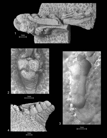
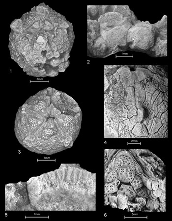
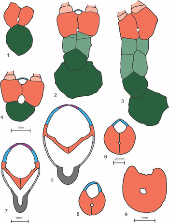
Present evidence shows that longitudinal feeding appendage canals, otherwise termed median canals (Sprinkle, Reference Sprinkle1973), exist in a diversity of blastozoans (gogiids, rhipidocystids, rhombiferans, blastoids) (Fay, Reference Fay1960; Sprinkle, Reference Sprinkle1973; Clausen et al., Reference Clausen, Jell, Legrain and Smith2009; Sumrall and Sheffield, Reference Sheffield and Sumrall2019b) (Figs. 2–4, 6.1–6.4, 6.6, 6.8, 6.9). However, the generally small scales of available material, and the tendency for diagenesis to obscure details with calcitic infilling or to eliminate them through moldic preservation that shows only plate exteriors, combine to contribute to the scarcity of data. Further, material with attached feeding appendages remains unavailable for many blastozoans, and even ambulacra on thecae are not commonly broken through in such a way that might reveal internal canals. Not surprisingly, no comprehensive study of blastozoan median canals is available, and none is documented for most taxa. Blastozoan median canals pass through floor plates and their extensions, the brachiolars, presumably to their tips. They are housed within floor plates, and in nearly all known cases, pass between opposing floor plate elements along the appendage midline. In one case, canals are encased in uniserial floor plates extending from the arms to the theca (Clausen et al., Reference Clausen, Jell, Legrain and Smith2009). The position of the canals and the fact that these seem to extend to the oral region suggest that they housed nerve branches extending from the circumoral ring. This latter anatomical configuration can be observed among living echinoderms.
Eumorphocystis multiporata Branson and Peck, Reference Branson and Peck1940, NPL 93144, large partial theca with two adjacent pseudo-arm stubs, one longer, one shorter: (1) coated oral view showing long arm stub (left), pseudo-arm cover plates, and brachiole facet (just above and to right and above scale); sharp keeled brachioles folded orally; one brachiole nearly complete to tip; proximalmost brachiolar short, wide; distal brachiolars with spinose processes; and tiny biserial cover plates, tapering to sharp termination, nearby thecal plates bearing dense diplopores; (2) coated end-on view of pseudo-arm in (1), broken at seventh floor plate beyond orals; tripartite arrangement; backing plate below; small ambulacral groove and cover plates above, larger ovate canal below; (3) immersed short pseudo-arm stub and nearby theca, broken at distal margin of ?third floor plate from oral, showing (from bottom to top), anchoring thecal plate with diplopores, first backing plate, two sets of two blocky buttress plates forming a solid construct, floor plates surrounding small dark pore (above), and brachiole stubs (see Fig. 6.3); (4) lateral view of pseudo-arm stub in (1) with backing plates (below), buttress plates filling wedge at thecal contact, large block-like, tumid, floor plates, and orally folded brachioles.

Previously reported blastozoan canals are circular openings, on the order of 0.1 mm2, using the area of a circle:
 $${\rm A} = {\rm \pi }{\rm r}^2$$
or smaller in section. These are housed within the floor plates or, in the case of brachiole-bearing blastozoans, the brachiolars. The roughly elliptical appendage cavities seen in Eumorphocystis are larger than those of other blastozoans, being ~1.3 mm2 in section, using the area of an ellipse:
 $${\rm A} = {\rm \pi ab}$$
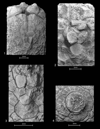
(Fig. 2.2 but see Fig. 3.6 for a much smaller canal). These cavities transition proximally to much smaller, more circular, canals, ~0.14 mm2 in section (Figs. 2.3, 3.2, 3.4, 3.5, 4.2, 4.3), on a scale like those of other blastozoans. These small proximal canals are often obscured by spar filling similar in color to adjacent spar-filled plates, but the implication is that they are very small (Figs. 2.3, 4.1). This narrowing occurs at the second to fourth ambulacrals distal to the orals, except in the C-ray where differentiated floor plates skirt the periproct region. However, in each case, this change takes place not far beyond the thecal wall. Topology in the proximal regions agrees with the most common blastozoan pattern in that the canals run along the perradial sutures (ambulacral midline) that form contacts between opposing floor plates. On the theca itself, canals proceed within floor plates just above, but not through, the thecal wall (Fig. 3.2, 3.5). Opposing floor plate walls each form a hemicanal or ‘half-pipe’ that can be traced proximally (Fig. 3.2, 3.5). These narrow canals were observed to reach the orals (Fig. 3.5). The larger than usual more distal canals could have accommodated expanded innervation supplying the dense array of brachioles.
Eumorphocystis multiporata Branson and Peck, Reference Branson and Peck1940, specimens showing pseudo-arm canal and adjacent morphology: (1, 2) OU 9049: (1) oral view of theca, with plates partly obscured by calcitic overgrowths, theca broken through C ambulacrum; arrow pointing to view of enlargement in (2); (2) magnified view showing exposed suture surface with normally concealed small hemicanal running down ambulacral midline ambulacral midline just above thecal interior (arrow); (3, 5) 1279TX126: (3) oral view of well-preserved theca, thecal shoulder broken through B ambulacrum (arrow) in (5); (5) magnified view of AB oral showing long interradial suture at center-right with vertical ‘half diplopores,’ short suture, BC oral; on left showing hemicanal, concealed in articulated examples, along midline of B ambulacrum (arrow); (4) OU 238157, deeply etched theca, E ray showing apparent small gap between floor plates, likely a weathered pore; (6) 1107TX2, AB interray view, showing face of pseudo-arm broken at fourth floor plate, small canal almost entirely within floor plates.

Eumorphocystis multiporata Branson and Peck, Reference Branson and Peck1940, showing pseudo-arms and stem facet: (1–3) specimens showing small canals near thecal juncture: (1) OU 9048, E ray, canal small, assumed to be spar-filled; (2) OU 238159, small individual lacking buttress plates (see Fig. 6.1); (3) 1404TX6, C ray, intermediate-sized individual with large buttress plates below and small wedge-shaped elements intercalated above, weathered (see Fig. 6.2 for interpretation); (4) OU 238157, stem facet with inset peg and groove crenularium.

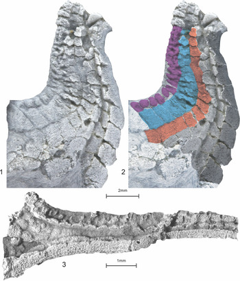
The situation among early crinoids (Figs. 5, 6.5, 6.7) is not comparable to that of Eumorphocystis or any other blastozoan (for detailed analysis, see Guensburg et al., Reference Guensburg, Mooi, Sprinkle, David and Lefebvre2010, Reference Guensburg, Sprinkle, Mooi, Lefebvre, David, Roux and Derstler2020). Unlike blastozoans, crinoid canals expand into the main body mass at the thecal shoulders, away from the peristome (Fig. 5.1–5.3). In addition, the arms themselves express secondary longitudinal grooves within the adoral brachial canals that extend out the arms. Subsequent evolutionary events led to the submergence of this secondary groove into the brachials, thereby transforming what began as grooves into intraplate canals (Guensburg et al., Reference Guensburg, Sprinkle, Mooi, Lefebvre, David, Roux and Derstler2020). Instances of the enclosed canal condition are known from as early as the Late Ordovician (e.g., Columbicrinus Ulrich, Reference Ulrich and Foerste1925; Guensburg et al., Reference Guensburg, Sprinkle, Mooi, Lefebvre, David, Roux and Derstler2020, fig. 7) and occur among all living crinoids. These canals house the brachial (aboral) nerve, part of the subepithelial system sensu Heinzeller and Welsh (Reference Heinzeller, Welsch, Harrison and Chia1994). Ontogeny of living crinoids recapitulates this change in the position of the nerve canal, which was originally only partly submerged into the brachials.
Arm-to-calyx transition of an early crinoid for comparison with Eumorphocystis morphology: (1, 2) Apektocrinus ubaghsi Guensburg and Sprinkle, Reference Guensburg and Sprinkle2009, 1983TX1, D ray arm trunk-to-calyx transition: (1) original image; (2) color overlay interpretation, color coding (also used in Fig. 6) for specific body wall regions; (3) Aethocrinus moorei Ubaghs, Reference Ubaghs1969, uncertain orientation; lateral plate field collapsed indicating expanding arm coelomic cavity merging with main thecal cavity; floor and cover plates visible above, brachials below. blue = axial, lateral cover plates; gray (light) = perforate extraxial laterals; gray (dark) = imperforate extraxial brachials; orange = axial floor plates; purple = axial, medial cover plates.

Color-coded feeding appendage cross sections: (1–4) Eumorphocystis multiporata Branson and Peck, Reference Branson and Peck1940 (see Figs. 2.2, 2.3, 4.2 and 4.3); (1–3) ontogenetic series (see Table 1), pseudo-arms broken off at theca-arm juncture, the second floor plate distal to the orals, small circular canals within floor plates: (1) OU 238159, see Fig. 4.2; (2) 1404TX6, see Fig. 4.3; (3) NPL 93144, see Fig. 2.3; (4) pseudo-arm broken, seven floor plates distal to the orals, large ovate canal; (5, 7) proximal arm cross sections of the crinoids (5) and Apektocrinus (7); (6, 8, 9) cross sections of blastozoan brachioles/arms with small canals: (6) rhipidocystid brachiole (after Sprinkle, Reference Sprinkle1973); (8) Gogia spiralis Robinson, Reference Robison1965 brachiole (after Sprinkle, Reference Sprinkle1973); (9) uniserial Cambrian blastozoan arm (after Clausen et al., Reference Clausen, Jell, Legrain and Smith2009). Green (light) = perforate extraxial buttress plates; green (dark) = perforate extraxial ‘radials’ and first backing plates; orange (light) = axial brachioles (floor plate extensions); other color coding as in Fig. 5.

It is important to note that the derived brachial canal condition in modern crinoids and certain fossils superficially resembles the situation found among blastozoans. In each case, a canal perforates the primary skeletal support elements of feeding appendages. However, comparative study of the nature and origin of the plate-bearing canals, using earliest crinoid anatomy as well as modern crinoid anatomy, reveals fundamentally different housing elements: extraxial brachials in crinoids, and axial floor plate and brachiolar canals in blastozoans (Fig. 6). Accordingly, proposed homology of Eumorphocystis feeding structures with those of crinoids becomes more conjectural, because there remains no plausible evidence for somatocoels. The polarity of the changes above does not rely on a priori reasoning, but on reciprocal illumination of the direct observation of conditions that have nothing to do with the nerve canals themselves, and that are congruent with the topology of the same tree that makes sense of these canal character transitions.
Tripartite pseudo-arms
See Summary of character analysis findings below for a definition of ‘pseudo-arm.’ A finding for homologous tripartite feeding appendages assumes that this condition arose through a Eumorphocystis ancestor to be inherited by earliest crinoids. Recently revealed evidence from earliest crinoids does not support such a series of events because the tripartite pattern is not the earliest condition for crinoid arms (Guensburg et al., Reference Guensburg, Sprinkle, Mooi, Lefebvre, David, Roux and Derstler2020). Instead, it simply represents an additional state observed among more crownward crinoids, a state not found among the common ancestor of that group. Earliest crinoid arms (Apektocrinus Guensburg and Sprinkle, Reference Guensburg and Sprinkle2009; Titanocrinus Guensburg and Sprinkle, Reference Guensburg and Sprinkle2003; Glenocrinus Guensburg and Sprinkle, Reference Guensburg and Sprinkle2003) are not tripartite because they express lateral fields of platelets extending from the cup along the arms between the brachials and floor plates, or in one case, all the way to the arm tips (e.g., Titanocrinus) (Guensburg et al., Reference Guensburg, Sprinkle, Mooi, Lefebvre, David, Roux and Derstler2020, fig. 10.5, 10.6).
Radials
Differentiated thecal plates lying at the base of the extraxial feeding appendage series in Eumorphocystis have been interpreted as homologous with similarly positioned crinoid radials (Sheffield and Sumrall, Reference Sheffield and Sumrall2019b). These, like those of crinoids, lie at the base of a uniserial, radially positioned plate column, occur at the juncture with the thecal wall, and express a distinct facet articulating with a more distal extraxial plate. The comparison of proposed Eumorphocystis radials fails when a more nuanced attempt to homologize this pattern with that of Early Ordovician crinoids is implemented. In early camerates (Guensburg and Sprinkle, Reference Guensburg and Sprinkle2003), fixed brachials continue upward from radials in a uniserial series within the body wall. However, in the case of Eumorphocystis, no uniserial body wall plates continue above putative radials (see Eumorphocystis backing series below). If the comparison is with cladids, the radial facets should express notches accommodating the passage of coeloms extending outward from the thecal shoulder, yet they do not. There is no opening leading distally into the feeding appendages from the ‘radials’ of Eumorphocystis. Plates extending distally from the proposed Eumorphocystis ‘radial’ confirm this anatomy. The first aboral plate beyond the ‘radial’ of Eumorphocystis lacks an adoral groove. Stated another way, the proposed coelomic canal (above) does not bound either the putative radials or the first two or three, proximalmost, putative brachials. Furthermore, diplopores are evident in the so-called radials in Eumorphocystis (Fig. 3.6), features unlike crinoid respiratory structures.
Ambulacral grooves
Ambulacral grooves of Eumorphocystis are narrow and shallow, only approximately one-quarter of the entire arm width. Those of early crinoids are nearly the entire arm width and deep, set within the adoral grooves of brachials, spanning the entire floor plate and arm width (Guensburg et al., Reference Guensburg, Sprinkle, Mooi, Lefebvre, David, Roux and Derstler2020, fig. 4). It is not until more crownward taxa within the Crinoidea that narrower ambulacral grooves are encountered. Accordingly, it is not parsimonious to hypothesize that Eumorphocystis ambulacral grooves represent the plesiomorphic morphology of the common ancestor of crinoids and blastozoans.
Floor plates
Like blastozoans in general, Eumorphocystis floor plates are massive, blocky elements lacking podial pores. They have large exposed surfaces, both on the theca and along the pseudo-arms, where they form the exposed tops and sides of the arms. In nearly all blastozoans, pseudo-arms do not include extraxial elements and form the entire aboral appendage surface. Each Eumorphocystis floor plate bears a specialized facet at its base that facilitated pivoting of the brachioles toward the peristome (Fig. 2.1, 2.4). The initial floor plate just beyond the orals is distinctly elongate. In contrast, early crinoid floor plates are delicate, largely internal, slat-like elements, with shared podial pores between sequential elements (Guensburg and Sprinkle, Reference Guensburg and Sprinkle2001, Reference Guensburg and Sprinkle2009; Guensburg et al., Reference Guensburg, Sprinkle, Mooi, Lefebvre, David, Roux and Derstler2020). Arm support in this case is provided by brachials, extraxial elements that form the aboral surface of the arm (Fig. 5). Although one could envision that thin crinoid floor plates were highly modified from the block-like versions seen in Eumorphocystis, the lack of podial pores makes this an unparsimonious proposition given the presence of pores much like those of early crinoids in other echinoderms (e.g., edrioasteroid-like forms, certain edrioasteroids) that have hitherto not been at all associated with blastozoans (Guensburg et al., Reference Guensburg, Sprinkle, Mooi, Lefebvre, David, Roux and Derstler2020). Extensive convergent evolution of podial pores would have to be proposed in the face of strong similarities among them, as well as broadly accepted phylogenetic evidence supporting blastozoan monophyly to the exclusion of all other major echinoderm clades (Guensburg et al., Reference Guensburg, Blake, Sprinkle and Mooi2016, Reference Guensburg, Sprinkle, Mooi, Lefebvre, David, Roux and Derstler2020).
Pinnules and brachioles
First, it should be recognized that blastozoan brachioles and crinoid pinnules, although superficially similar, are only partly homologous structures in which respective nonhomologous portions indicate significantly different soft-tissue anatomies. Both express cover plates (axial constructs) over the ambulacra. However, primary supporting skeletal structures are nonhomologous. Blastozoan brachiolars are axial extensions from ambulacral floor plates; crinoid pinnulars are extraxial extensions from the thecal body wall (Mooi and David, Reference Mooi and David1997, Reference Mooi and David1998). Brachioles were a diagnostic blastozoan apomorphy from the onset of their origination during the early Cambrian (Sprinkle, Reference Sprinkle1973). On the other hand, all known earliest crinoids were apinnulate (Guensburg and Sprinkle, Reference Guensburg and Sprinkle2003; Guensburg, Reference Guensburg2012; Guensburg et al., Reference Guensburg, Sprinkle, Mooi, Lefebvre, David, Roux and Derstler2020). It is widely agreed among crinoid workers that pinnules evolved independently, and at different times, among camerate, cladid, and disparid crinoids (Ausich, Reference Ausich1988; Sheffield and Sumrall, Reference Sheffield and Sumrall2019b). Modern crinoid anatomy shows the same basic anatomy in arms and pinnules, including the presence of left and right somatocoel extensions (Mooi and David, Reference Mooi and David1997, Reference Mooi and David1998). There is no evidence that would support a different conclusion in the various fossil crinoids.
Thecal and feeding appendage floor plates of Eumorphocystis each bear a uniserial brachiole (Fig. 2.1). These formed a dense filtration fan superficially much like pinnulate crinoid arms (Parsley, Reference Parsley and Sprinkle1982) (Fig. 1). The suggestion that a Eumorphocystis-like ancestor first lost brachioles, then later evolved pinnules, after crinoids themselves had evolved (Sheffield and Sumrall, Reference Sheffield and Sumrall2019b), requires the loss of brachioles in a precrinoid phase, followed by development of pinnules after the origination of crinoids. The added complexity for this posited sequence of events undermines a supposed homology between the feeding appendages of Eumorphocystis and the arms of any of the pinnulate crinoids, whether pinnules evolved more than once or not.
Cover plates
Arm cover plates of the earliest crinoids are arranged in a two-tiered pattern (Guensburg et al., Reference Guensburg, Sprinkle, Mooi, Lefebvre, David, Roux and Derstler2020). Eumorphocystis appendage cover plates are arranged in a single-tiered alternating biseries (Fig. 2.1). This simple biserial pattern occurs consistently among early blastozoan brachioles (e.g., the Cambrian imbricates or lepidocystids, and gogiids; Sprinkle, Reference Sprinkle1973). Derivation of a two-tiered early crinoid-type arm cover-plate pattern from this single blastozoan brachiole or arm biseries necessitates novel development from the simpler blastozoan pattern. This option is not parsimonious given that complex cover-plate configurations with an incipient two-tiered early crinoid pattern potentially originated where available among Cambrian edrioasteroid-like taxa (Smith and Jell, Reference Smith and Jell1990, fig. 4a; Zhao et al., Reference Zhao, Sumrall, Parsley and Peng2010, fig. 6.7; Zamora et al., Reference Zamora, Lefebvre, Hosgör, Franzen, Nardin, Fatka and Alvaro2015) and Early Ordovician edrioasterid echinoderms (Sprinkle and Sumrall, Reference Sprinkle and Sumrall2015; Guensburg et al., Reference Guensburg, Blake, Sprinkle and Mooi2016).
Eumorphocystis backing series
Exterior views of the uniserial backing plates of Eumorphocystis appear crinoid-like in external view. However, their interiors (oral surfaces) do not. The adoral groove of early crinoids is much larger and deeper than the groove forming the bottom of the pseudo-arm canal in Eumorphocystis, and it bears a secondary groove (Guensburg et al., Reference Guensburg, Sprinkle, Mooi, Lefebvre, David, Roux and Derstler2020, figs. 4.5–4.7, 7.1–7.3) (Fig. 6).
Pseudo-arm buttressing
Eumorphocystis pseudo-arms are buttressed by a solid wedge of plates underlying the floor plates (Figs. 3.4, 4.1, 4.3). Additions to this plate wedge are inserted below the floor plates during ontogeny (Figs. 1–3, 6). Crinoid arms expand approaching the theca and this expanded region is hollow; the expanding space is bounded aborally to cover the floor plates by brachials and lateral plate fields (Fig. 5; Guensburg et al., Reference Guensburg, Sprinkle, Mooi, Lefebvre, David, Roux and Derstler2020, figs. 7.1–7.3, 10.1, 10.6). No known intermediates link these disparate morphologies.
Summary of character analysis findings
Detailed anatomical analysis does not support somatocoelar, radial, or brachial homologies linking Eumorphocystis with crinoids. To signify these essential differences between the feeding appendages in blastozoans and the ‘true arms’ of crinoids, we refer to those seen in Eumorphocystis and other blastozoans with similar configurations as ‘pseudo-arms’ (referred to as exothecal ambulacra by Sprinkle et al., Reference Sprinkle, Parsley, Zhao and Peng2011). Thecal plate and respiratory systems, and stem/stalk morphology, concur with these findings (Guensburg and Sprinkle, Reference Guensburg and Sprinkle2007; Guensburg et al., Reference Guensburg, Mooi, Sprinkle, David and Lefebvre2010, Reference Guensburg, Blake, Sprinkle and Mooi2016).
Testing the claim of crinoid sister-group status for Eumorphocystis
The finding of a sister group relationship of Eumorphocystis, a blastozoan, and crinoids was accompanied by a phylogenetic analysis (Shefffield and Sumrall, Reference Sheffield and Sumrall2019b). A more recent study that used Eumorphocystis and other taxa from this study recovered different results—that crinoids arose independently from pentaradiate echinoderms apart from blastozoans (Guensburg et al., Reference Guensburg, Sprinkle, Mooi, Lefebvre, David, Roux and Derstler2020).
The character list of Guensburg et al. (Reference Guensburg, Sprinkle, Mooi, Lefebvre, David, Roux and Derstler2020) is here increased from 34 to 39 characters:
(1) Left and right somatocoels: left and right somatocoels underlie ambulacra along their entire length (0); somatocoels restricted to thecal interior (1). State (0) includes those arm-bearing taxa with cavities extending uninterrupted from the thecal shoulders. This trait, from a practical standpoint, highlights a key difference in feeding appendage construction. State (1) includes cases in which cavities do not extend uninterrupted from the theca, e.g., Eumorphocystis. Here, this relationship is considered similar to that of paracrinoids in which such a cavity has been referred to as a lumen (Parsley and Mintz, Reference Parsley and Mintz1975).
(2) Podial pores or basins: present (0); absent (1). Determining the existence of podial pores or podial basins is crucial to assessing relationships among early crinoids, as well as with other early echinoderm groups. The fossils can be difficult to interpret when weathering and diagenesis have obscured plate boundaries as in the fossils treated here (see Taphonomy and preparation above). The best supported interpretation, obtained by coated, submersed, and dry images, is that there are at least podial basins if not actual pores in basins that extend to water vascular elements inside the coelom, internal to the floor plates. Although not documented in later Paleozoic crinoids, these structures can be seen in Aethocrinus Ubaghs, Reference Ubaghs1969, Athenacrinus Guensburg et al., Reference Guensburg, Sprinkle, Mooi, Lefebvre, David, Roux and Derstler2020, Apektocrinus, Titanocrinus, and possibly Glenocrinus (Guensburg et al., Reference Guensburg, Sprinkle, Mooi, Lefebvre, David, Roux and Derstler2020, figs. 4.4, 4,6, 10.3, 10.4).
(3) Floor plates on the theca: short, relatively wide (0); long, relatively narrow (1). This trait does not code for appendage morphology.
(4) Floor plates in appendages: thin, slat-like, not providing primary appendage supports (0); thick, blocky, forming primary appendage skeletal supports (1).
(5) Ambulacral cover plates: arranged in lateral and medial tiers (0); arranged in a single biseries of lateral plates (medial tier not expressed) (1). Medial and lateral tiers were previously referred to as primary and secondary cover plates (Paul and Smith, Reference Paul and Smith1984). Single cover plate tiers can be arranged in an alternating double or other multiple series, but essentially forming one level. This differs from the two-tiered pattern in which cover plates form two distinct levels (Guensburg et al., Reference Guensburg, Sprinkle, Mooi, Lefebvre, David, Roux and Derstler2020). Patterns can be difficult to interpret in plesiomorphic Cambrian forms in which the plates are more irregular, but an incipient two-tiered pattern can be discerned (Smith and Jell, Reference Smith and Jell1990; Zhao et al., Reference Zhao, Sumrall, Parsley and Peng2010).
(6) Medial cover plates: overlapping elements diminishing in size as they arch over the perradial suture (0); an alternating double biseries (1). This character requires medial cover plates and was scored inapplicable for those taxa lacking medial cover plates.
(7) Hinging of thecal (non-appendage) cover plates: hinged, capable of opening and closing (0); fixed, forming closed ambulacral tunnels (1).
(8) Axial orals: absent (0); expressed as differentiated interradial elements surrounding the peristome in all interrays and forming junctions of ambulacra (1). Axial orals are not regarded as homologous with similarly positioned, extraxial, oral-like plates as in modern crinoids or Hybocrinus nitidus Sinclair, Reference Sinclair1945 and Carabocrinus treadwelli Sinclair, Reference Sinclair1945 (for supporting argumentation, see Guensburg et al., Reference Guensburg, Blake, Sprinkle and Mooi2016). Further, earliest hybocrinids lack orals entirely, suggesting independent acquisition from (and therefore not homologous with) the orals, seen in blastozoans, e.g., Eumorphocystis (Guensburg and Sprinkle, Reference Guensburg and Sprinkle2017). The plating of the oral region of Stromatocystites pentangularis Pompeckj, Reference Pompeckj1896 includes oral-like plating in AB and EA interrays. The latter state is autapomorphic among the taxa studied and was omitted from the analysis.
(9) Brachioles: absent (0); present (1). Brachioles are entirely axial in construction whether uniserial or biserial; their primary support structures arise from (axial) floor plates, except in a few derived taxa.
(10) Fixed rays: contacted entirely by nonstandardized plating (0); contacted by standardized circlet(s) in part or entirely (1). Fixed rays are the uniserial series in continuity with the primary appendage support-plate series. This character was scored inapplicable for those taxa lacking true arms sensu David and Mooi (Reference David and Mooi1999, p. 92) and David et al. (Reference David, Lefebvre, Mooi and Parsley2000, p. 354).
(11) Respiratory pores: epispires (0); absent (1); diplopores (2). State (1) includes taxa with a thin, often corrugated, stereom at the plate corners.
(12) Thecal base circlet: absent (0); several irregular plates (1); five infrabasal plates (2); four plates (3); single fused element (4); three plates (5). State (1) consists of a ring of larger thecal plates above a narrower, pinched, pedunculate zone.
(13) Dorsal cup: conical (0); bowl-shaped (1). The term ‘dorsal cup’ requires left and right somatocoels extending from the thecal shoulders; see (1) above. This character was scored inapplicable for those taxa lacking true arms according to David and Mooi (Reference David and Mooi1999, p. 92) and David et al. (Reference David, Lefebvre, Mooi and Parsley2000, p. 354); see (19).
(14) CD interradius elevation: not expressed except for periproct or anal cone (0); long cylindrical sac (1).
(15) CD interradial gap plate: present (0); absent (1). This character requires the presence of true arms. State (0) requires extension of the CD interray gap to the stem/stalk, i.e., they interrupt the cup base circlet. Gap plates are relatively small and interrupt the thecal base circlet; see (12).
(16) True basals: absent (0); expressed as a differentiated midcup circlet between infrabasals, if present, and true radials (1). State (1) requires the presence of true arms and is therefore not applicable in cases in which true arms are absent; see (19).
(17) Secondary median groove: absent (0); expressed in feeding appendages (1). State (1) refers to a subsidiary channel along the interior aboral surface of the presumed coelomic channels in feeding appendages and extending from the theca. This groove could have housed the brachial nerve.
(18) True radials: absent (0); present (1). A true radial represents the proximalmost extraxial plate of a true arm ray series. These support free arms at least early in ontogeny. This character requires the presence of true arms and was therefore scored inapplicable in cases lacking true arms. Eumorphocystis expresses extraxial elements superficially similar to true radials of the type seen in derived crinoids in which radials form the cup top. Unlike crinoids, the posited Eumorphocystis radials are not located at the cup top (see Sheffield and Sumrall, Reference Sheffield and Sumrall2019b), and facets have no coelomic notches or other evidence of any communication to the thecal interior.
(19) Left and right somatocoels extended off the theca in feeding appendages, thus forming true arms: absent (0); present (1).
(20) True arm branching pattern: true arms atomous, nonbranching (0); isotomously branching (1); endotomously branching (2). This character was scored inapplicable for taxa lacking true arms and refers to the distalmost branching pattern.
(21) Brachials: absent (0); present (1). Brachials, when expressed, constitute primary skeletal supports for the feeding appendages. This character requires true arms and was scored inapplicable for taxa lacking true arms. Eumorphocystis expresses uniserial backing plates superficially resembling brachials, but these do not form primary appendage supports and do not contain a through-going coelomic canal.
(22) Extraxial laterals: present, accompanying extended thecal wall out arms (0); absent. Extraxial laterals, when present, occupy aboral arm surfaces aside from brachials. State (0) requires true arms and was scored inapplicable for taxa lacking true arms.
(23) Platelet webs at branchings: present (0); absent (1). These plate fields are most parsimoniously regarded as extensions of extraxial lateral plating; see (22). This character requires true arms and was scored inapplicable for taxa lacking true arms.
(24) Fixed brachials: present (0); absent (1). Fixed brachials are ray plates that extend aborally from true radials and are embedded in the cup; they articulate laterally with interradial plates. This character requires true arms and was scored inapplicable for taxa lacking true arms.
(25) Cup-like fixed brachials: three or more in all rays (0); none to two in all rays (1); cup-like fixed brachials in C or E rays only (2). Cup-like indicates plates embedded in the cup with margins flush with adjacent cup plates, much like radials. This character requires true arms and was therefore scored inapplicable for taxa lacking true arms. Polarity was established by the known crinoid record.
(26) One or more brachial pairs in lateral union above branchings: present (0); absent, not paired above branchings (1). This character requires true arms and was therefore scored inapplicable for taxa lacking true arms.
(27) Interradial plate fields separating multiple fixed primibrachials: much wider than fixed rays (0); not as wide as fixed rays (1); absent (2). Width was assessed across the widest portion of the field and compared with the widest fixed brachial. This character requires true arms and was therefore scored inapplicable for taxa lacking true arms.
(28) CD interradius: extending downward to the base of the thecal cavity (0); ending at the true radials (1). State (0) indicates that the radial circlet is interrupted across the CD interradius, and state (1) indicates that the radials are contiguous below the CD interradius. This character requires true arms and was therefore scored inapplicable for taxa lacking true arms.
(29) Radianal(s) and anal X plates: absent (0); present (1). State (1) consists of differentiated plates occupying the space below and to the left of a ‘raised’ C radial. The radianal can be absent in later, more derived taxa, but not in those treated here. This character requires presence of true arms and are therefore scored as not applicable for those taxa lacking true arms.
(30) Anibrachial plate: absent (0); present (1). This character requires true arms and was therefore scored inapplicable for those taxa lacking true arms.
(31) Peduncle, stem, or stalk: absent or only slightly developed as an attachment structure (0); anisotropic, imbricate, plated peduncle (1); irregularly tessellated peduncle with pinched demarcation at the base of the theca (2); monomeric (holomeric) stem (3); pentameric stalk or stem (4). Carabocrinus treadwelli and Hybocrinus nitidus pentameres are inconspicuous (see Sprinkle, Reference Sprinkle and Sprinkle1982, figs. 45D, 46H). Note: The presence of a stem has traditionally been used as a key feature linking blastozoans and crinoids, together comprising the pelmatozoans. Stems are now known among edrioasteroids as well as blastozoans and crinoids (Guensburg and Sprinkle, Reference Guensburg and Sprinkle2007; Guensburg et al., Reference Guensburg, Mooi, Sprinkle, David and Lefebvre2010), therefore, it is parsimonious to assume that stems/stalks evolved more than once (Sprinkle, Reference Sprinkle1973). Here, we identify types of stems in which, at least among treated taxa, a pattern emerges whereby blastozoan and earliest crinoid stems are distinguishable. This approach does not apply to later, more crownward taxa in which homoplasy presumably results in more similar constructs.
(32) Stalk/stem lumen: lacking (0); round or irregularly trilobate in cross section (1); pentalobate in cross section (2). States (1) and (2) require a stalk or a stem; state (0) indicates inapplicable for those forms lacking a meric stalk/stem.
(33) Ray length on theca: long, approaching the perforate/imperforate boundary in the extraxial body wall (0); short, restricted to the region around the peristome and not approaching the boundary between the perforate and imperforate extraxial body wall (1).
(34) Extraxial ‘orals’: absent (0); present (1). The interradial circlet bordering the peristome of Hybocrinus nitidus and Carabocrinus treadwelli is considered extraxial and homologous among these and a few other crinoids (e.g., Porocrinus Billings, Reference Billings1857; Palaeocrinus Billings, Reference Billings1859). These are all characterized by nearly flat tegmens of few plates and a hydropore within a single posterior ‘oral.’
(35) Gonopore: undifferentiated from the hydropore (0), a slit apart from the hydropore (1). State (1) requires an opening in the CD interray separate from the hydropore.
(36) Hydropore or combined hydropore-gonopore: an interplate pore bordered by small platelets (0); a slit shared across two plates separate from the hydropore (1); an intraplate pore (2); a subcircular pore shared across two plates (3).
(37) Pinnules: absent (0); present (1). This character requires true arms. Pinnules are supported by extraxial elements, and are constructed nearly identically to true arms, including containing the coeloms characteristic of arms. Pinnules are not homologous with brachioles, which can nonetheless superficially resemble pinnules, as in Eumorphocystis.
(38) Ray branching in dorsal cup: no branching (0); branching from a fixed brachial on the theca (1).
(39) Uniserial posterior plate column: absent (0); present (1).
Results of the phylogenetic analysis
We are acutely aware that merely piling up evidence that crinoids are different from blastozoans is insufficient to falsify the idea that crinoids are derived from within the blastozoan clade, let alone the diploporites, which appear to be nonmonophyletic in any case (Sheffield and Sumrall, Reference Sheffield and Sumrall2019a). However, unlike previous analyses, we allow for the strong likelihood that morphologies in feeding and other structures are merely superficially similar in blastozoans and crinoids, and lack phylogenetic signal because of homoplasy. For us, the key to uncovering this homoplasy is detailed study of the fossils themselves, in addition to broader comparisons with early taxa of both crinoid and blastozoan clades in which these features have very different expressions from those in more crownward forms. Even if it could be shown that Eumorphocystis was a sister to crinoids, this is insufficient to place Crinoidea within any more inclusive blastozoan taxon, without trying to explain why crinoids lack so many of the apomorphies of blastozoans, as also discussed by Guensburg et al. (Reference Guensburg, Sprinkle, Mooi, Lefebvre, David, Roux and Derstler2020).
Nevertheless, our study of material adequate to close data gaps evident in the Sheffield and Sumrall (Reference Sheffield and Sumrall2019b) analysis are not consistent with the suggestion that Eumorphocystis is relevant to the question of a crinoid relationship with blastozoans. The latter is a monophyletic assemblage exclusive of the Crinoidea. No blastozoans, let alone the highly derived Eumorphocystis, are more closely related to crinoids than they are to other blastozoans (see Sheffield and Sumrall, Reference Sheffield and Sumrall2019a, for a recent treatment of diploporites, in which Eumorphocystis has traditionally been included).
Results generally mirror those of Guensburg et al. (Reference Guensburg, Sprinkle, Mooi, Lefebvre, David, Roux and Derstler2020) and, even with additional taxa, are not surprising given the disparate morphological interpretations relative to those of the opposing view (Sheffield and Sumrall, Reference Sheffield and Sumrall2019b). This phylogenetic analysis recovered 660 most parsimonious trees of length 75, consistency index (CI) 0.675, retention index (RI) 0.845, rescaled consistency index (RC) 0.594, and homoplasy index (HI) 0.307. Strict 50% and majority rule consensus trees are shown in Figure 7, along with the results of the bootstrap analysis. Eumorphocystis branches high in the blastozoan lineage, distantly related to crinoids. Crinoids branched outside blastozoans, deep in pentaradiate echinoderm phylogeny. Blastozoan history began during the early Cambrian, already separate from the precursors to crinoids.
Strict and 50% majority rule consensus trees for parsimony analysis of data matrix in Table 3. All node frequencies occurred in 100% of trees except where indicated, and bootstrap values are indicated by numbers in parentheses.

Crinoids are first recognized during the Early Ordovician (e.g., Guensburg and Sprinkle, Reference Guensburg and Sprinkle2003, Reference Guensburg and Sprinkle2009). Arm morphology in both modern and fossil crinoids indicates an origin from nonblastozoan pentaradiate echinoderms (David and Mooi, Reference David and Mooi1999; Mooi and David, Reference Mooi and David2000; Guensburg and Sprinkle, Reference Guensburg and Sprinkle2007; Guensburg et al., Reference Guensburg, Mooi, Sprinkle, David and Lefebvre2010, Reference Guensburg, Blake, Sprinkle and Mooi2016, Reference Guensburg, Sprinkle, Mooi, Lefebvre, David, Roux and Derstler2020; Guensburg, Reference Guensburg2012). Incorporation of the camerate crinoid taxa Proxenocrinus inyoensis and Gaurocrinus nealli in the expanded dataset here (characters 35–39) produced a different overall crinoid topology from an earlier iteration (Guensburg et al., Reference Guensburg, Sprinkle, Mooi, Lefebvre, David, Roux and Derstler2020) with disparid and traditional camerate clades as sister taxa. This result contrasts with recent findings (Ausich et al., Reference Ausich, Kammer, Rhenberg and Wright2015a, Reference Ausich, Kammer, Wright, Cole, Peters, Rhenberg, Zamora and Romanob, Reference Ausich, Wright, Cole and Sevastopulo2020) but is essentially that suggested by another author (Gahn, Reference Gahn, Zamora and Rabano2015). The inclusion of pinnulation (character 37) is interesting because it appears to provide phylogenetic signal only early in crinoid history with their earliest occurrence among late Floian camerate crinoids. Pinnules are first recorded among ?cladids and disparids during the Late Ordovician.
Acknowledgments
D. Quednau ably assisted in the preparation of figures. We gratefully acknowledge the loan of Eumorphocystis material by R. Burkhalter, Sam Noble Museum, University of Oklahoma. C. Paul and an anonymous reviewer provided thoughtful and constructive reviews.