Amyloidosis is a relatively rare disease in which insoluble extracellular protein fibrils in β-pleated sheets infiltrate multiple organs, causing organ dysfunction and failure. Although cardiac involvement is frequently seen, cardiac amyloidosis presented with normal interventricular septum is extremely rare, and in these cases, the diagnosis can be difficult. We hereby report a unique case of isolated cardiac amyloidosis with normal interventricular septum.
Case report
A 44-year-old woman presented with gradually progressive breathlessness and pedal oedema for 10 months with acute worsening of symptoms. There was associated orthopnoea and paroxysmal nocturnal dyspnoea. Oedema initially started in the ankles and later involved the whole lower limbs. She had no significant illnesses in the past, and there was no similar illness among her family members.
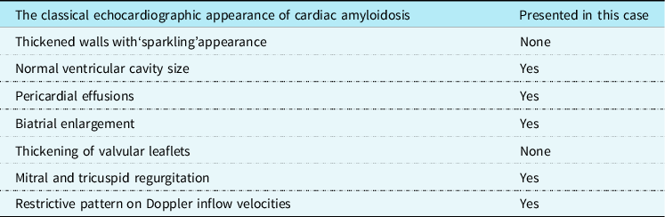
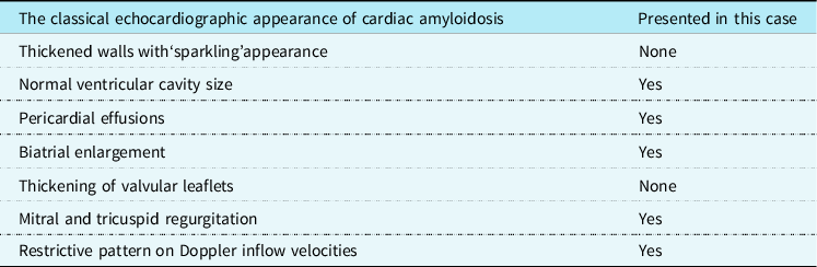
On physical examination, the patient appeared fatigued but in no apparent distress. Her blood pressure was 80/57 mmHg with pulse rate of 68 beats per minute (bpm). Marked jugular venous distention, moderate hepatomegaly, and lower extremity oedema were noted. Cardiac and respiratory examination showed muffled heart sounds and an absence of murmurs, gallops, and clear lung fields. Most laboratory tests that included cardiac troponin were normal or negative. N-terminal fragment brain natriuretic peptide was 3035 pg/ml (Normal<125pg/ml). Electrocardiography revealed normal sinus rhythm with criteria for low voltage, QS waves in precordial, and inferior leads. Echocardiography showed a ejection fraction of 52% with a restrictive pattern of transmitral flow (mitral E wave velocity =0.79 m/s, A wave = 0.31 m/s, medial E/E’ =20), which indicated coincident diastolic dysfunction. Biatrial enlargement, mild mitral and tricuspid regurgitation, and mild pericardial effusion without feature of pericardial thickening were noted. End-diastolic thickness of the interventricular septum was normal without granular sparkling appearance (comparison of the classical echocardiographic appearances of cardiac amyloidosis with this case ise listed in Table 1).
Comparison of the classical echocardiographic appearance of cardiac amyloidosis with this case
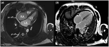
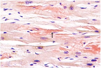
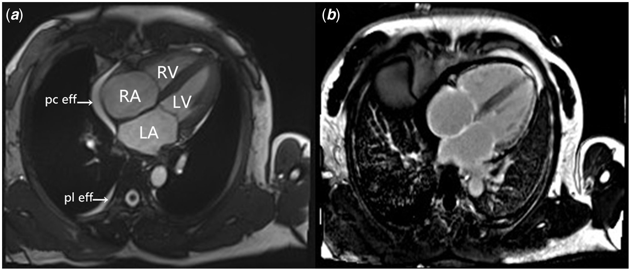
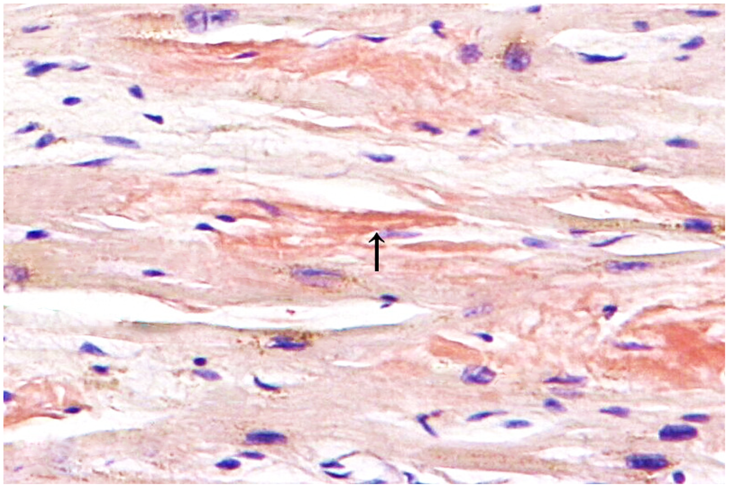
In view of the above findings, investigation for amyloidosis was initiated. Results of serum electrophoresis showed a mild increase in gamma globulin with a polyclonal band. Further biochemical analysis showed an elevated level of free immunoglobulin λ light chains in serum (700 mg/dl, reference range 298 mg/dl to 665 mg/dl) and a normal concentration of κ-serum-free light chains (523 mg/dl, reference range 598 mg/dl to 1329 mg/dl). In addition, the κ/λ was abnormally low. The patient underwent abdominal fat and rectum biopsies that were negative for amyloid deposits. Since the available data and the clinical picture were suggestive for amyloidosis, cardiac MRI was also pursued. Cardiac MRI revealed diffuse infiltrative changes and which increased the possibility of amyloidosis (Fig 1). The diagnosis of cardiac amyloidosis was finally established after endomyocardial biopsy in which the amyloid deposits were confirmed (Fig 2). The patient was sent to department of haematology to begin chemotherapy involving oral administration of melphalan and dexamethasone (six cycles). At the time of this writing, the patient has completed 3rd phase of chemotherapy. She is stable and symptoms have relieved.
( a ) Four-chamber steady-state free precession image shows normal thickness of left ventricular wall (10 mm) and IVS (11 mm), apparent biatrial dilatation and pericardial effusions (Pc eff), which are consistent with results of echocardiography. Mild pleural effusions (Pl eff) is also observed. ( b ) Four-chamber view from postgadolinium delayed enhancement images show biatrial, tricuspid valve, mitral valve, right ventricle and left ventricle line-, granular- or patchy-like enhancement.
Amyloid deposits are confirmed by a positive Congo red stain, which gives the characteristic salmon-pink color (arrow) (Congo red, 400x).
Discussion
Amyloidosis refers to a collection of conditions in which abnormal protein folding results in insoluble fibril deposition in tissues. The major types of amyloidosis, classified on the basis of their precursor protein, include light chain, senile systemic (wild-type transthyretin), hereditary (mutant transthyretin), and secondary (AA) disease. It is a multisystemic disease. The frequency of cardiac involvement varies among the types of amyloidosis and is common with light chain (AL) disease (in up to 50% of cases). Reference Martinez-Naharro, Hawkins and Fontana1
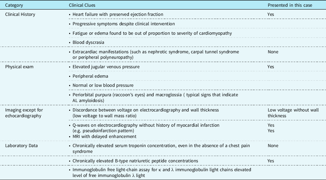
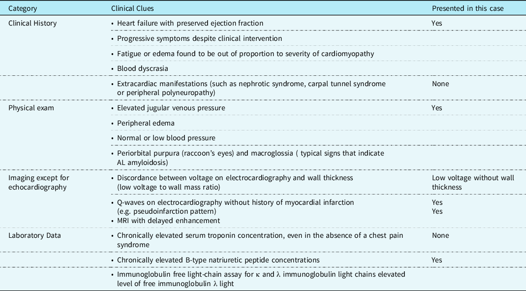
Although cardiac involvement is frequently seen, isolated cardiac involvement, as in our patient, is rare and was described in only 4% of a series of 232 patients who had AL amyloidosis. Reference Dubrey, Cha, Anderson and Chamarthi2 In these cases, the diagnosis can be difficult due to minimal or absent extracardiac features. In order to facilitate the identification of a suspected cardiac amyloidosis, it is important to integrate and reconcile findings from clinical features, electrocardiography, electrocardiogram, and cardiac MRI. Table 2 highlights the clinical clues, imaging and laboratory data that should raise concern for the presence of cardiac amyloidosis. In a selected group of patients, a myocardial biopsy is imperative to reach a conclusive diagnosis.
Clinical clues that may prompt evaluation for cardiac AL amyloidosis and presented in this case
Echocardiography should be the first noninvasive test performed to evaluate for cardiac amyloidosis, although imaging alone cannot confirm the diagnosis. Clinical guidelines suggest increased interventricular septum thickness with granular or sparkling appearance by echocardiography, in conjunction with a positive non-cardiac biopsy, is sufficient to define cardiac involvement in amyloidosis. However, in our case, there was neither thickened of interventricular septum nor granular appearance of myocardium. Amyloid infiltration may result in increased echogenicity or granular appearance of myocardium and which may often appear in the late stages of disease. In addition, with advances in digital image analysis techniques (particularly harmonic imaging), myocardial speckling has a low sensitivity and specificity for diagnosis of cardiac amyloidosis. Therefore, this finding is merely suggestive, but cannot be used in isolation for the diagnosis of cardiac amyloidosis. On very rare occasions, cardiac amyloidosis presented with normal interventricular septum thickness have been found. In a retrospective case–control study, Rahul Suresh et al found only 7 (3%) of 255 patients diagnosed cardiac amyloidosis by endomyocardial biopsies had a normal interventricular septum thickness. Reference Koyama, Ikeda and Ikeda3 Our patient with histologically confirmed cardiac amyloidosis have normal interventricular septum thickness, and there are several theoretical possibilities to explain the absence of increased interventricular septum. The first potential explanation is that patients with AL amyloidosis tend to become symptomatic earlier on in the disease course, sometimes before left ventricle walls become severely hypertrophied, and diastolic dysfunction (pseudonormalization or restrictive filling) is the norm. Elevated biventricular filling pressures, as well as direct atrial infiltration by amyloid protein, lead to atrial dilation. The second potential explanation is that total myocardial amyloid deposition and extent of cardiomyocyte hypertrophy in the patients with normal interventricular septum thickness may be lower as compared to those with increased interventricular septum thickness. Another possible explanation for the findings of normal interventricular septum thickness is that baseline wall thickness, prior to amyloid deposition, may have been at the lower end of normal such that, despite significant amyloid infiltration, the wall thickness measurements remained within the normal range.
Except for thickened ventricular and septal walls with refractile myocardium, there are other echocardiographic clues to the presence of cardiac amyloid such as prominent biatrial dilatation with a normal or small ventricular size, right ventricular free wall thickening (rarely seen in hypertensive or valvular disease), diffuse valve thickening and restrictive haemodynamics with a relatively preserved ejection fraction. Reference Koyama, Ikeda and Ikeda3 In addition to echocardiography, electrocardiographic findings can also provide further evaluation for amyloid. Low QRS voltages (all limb leads<5 mm in height) with poor R wave progression in the chest leads (pseudoinfarction pattern) occur in up to 50% of patients with cardiac AL amyloidosis. The combination of low voltage with concentrically increased wall thickness is highly suspicious for cardiac amyloidosis. Other findings include first-degree atrioventricular block (21%), non-specific intraventricular conduction delay (16%), second- or third-degree atrioventricular block (3%), atrial fibrillation/flutter (20%), and ventricular tachycardia (5%). Left and right bundle branch block can also occur. Reference Suresh, Grogan and Maleszewski4
Cardiac MRI may provide an alternative, non-invasive option to screen for amyloid involvement and should be considered in patients with cardiomyopathy of unclear aetiology. The appearance of global, subendocardial late gadolinium enhancement is highly characteristic of cardiac amyloid and correlates with prognosis. Reference Maceira, Joshi and Prasad5 In normal myocardium, gadolinium is not retained after administration, a phenomenon known as “nulling of myocardium.” In amyloid heart, the distribution kinetics of gadolinium are altered due to extracellular deposition of amyloid, leading to retained contrast that produces the characteristic late gadolinium enhancement.
In conclusion, our case suggests that patient who present with heart failure of uncertain aetiology may has amyloidosis despite normal interventricular septum thickness, warranting increased vigilance for this disease in such patient. A combination of clinical, electrocardiographic, other echocardiographic findings common in amyloidosis and delayed enhancement pattern of gadolinium uptake in the myocardium by cardiac MRI may provide important clues to suggest further workup.
Conflicts of interest
Authors have no interests to disclose. The authors report no financial relationships or conflicts of interest regarding the content herein.