INTRODUCTION
Species of Polydora and related genera (Polychaeta: Spionidae) are collectively called polydorids (i.e. Dipolydora, Boccardia, Boccardiella, Carazziella and Pseudopolydora), each of them having modified fifth chaetigers (Blake, Reference Blake and Blake1996). Many polydorid species are widely known for their boring activities into calcareous substrates, and they have been reported in various types of living and non-living calcareous structures including mollusc shells, corals, and coralline algae (Blake & Evans, Reference Blake and Evans1973; Sato-Okoshi & Okoshi, Reference Sato-Okoshi and Okoshi1997; Sato-Okoshi, Reference Sato-Okoshi1999, Reference Sato-Okoshi2000; Sato-Okoshi & Takatsuka, Reference Sato-Okoshi and Takatsuka2001). Polydorids may cause several effects to their calcareous hosts, e.g. reduce the commercial value, growth rate and meat yield or induce their mortality. Therefore, the infestation of commercially important shells is regarded as a serious problem in aquaculture (Okoshi & Sato-Okoshi, Reference Okoshi and Sato-Okoshi1996; Martin & Britayev, Reference Martin, Britayev and Ansell1998). Polydorid infestation in various molluscs, such as oysters (Haswell, Reference Haswell1885; Lunz, Reference Lunz1941; Loosanoff & Engle, Reference Loosanoff and Engle1943; Handley & Bergquist, Reference Handley and Bergquist1997), pearl oysters (Takahashi, Reference Takahashi1937; Mizumoto, Reference Mizumoto1964; Mohammad, Reference Mohammad1972), mussels (Kent, Reference Kent1979; Ambariyanto & Seed, Reference Ambariyanto and Seed1991), scallops (Evans, Reference Evans1969; Sato-Okoshi et al., Reference Sato-Okoshi, Sugawara and Nomura1990), clams (Caceres-Martinez et al., Reference Caceres-Martinez, Tinoco, Bustamante and Gomez-Humaran1999) and abalone (Kojima & Imajima, Reference Kojima and Imajima1982; Lleonart et al., Reference Lleonart, Handlinger and Powell2003; Vargas et al., Reference Vargas, Quijón and Bertrán2005) have been reported over a long historical period.
Although there are some taxonomic studies on polydorid species in Australian and adjacent seas (Rainer, Reference Rainer1973; Read, Reference Read1975; Blake & Kudenov, Reference Blake and Kudenov1978; Blake, Reference Blake1983; Hatchings & Turvey, Reference Hutchings and Turvey1984), they were conducted chiefly in south-eastern waters and there is little information for the polydorid polychaetes inhabiting south-western Australia. On the other hand, polydorids are being increasingly regarded as invasive boring species in the field of mollusc aquaculture and environment science, as they damage the health and commercial value of their hosts (Whitlegge, Reference Whitlegge1890; Lleonart et al., Reference Lleonart, Handlinger and Powell2003). Particular examples are the Sydney rock oyster Saccostrea commercialis, which harvesting was impacted upon economically by a heavy polydorid infestation (Smith, Reference Smith1984) and the abalone. Accordingly, since the mid-1990s, polydorid infestations have been considered responsible for the mortality of cultured abalone, mainly from sea-based farms in southern Tasmania (Lleonart et al., Reference Lleonart, Handlinger and Powell2003). To date, Polydora hoplura Claparède, 1870 and Boccardia knoxi Rainer, Reference Rainer1973 have been reported to cause severe damage to abalone and oysters and considerable infestation rates even leading to mortality in abalone. Polydora hoplura and B. knoxi have also been reported from sea-based systems in New Zealand, as well as from land-based systems in Tasmania, South Australia and Chile (Handley, Reference Handley1995; Handley & Bergquist, Reference Handley and Bergquist1997; Radashevsky & Olivares, Reference Radashevsky and Olivares2005; Vargas et al., Reference Vargas, Quijón and Bertrán2005).
More recently there has been a dramatic increase of mollusc aquaculture activities, resulting in a quick and easy worldwide transport of some of the more commercially important molluscs (Cohen & Carlton, Reference Cohen and Carlton1998). Consequently, some of their associated organisms such as sessile and boring species also spread from their native regions (Bailey-Brock, Reference Bailey-Brock2000).
In Australia, mollusc aquaculture has been expanding, not only in the eastern states, but also in the western parts. Mollusc aquaculture is less economically relevant in western than in eastern Australia. However, it is very important to survey the presence of boring polydorid species in molluscs from south-western waters during this early stage of the industry. Accordingly, the boring polydorids from shells of both cultured and native living molluscs were examined to determine whether they are native or introduced. Their morphological and ecological characteristics were examined to better understand the occurrence and potential impacts of polydorid polychaetes in south-western Australian waters.
The specimens collected have been deposited in the Australian Museum.
MATERIALS AND METHODS
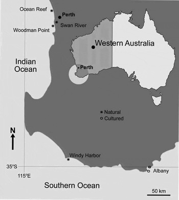
Polydora, Dipolydora and Boccardia species were collected at six sites from south-western Australian waters in August 2005 and January 2006 (Figure 1). Polydorid species were extracted from the shells of both native (Haliotis roei: shell length (SL) 62–91 mm, H. conicopora: SL 110 mm, Chlamys australis: shell height (SH) 20–99 mm, Thais orbita: SH 60–68 mm, Turbo torquatus: SH 60–75 mm, Patelloida alticostata: SL 15 mm) and cultured molluscs (Haliotis laevigata: SL 50–91 mm, H. roei: SL 57–67 mm, Saccostrea commercialis: SH 55–97 mm). Native molluscs were collected from intertidal to subtidal rocky shores by diving. Cultured abalone (H. laevigata and H. roei) were obtained from land-based tanks and cultured oysters (S. commercialis) were collected from shallow water net baskets, both in Albany.
Sampling sites in south-western Australia.

Living worms were extracted by fracturing the shells with cutting pliers and a hammer and their morphological characteristics, state of sexual maturity, and presence and condition of egg capsules and larvae were assessed. The specimens were then fixed with 10% formalin. Complementary morphological observations were also conducted after fixation.
RESULTS
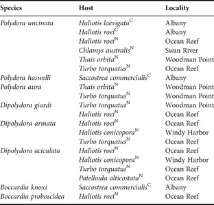
Eight polydorid species (3 Polydora, 3 Dipolydora and 2 Boccardia) were extracted from the shells of cultured and native molluscs (Table 1). Seven boring polydorid species were found to make self-excavated burrows inside the calcareous substrates, while Boccardia proboscidea inhabited mud within shell crevices.
Polydora, Dipolydora and Boccardia species extracted from mollusc shells in south-western Australian waters.

C, cultured; N, natural.
Polydora uncinata was the most commonly extracted species among various natural and cultured mollusc shells during the survey, being especially numerous in shells of cultured abalone and forming aggregates in scallop shells. Polydora aura, Dipolydora armata, D. giardi and D. aciculata were commonly observed inhabiting the natural molluscs. Boccardia knoxi was very abundant in shells of the cultured oysters, and was never observed in other native or cultured molluscs. Polydora haswelli and B. proboscidea were seldom found during this survey. No worm was observed to bore into the shells of the scallop Amusium balloti from the Indian Ocean off Fremantle.
The finding of Polydora uncinata and P. aura represents the first report for the whole Australian waters, while Boccardia knoxi and D. aciculata were new for the south-western waters.
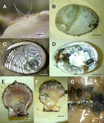
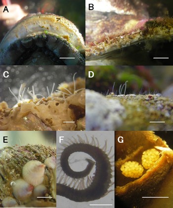
Polydora uncinata. (A) Palps with black bars protruding from mucous–muddy tube openings, stemming from self-excavated burrows on the outer surface of cultured Haliotis laevigata; (B) outer surface of a cultured H. laevigata shell showing mucous–muddy tubes along the edge, around the respiratory pores and near the apex; (C) inner surface of a natural Haliotis roei shell, showing some thin layers of a dark organic substance deposited as a protective barrier at the point of contact with polydorids; (D) inner surface of a cultured H. laevigata shell showing a heavy infestation by polydorids and the marks of the extra organic and calcareous materials secreted to prevent further penetrations. Black and/or brown organic deposits and calcareous shell materials deposited on the organic materials were observed; (E) inner surface of a natural Chlamys australis shell showing thin black layers along the polydorid burrows; (F) broken shell of natural C. australis with crack along the polydorid burrows; and (G) magnified C. australis shell area showing the black and brown protective depositions. Scale bars 3 mm for A, 15 mm for B–F, 1 mm for G.

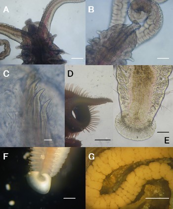
Polydora uncinata. (A) Dorsal view of anterior end; (B) ventral view of anterior end; (C) magnified modified spines of chaetiger 5 alternating with pennoned companion chaetae; (D) posterior chaetigers with pygidium and long branchiae continuing to almost end of body; (E) posterior chaetigers with pygidium, and recurved hook and long capillaries on each side of notopodium; (F) posterior chaetigers with glittering recurved hooks; and (G) egg string divided into many capsules containing developed larvae and nurse eggs. Scale bars 0.3 mm for A & B, 0.01 mm for C, 1.5 mm for D, 0.1 mm for E, 0.2 mm for F, 1 mm for G.

Morphology of new material
Size up to 25 mm long. Prostomium weakly incised anteriorly, caruncle extending to middle of chaetiger 3 in large specimens, short occipital tentacle present, four eyes present. Palps crossed by 8 to up to 11 black bars and, occasionally, palps bordered by slight dark pigmentation or without pigmentation. Dark pigmentation along caruncle. Branchiae present from chaetiger 7, continuing to almost end of body in large specimens and to the last 10–20 chaetigers in small specimens (less than 40 chaetigers). Setiger 1 without notochaetae, only capillary neurochaetae. Setigers 2–4 with winged capillary neuro- and notochaetae.
Modified spines of chaetiger 5 falcate with lateral flange, and few young spines buried inside, not yet protruding, major spines alternating with pennoned companion chaetae; 4–5 short dorsal and ventral unilimbate chaetae present. Bidentate hooded hooks in neuropodia from chaetiger 7, hooks with constriction on shaft and main fang at right angle to shaft, with acute angle between main fang and apical tooth, continuing to almost end of body. Single, occasionally two, recurved hooks and a few long capillaries present on posterior notopodia. Only the last 2–5 chaetigers possess recurved hooks in smaller specimens (less than 40 chaetigers long).
Pygidium a flaring disc, dorsally wide open.
Reproduction and larval development
Mature females with oocytes in the coelom and egg capsules in their burrows, this supporting an iteroparous reproductive behaviour. Size of the oval oocyte was 150–180 µm, with 170 µm in peak, in long axis. Size of the oval egg was 175 µm × 125 µm in average. Thirty to 50 capsules were observed joining each other to form one string. There were few developing larvae and numerous nurse eggs in each capsule, showing entirely exolecithotrophy and adelphophagy. Various developmental stages were observed: e.g. cleavage, 1–3 chaetiger–larvae, late larval stages showing palps on anterior region. Mature males were absent during the period of study.
Ecology
Large specimens were conspicuously observed to bore into the shells of land-based cultured abalone Haliotis laevigata and H. roei. Thirty to more than 50 specimens inhabited one large shell (mean length: 83 mm; mean wet weight: 32 g), showing a bimodal population structure of mean size for the two modes of 0.55–0.60 mm and 1.1–1.2 mm in width of the 5th chaetiger, respectively. Numerous individuals frequently infested the surface of the shell around the respiratory pores, along the edge of the shell, and near the apex (Figure 2B–D). Heavy infestations induced the host to secrete extra organic and calcareous shell materials, to prevent penetration of the worms through the shell. As a result, black and/or brown deposits and calcareous shell materials deposited on the organic materials were observed on the inner surface of the shells (Figure 2C–D).
On the other hand, the native abalone (Haliotis roei), scallop (Chlamys australis), and gastropods (Thais orbita and Turbo torquatus) were also infested by Polydora uncinata. Less than 10 ind/shells with apparently two different size-generations were observed to bore into H. roei in August. In the case of the scallop, there were five to 10 individuals per shell, distributed continuously between 0.6 to 1.2 mm in width of the 5th chaetiger in size with three different size-generations in August, each mode being 0.65 mm, 0.8 mm, and 1.2 mm, respectively. The finding of about 90% of the larger individuals of the latter two modes having both intracoelomic oocytes and egg capsules supported the iteroparity of the species and pointed to its high reproductive potential.
Morphology of new material
Up to 15 mm long. Prostomium anteriorly widely bilobed, curved downward; caruncle extending to middle of chaetiger 3. Palps short, with dark pigmentation along edge. Four eyes arranged trapezoidally. Branchiae present from chaetiger 7, fairly long and continuing to the last 10–15 chaetigers. Setiger 1 lacking notochaetae.
Modified spines of chaetiger 5 falcate, with lateral tooth alternating with pennoned companion chaetae; dorsal and ventral chaetae present.
Pygidium a flaring disc with distinct dorsal notch.
Reproduction and larval development
Coelomic oocytes and independent egg capsules were found in self-excavated burrows in August. No nurse eggs were found, and all embryos developed simultaneously in each capsule, most of them being at the developmental stage from cleavage to 1-chaetiger larvae in August.
Ecology
The few specimens found were only extracted from the shells of the cultured oyster Saccostrea commercialis.
Morphology of new material
Up to 12 mm long. Body and palps light orange, transparent in life. Prostomium anteriorly weakly incised, caruncle extending to chaetigers 2–3, short occipital tentacle present. Eyes present or absent. Branchiae beginning from chaetiger 7, continuing to end of body.
Modified spines of chaetiger 5 falcate with lateral flange, alternating with pennoned companion chaetae.
Special notochaetae present in posterior chaetigers, consisting of tight cylindrical bundles of short needles, mostly of equal length, bundles not protruding through cuticle; these accompanied by long and short extended capillaries.
Pygidium a wide, flaring disc.
Larval development
All egg capsules independent, without nurse eggs, and all embryos developing simultaneously. A single batch of egg capsules was observed in August.
Ecology
One to five individuals were extracted from the large gastropods Thais orbita (SH: 60–68 mm) and Turbo torquatus (SH: 73 mm), found to burrow mainly around the outer lip of the aperture of the shell. Polydora aura was absent from cultured molluscs.
Remarks
There was no occipital tentacle description of the species by Sato-Okoshi (Reference Sato-Okoshi1998), originally, short occipital tentacle was observed in Japanese specimens until now.
New to Australia.
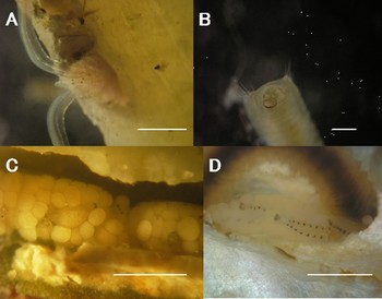
Dipolydora aciculata. (A) Dorsal view of anterior end, protruding from the opening of its self-excavated burrow; (B) posterior end with pygidium, which divided into four lobes, a dorsal pair smaller and a ventral pair larger. Long and short notochaetae present on posterior notopodium; (C) egg string with 4-eye-spotted larvae and many nurse eggs; and (D) egg string with succeeded larvae of more than 10 chaetigers and co-existing nurse eggs. Scale bars 1 mm for A,C & D, 0.35 mm for B.

Morphology of new material
Up to 10 mm long. Colour in life tan, with faint brown lines along the palp edge and in some specimens, a faint dark pigmentation around prostomium and caruncle. Palp transparent. Prostomium anteriorly round, caruncle extending to end of chaetiger 3. Eyes absent or if present, 4 eyes arranged trapezoidally. Branchiae beginning from chaetigers 7 or 8 and continuing to half of body.
Modified 5th chaetiger with superior dorsal chaetae and a fascicle of ventral chaetae; major spines falcate with lateral flange and bristles on convex side of curved end, alternating with companion chaetae.
Special thick and short notochaetae present in posterior chaetigers accompanying fairly long capillaries, continuing to the very end. Posteriorly, short neuropodial spines displaced the hooded hooks.
Pygidium small, divided into four lobes, the dorsal pair smaller than the ventral one.
Reproduction and larval development
Few elongated egg strings without partitioning into small capsules were observed in August. Many nurse eggs co-existed with developing embryos, suggesting exolecithotrophy and adelphophagy. Larval developing stage of four-eye-spotted one- to two-chaetiger was observed in one string. Larger larvae, more than 10-chaetiger long, observed in another, having a pair of black dots on each dorsal segment.
Ecology
Large individuals were extracted from the shells of the native abalone Haliotis roei and H. conicopora, as well as from the native gastropods Turbo torquatus and Patelloida alticostata.
Remarks
Morphologically, the Australian specimens coincided with the original description (Blake & Kudenov, Reference Blake and Kudenov1978), except for the presence of eyes and the beginning chaetiger possessing branchiae. Although branchiae were described to start from chaetiger 9 in the original description, chaetigers 7 and 8 possessed small branchiae in new materials.
New to south-western Australia.
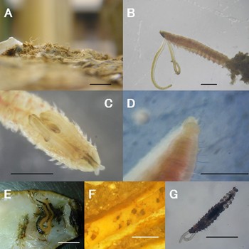
(A–D) Dipolydora armata, (D–E) Dipolydora giardi and (F–G) Polydora haswelli. (A) Traces of infestation along the edge of a Haliotis conicopora shell; (B) cross-section of a H. conicopora shell showing many burrows, mucous–muddy tubes and palps protruding from the openings of the burrows; (C) anterior ends of polydorids protruding from self-excavated burrows in a H. conicopora shell; (D) patches of palps of Dipolydora armata and D. giardi (co-exist in a Turbo torquatus shell), protruding from the burrows; (E) numerous palps protruding from polydorid burrow openings in a T. torquatus shell; (F) posterior chaetigers with flared pygidium and long branchiae lasting to almost end of body; and (G) independent egg capsules with all embryos developing simultaneously. Scale bars 1 cm for A & B, 1 mm for C,F & G, 2 mm for D & E.

Morphology of new material
Up to 4 mm long. Colour in life tan, without pigmentation. Prostomium anteriorly bilobed, caruncle extending to end of chaetiger 2. Eyes absent. Branchiae beginning from chaetiger 7, continuing only to chaetiger 11.
Modified spines of chaetiger 5 bidentated with tooth connected by lateral hood, no companion chaetae.
Special notochaetae present in posterior chaetigers, consisting of a cone-shaped bundle of thick acicular spines protruding through cuticle, accompanying capillaries.
Pygidium small and cylindrical, cuff-shaped with dorsal notch.
Reproduction
More than 50% of individuals had oocytes in their coelom from chaetiger 11, but no egg capsule was observed in August.
Ecology
Numerous individuals were observed to bore patchily in shells of the native gastropods Turbo torquatus and Haliotis roei, particularly around the shell areas covered with coralline algae. Moreover, a high number of specimens (over 1000 from a single shell measuring 110 mm long and with a dry weight of 82 g) were extracted from the shells of the subtidal abalone H. conicopora at Windy Harbor in January. The worms were small and remain sticky when alive.
Morphology of new material
Up to 5 mm long. Colour in life light tan, without pigmentation. Eyes absent. Caruncle extending to end of chaetiger 3. Branchiae appeared from chaetiger 9.
Pygidium disc-like with one large ventral lobe and two small dorsal ones, occasionally the three lobes continuous and inconspicuous.
Reproduction and larval development
Although numerous individuals were extracted from the shells of native gastropods, intracoelomic gametes and egg capsules were absent in August.
Ecology
Numerous specimens were observed to bore patchily in areas covered with coralline algae on shells of the native abalone Haliotis roei, as well as in the shells of the larger intertidal gastropod Turbo torquatus. Dipolydora giardi co-inhabited the same shells with D. armata. Worms were very small and remain sticky when extracted.
Boccardia knoxi. (A) Palps protruding from the mucous–muddy tubes on a cultured Saccostrea commercialis shell; (B) anterior chaetigers protruding from its tube, showing the palps with slight black pigmentation along edge and crossed by white bars; (C) dorsal view of anterior chaetigers, showing the conspicuous black pigmentation on prostomium; (D) posterior chaetigers showing the two transversely subequal pygidial lobes; (E) inner view of a burrow, showing a long egg string without egg capsules; (F) developed larvae with black pigmentation on their dorso-central region and pygidium, coexisting with many nurse eggs co-exist in a string; and (G) crawling 15-chaetiger larvae without modified fifth chaetiger on an oyster shell. Scale bars 1 cm for A & E, 1 mm for B–D & F, 0.5 mm for G.

Morphology of new material
Up to 20 mm long. Colour in life tan, with conspicuous black pigmentation on prostomium. Prostomium long and bilobed, curved downward anteriorly, and prominent in lateral view; occipital cirri absent; caruncle extending to middle of chaetiger 3. Four eyes arranged trapezoidally. Palps transparent with slight black pigmentation along edge and irregular colourless spots crossing transversely, giving the appearance to be crossed by white bars in lateral view. Branchiae present on chaetigers 2, 3, 4, 6 and succeeding ones, to one-quater of body. Chaetiger 1 with both noto- and neurochaetae. No special posterior notochaetae.
Chaetiger 5 modified, bearing two types of spines (falcate and distally expanded, with a concavity and a central cone) arranged in two rows; accompanying chaetae absent. Only having the ventral fascicle. Bidentate hooded hooks present from chaetiger 7, without accompanying capillary chaetae.
Pygidium small, with two transversely subequal lobes.
Reproduction and larval development
Approximately 30% of individuals possessed egg strings in their burrows in August, most of them also with orange intracoelomic oocytes, present from chaetiger 18. Numerous nurse eggs co-existed with developing embryos, showing adelphophagy; egg string elongated, without partitioning into small capsules. Colour of developing larvae dark black; very conspicuous black in dorso-central region and around pygidium. Both crawling larvae 15 to 16-chaetiger long without modified spines on chaetiger 5 and juveniles already having these spines were extracted from mud deposits in crevices of shells of the cultured oyster Saccostrea commercialis.
Ecology
This species was only observed in association with the shells of the shallow water net cultured oyster Saccostrea commercialis in Albany. Mud deposits were conspicuous among the imbricate structures of the outer prismatic layer of the shell. Mud tubes were observed to protrude from their burrows on the outer shell surface. The upper right valve was more heavily infested than the left one, usually with less than 10 individuals per valve, occasionally more than 50. Large, medium and small sized individuals were observed on the same shell in August, the small ones seeming to be recently settled juveniles. Both crawling larvae (with 15 chaetigers or more) and juveniles were extracted from mud deposits among imbricate shell structures. Living worms remained very sticky when extracted.
Remarks
Morphologically, the present material coincided with the original (Rainer, Reference Rainer1973) and posterior (Read, Reference Read1975) descriptions, except for the pigmentation (new south-western materials showed conspicuous black pigmentation on caruncle and white transverse bars on palps), number of eyes (four arranged trapezoidally), and the first chaetiger possessing neuropodial hooded hooks (chaetiger 7 instead of chaetiger 8, as previously described).
New to south-western Australian waters.
Morphology of new material
Up to 10 mm long. Colour in life greenish tan, with black pigmentation along both sides of caruncle. Prostomium anteriorly weakly incised, caruncle extending to end of chaetiger 3. Four eyes present. Branchiae on chaetigers 2, 3, 4, 6 and succeeding ones to almost end of body. Setiger 1 with both noto- and neurochaetae.
Setiger 5 modified, bearing two types of modified spines arranged in two rows; no companion chaetae; only the ventral fascicle present. Bidentate hooded hooks from chaetiger 7, with accompanying capillary chaetae.
Pygidium with four subequal lobes.
Reproduction and larval development
Neither intracoelomic gametocytes nor egg capsules were observed in August.
Ecology
The few individuals observed during this survey occurred only in the shells of native abalone Haliotis roei.
DISCUSSION
Infestation
From an aquaculture point of view, the damage caused by the boring habits of Polydora uncinata and Boccardia knoxi is a serious issue. On the external surface of the shell, these species occur as numerous protruding mucous–mud tubes and/or paired burrow openings. On the internal side of the shell, both the abalone and oysters secreted a thin layer of dark and/or black organic and calcareous material at the point of contact with the boring polydorid worms, which acts as a physical protective barrier to prevent further contact of the worms and thus, prevent further invasion. Nonetheless, some worms continue penetrating the inner surface of the thin and weakened shell, thus inducing further secretion by the mollusc (Figure 2D).
The extra secretion of materials such as organic substances, calcitic and/or aragonitic shells to prevent further polydorid penetration, requires additional energy and nutrients that must be redirected from growth and reproduction, thereby having long term impacts on the health of the host. Polydorid infestation may not always be a direct lethal factor for cultured molluscs, but may be enough to reduce the growth rates of both shell and meat yield during the infestation. Moreover, due to the stress caused by the infestation, the molluscs are more susceptible to adverse environmental changes, likely resulting in an increased mortality. The protective secretions deposited by the molluscs also spoil the appearance of the shell, reducing its commercial value. All these factors must be accounted for in aquaculture systems, as they may produce a strong impact upon production efficiency.
Reproductive traits
Polydorids are recognized to deposit three kinds of egg capsules in the burrow: namely, small capsules joined in a beadlike string, a series of single independent capsules, and un-capsulated cylinder-like string. The larvae have been observed to develop as lecithotrophic and/or planktotrophic. Various larval developmental patterns have been reported even within the same polydorid species (Blake, Reference Blake and Blake1996).
Polydora uncinata from south-western Australian waters showed exolecithotrophic development and adelphophagy similar to Japanese populations (Sato-Okoshi, Reference Sato-Okoshi, Hayashi and Nakao2005, Reference Sato-Okoshi and Fukuda2006). In Japanese specimens, the average length of the long axis of both maximum intracoelomic oocytes and oval eggs from egg capsule measured ~170 µm (Sato-Okoshi, Reference Sato-Okoshi1999). In south-western specimens, the length of the long axis of oval eggs in egg capsule was 175 µm on average. The size of the eggs of the Chilean specimens extracted from land-based cultured abalone was reported by Radashevsky & Olivares (Reference Radashevsky and Olivares2005) to be 170–175 µm × 133–138 µm in diameter. So the size of the eggs of the species seemed to be 170–175 µn in long axis.
The elongated larvae with short palps still remain inside the capsules suggesting that larval development occurs almost entirely within the capsule. The larvae of the same chaetiger were also found swimming and crawling for a very short period or crawling and settling just after hatching. Worms' size was found to vary from smaller juveniles to larger adults in the south-western population in August, which may indicate the possibility of its long life span. Moreover, many females possessed both egg capsules and larger intracoelomic oocytes so that P. uncinata apparently keeps the potential to produce different batches of egg capsules during one reproductive season. These characteristics are common to symbiotic (e.g. parasitic) polychaetes colonizing restricted habitats (the mollusc shells in this case) as the infestation rates may quickly increase even from a single initial colonizer (Martin & Britayev, Reference Martin, Britayev and Ansell1998) and, from the point of view of the polydorids would be particularly advantageous in a closed ecosystem, such as land-based cultures, as they would increase population size and survivorship. Since planktotrophic larvae need to feed during development, the larvae of P. uncinata developed in the capsule feed on nurse eggs instead, thereby omitting the stress of the loss of survivorship.
The development of Boccardia knoxi is somewhat similar to that of Polydora uncinata. It also exhibited exolecithotrophy and adelphophagy, and bimodal populations were observed in August. The south-western Australian population produced long un-capsulated egg strings containing numerous nurse eggs. Conversely, the population from shells of Tasmanian abalone produced a string of egg capsules without nurse eggs, and larvae developing simultaneously until hatching (Handley, Reference Handley2000). The finding of crawling and recently settled larvae allows us to suggest that hatching occurs at the 15 or 16-chaetiger larval stage in the south-western population. Like P. uncinata, the south-western B. knoxi seems to be well adapted to quickly increase its population densities within a closed ecosystem.
Polydorid species and their distribution
Eight polydorid species were found in shells of both natural and cultured molluscs from south-western Australian coasts. Among them, only Polydora uncinata was common both as a native and land-based cultured mollusc. Their numerous individuals extracted from the shells of intertidal and, especially, subtidal (e.g. Chlamys australis) molluscs, suggests that this species may be native from south-western Australian waters. However, its intensive infestation of molluscs from the land-based culture systems may also suggest a possible spreading due to artificial transportation.
Polydora haswelli and Boccardia knoxi were extracted only from the shells of the cultured oyster and never from the natural ones. Boccardia knoxi, and small numbers of Polydora haswelli, was only extracted from the cultured oyster Saccostrea commercialis, suggesting that they were transported artificially, either from offshore or from eastern Australian coasts. Ostrea angasi is a native south-western Australian oyster, while S. commercialis originates from eastern Australia. Although only a few young O. angasi were observed to attach on the shells of cultured S. commercialis, no polydorids were extracted from the shell of O. angasi in this study. This study is the first reporting heavy B. knoxi infestations in shells of S. commercialis cultivated in a shallow water system in south-western waters. Previously, B. knoxi was only known to infest the introduced intertidal oyster Crassostrea gigas and cultured abalone from Tasmania. Therefore, B. knoxi may be an introduced species in south-western Australia, and the characteristics of its infestation should be monitored to forecast future influences on the affected cultured and native molluscs.
The other five polydorid species were observed to inhabit only the shells of the five natural gastropods and the single bivalve species examined during the study. Polydora aura, Dipolydora giardi, D. armata, and D. aciculata were easily found, and are likely to be native inhabitants of the intertidal and subtidal in south-western Australian waters.
Polydora uncinata was first discovered and described from the shells of the oyster Crassostrea gigas cultivated along the Pacific coast of Japan (Sato-Okoshi, Reference Sato-Okoshi1998). The species was then extracted from the shells of the abalone Haliotis discus hannai in a sea-based culture system in western Japan in 1999 (Sato-Okoshi, Reference Sato-Okoshi, Hayashi and Nakao2005). However, it was not common in natural or cultured mollusc shells, occurring only in restricted areas with a patchy distribution along Japanese waters. In 2004, the Polydora uncinata was unexpectedly extracted from shells of the hanging cultured oyster C. gigas in South Korea, where this species has not been found since 1999 (Sato-Okoshi, Reference Sato-Okoshi, Hayashi and Nakao2005; unpublished); and from introduced H. discus hannai in Chilean land-based cultures (Radashevsky & Olivares, Reference Radashevsky and Olivares2005). Although the origin of P. uncinata remains unknown, it seems evident that the species is becoming widespread in aquaculture systems, possibly as a result of the transportation of the cultured molluscs.
Polydora uncinata morphologically resembles P. hoplura as mentioned in the original description (Sato-Okoshi, Reference Sato-Okoshi1998), the latter being also reported as a pest for eastern Australian cultured molluscs (Lleonart et al., Reference Lleonart, Handlinger and Powell2003). Further investigations based on different characteristics (viz. morphology, ecology and reproduction) should address whether the south-western and eastern Australian populations are the same species or not. Not only P. uncinata, but B. noxi also needs intensive investigations to analyse the populations of south-western and eastern waters as the south-western characteristics suggested the possibility of the different species. In addition to this systematic approach, special efforts should be addressed to monitor the potential impact of P. uncinata and B. knoxi in aquaculture, not only in south-western Australian waters but in marine environments across the globe.
ACKNOWLEDGEMENTS
We express our appreciation to Drs John Webb and David Macey of Murdoch University for providing the opportunity to carry out this survey in Australia. We are grateful to all the staff in the Division of Science and Engineering, Murdoch University, especially Mr Edward Stockdale for providing facilities and support when carrying out this survey. Special thanks are given to Mr Mark Maddern for his great support in collecting molluscs. We are indebted to Dr D. Martin of Centre d'Estudis Avancats de Blanes (CSIC), Spain for his critical reading and constructive comments on the manuscript.