




No CrossRef data available.
Article contents
Cysteine proteinase activities in the fish pathogen Philasterides dicentrarchi (Ciliophora: Scuticociliatida)
Published online by Cambridge University Press: 06 May 2004
Abstract
This study investigated protease activities in a crude extract and in vitro excretion/secretion (E/S) products of Philasterides dicentrarchi, a ciliate fish parasite causing economically significant losses in aquaculture. Gelatin/SDS–PAGE analysis (pH 4, reducing conditions) detected 7 bands with gelatinolytic activity (approximate molecular weights 30–63 kDa) in the crude extract. The banding pattern observed in analysis of E/S products was practically identical, except for 1 low-molecular-weight band detected in the crude extract but not in the E/S products. In assays with synthetic peptide p-nitroanilide substrates, the crude extract hydrolysed substrates characteristic of cysteine proteases, namely Z-Arg-Arg pNA, Bz-Phe-Val-Arg pNA and Z-Phe-Arg pNA. These activities were strongly inhibited by the cysteine protease inhibitor E-64 and by Ac-Leu-Val-Lys aldehyde, a potent inhibitor of cysteine proteases of the cathepsin B protease subfamily. The proteases present in the crude extract degraded both type-I collagen and haemoglobin in vitro, consistent with roles in tissue invasion and nutrition respectively. Again, E-64 completely (collagen) or markedly (haemoglobin) inhibited this degradation. Finally, the histolytic activity of the ciliate in turbot fibroblast monolayers was strongly reduced in the presence of E-64, confirming the importance of secreted cysteine proteinases in the biology of Philasterides dicentrarchi.
Keywords
Information
- Type
- Research Article
- Information
- Copyright
- 2004 Cambridge University Press
INTRODUCTION
The protozoan parasite Philasterides dicentrarchi is a histophagous ciliate first described as an opportunistic parasite causing systemic infection in Dicentrarchus labrax in Mediterranean coastal lagoons (Dragesco et al. 1995), and subsequently from turbot, Scophthalmus maximus, farmed on the coast of Galicia in northwest Spain (Iglesias et al. 2001). In turbot, P. dicentrarchi causes severe disease, behaving as an endoparasite with multiorgan distribution that divides rapidly and feeds on cells (mainly erythrocytes) and other tissue components, causing the death of the host (Iglesias et al. 2003b).
Though it is not currently known exactly how this ciliate invades the host in nature, it has been demonstrated experimentally that these ciliates penetrate readily through minute superficial lesions, giving rise to severe infections (Paramá et al. 2003). One possibility is that culture tanks often show oxygen supersaturation, which may provoke the formation of small lesions in the skin, the eyes and the gills (Speare, 1998). Small lesions may also be accidentally produced during the routine manipulation of fish. Alterations in water temperature have also been suggested as a key factor in the epidemiology of scuticociliatosis in crustaceans and fishes (Cawthorn, 1997; Munday et al. 1997; Iglesias et al. 2001). During infection of turbot, P. dicentrarchi is frequently found in the bloodstream and in the perivascular and perineural connective tissue, suggesting that it uses these tissues as routes of access to the internal organs (Iglesias et al. 2001).
To achieve entry into the host and subsequent rapid dissemination through tissues, P. dicentrarchi must secrete proteolytic enzymes. Proteases are known to play a role in the invasion of cells and tissues by many species of pathogenic protozoans, in the destruction of mediators of the immune response, and in the degradation of host proteins in the course of feeding (Rosenthal, 1999). Here we present evidence of the presence of enzymes with proteolytic activity in cell extracts of this parasite, and in excretion/secretion products released in vitro into the culture medium. Our results indicate that the major proteolytic activity is of the cysteine proteinase type. In addition, we report preliminary findings on the possible role of these enzymes in the biology of this important fish pathogen.
MATERIALS AND METHODS
Parasite culture
Ciliates were isolated from ascitic fluid from the body cavity of naturally infected turbot (i.e. turbot showing scuticociliatosis, from a Galician fish farm affected by an outbreak of this disease). The ciliates were then cultured axenically at 18 °C in ‘complete’ L-15 medium (Leibovitz, 10% salinity, pH 7·2), as described by Iglesias et al. (2003a), which contains 90 mg/l each of adenosine, cytidine and uridine, 150 mg/l of guanosine, 5 g/l of glucose, 400 mg/l of L-α-phosphatidylcholine, 200 mg/l of Tween 80, 10% heat-inactivated foetal bovine serum (FBS) and 10 ml/l of 100× Antibiotic Antimycotic Solution (=100 units/ml of penicillin G, 0·1 mg/ml of streptomycin sulfate and 0·25 mg/ml of amphotericin B) (all from Sigma–Aldrich, USA).
Crude extract and excretion/secretion products
The crude extract (CE) was obtained as described by Williams & Coombs (1995), with some modifications. Briefly, the ciliates were washed 3 times by centrifugation at 650 g for 5 min in 0·25 M sucrose at 4 °C. The cells were then disrupted ultrasonically (Sonifier II, 250 W, Branson, USA) at 4 °C. The homogenate was centrifuged at 15000 g for 15 min at 4 °C and the resulting supernatant fraction stored in aliquots at −80 °C. Protein concentration in the extract was determined by the method of Bradford (1976) using a Bio-Rad Protein Assay kit (Bio-Rad Laboratories, Germany), with bovine serum albumin (Sigma–Aldrich) as standard.
Excretion/secretion products (E/S) were obtained by a method similar to that described by Karanu et al. (1993). Briefly, 2×10⁶ cells/ml where incubated for 24 h at 18 °C in ‘incomplete’ L-15 medium (like ‘complete’ L-15 medium but without nucleosides, glucose, lipids and FBS). The suspension was filtered through 0·22 μm sterile filters (Iwaki, Japan), and the filtrate concentrated 25 times using YM-10 Microcon centrifugal filter devices (Millipore, USA). The resulting concentrate (E/S) was stored at −80 °C.
Gelatin/SDS–PAGE
Proteases present in the 2 extracts (CE and E/S) were analysed by sodium dodecyl sulfate polyacrylamide gel electrophoresis (SDS–PAGE) in 10% polyacrylamide minigels containing a final concentration of 0·1% (w/v) gelatin, following a method similar to that described by North (1997). Electrophoresis was performed in a mini-Protean system (Bio-Rad Ltd, UK) under non-reducing conditions and without heating the samples. After electrophoresis, the gels were washed in a 2·5% (v/v) Triton X-100 solution at room temperature for 30 min with continuous shaking to eliminate SDS and renaturalize the proteins. After washing, the gels were cut into strips, incubated at 37 °C for 12 h in 10 ml of 0·1 M citrate buffer, pH 4, 0·1 M phosphate buffer, pH 6 or 0·1 M Tris–HCl buffer, pH 9 (CE), or in 0·1 M citrate buffer, pH 4 (E/S). Assays were performed with or without 1 mM dithiothreitol (DTT; Sigma–Aldrich) present in the buffer.
The proteolysis bands appeared as clear bands on a blue background after staining with Coomassie G-250 (0·06% Coomassie Blue, 10% acetic acid) for 30 min with continuous shaking, and subsequent destaining with a mixture of 10% acetic acid and 5% methanol. Proteolytic activities were visualized with the aid of a transilluminator. Molecular weights were estimated by comparison with low-range prestained SDS–PAGE standards (Bio-Rad).
To determine the inhibitory effect of the cysteine protease inhibitor trans-epoxysuccinyl-leucylamide-(4-guanidine) butane (E-64) on the proteolytic activities detected, the gel strips were incubated in the corresponding buffer in the presence of 10 μM E-64 (Sigma–Aldrich) and 1 mM DTT. The gels were stained and destained as described above.
Colorimetric assays of proteolytic activity
Protease activities in the crude extract of P. dicentrarchi were analysed in triplicate in 96-well microtitration plates (Bibby Sterilin, UK), by quantification of the hydrolysis of 7 peptide p-nitroanilides (Rawlings & Barrett, 1994), as follows. (1) As cysteine protease substrates (a) N-benzyloxycarbonyl-arginyl-arginine pNA (Z-Arg-Arg pNA, Bachem AG, Switzerland), (b) L-arginine pNA (L-Arg pNA, Sigma–Aldrich), (c) N-benzyloxycarbonyl-phenylalanyl-arginine pNA (Z-Phe-Arg pNa, Bachem), and (d) N-benzoyl-phenylalanyl-valyl-arginine pNA (Bz-Phe-Val-Arg pNA, Sigma–Aldrich). (2) As serine protease substrates (e) succinyl-L-phenylalanine pNA (Suc-Phe pNA, Sigma–Aldrich), (f) succinyl-alanyl-alanyl-prolyl-leucine pNA (Suc-Ala-Ala-Pro-Leu pNA, Sigma–Aldrich), and (g) N-α benzyloxycarbonyl-arginine pNA (Z-Arg pNA, Sigma–Aldrich). Stock solutions of Z-Arg-Arg pNA and L-Arg pNA were prepared at a concentration of 4 mM in distilled water; the other stock solutions were prepared in 8 mM dimethyl sulfoxide (Me2SO, Sigma–Aldrich). The final Me2SO concentration in the reaction mixture in no case exceeded 10%.
In each assay the reaction mixture (165 μl per well) consisted of 15 μl of crude ciliate extract, the volume of substrate stock solution required for a final substrate concentration of 0·5 mM, and reaction buffer. The buffers used were 0·1 M phosphate buffer, pH 6, containing 5 mM DTT for assays with cysteine protease substrates, or 0·1 M Tris–HCl buffer, pH 7·6, containing 20 mM CaCl2 for assays with serine protease substrates. Before addition of the substrate to the reaction mixture, the buffer and ciliate extract were incubated for 30 min at 37 °C in a humid chamber to activate the enzymes. After addition of the substrate, the mixture was incubated at 37 °C for 15 h. Release of p-nitroaniline was determined at 405 nm using a Multiskan RC device (Labsystems, EU); an increase in optical density of 0·1 units corresponded to 39·3 nmol of p-nitroaniline released. The results are expressed as nmol of p-nitroaniline released per mg of protein in the ciliate extract per minute. As a control for possible spontaneous release of p-nitroaniline by the pNA substrates, assays were performed with substrate and buffer only (no ciliate extract); as a background activity control, assays were performed with buffer and extract only.
For inhibition assays, we used the same assay procedure, except that before addition of the substrate (Z-Arg-Arg pNA, Bz-Phe-Val-Arg pNA, or Z-Phe-Arg pNA), the ciliate extract was incubated with the corresponding inhibitor for 30 min at 37 °C. The inhibitors assayed were E-64 (50 μM), pepstatin A (10 μM), phenylmethane sulfonyl fluoride (PMSF, 1 mM), N-α-p-tosyl-L-lysine chloromethyl ketone (TLCK, 10 μM), N-tosyl-L-phenylalanine chloromethyl ketone (TPCK, 200 μM), ethylenediaminetetraacetic acid (EDTA, 10 mM), all from Sigma–Aldrich, and acetyl-leucyl-valyl-lysine aldehide (Ac-Leu-Val-Lys aldehyde, Bachem; 500 μM). All inhibition assays were performed in 0·1 M phosphate buffer. The activity of each inhibitor was expressed as percentage inhibition (%I) calculated as follows:

where Ai− and Ai+ are activities without and with inhibitor respectively.
Protein degradation assays
Stock solutions of haemoglobin (32 mg/ml; Sigma–Aldrich) and type-I collagen (3 mg/ml; Sigma–Aldrich) were prepared in 0·1 M phosphate buffer, pH 6, and 5 mM DTT. The collagen solution was prepared from 6 mg of collagen in 1 ml of 0·1 M acetic acid, with continuous agitation in the dark at room temperature for 3 h until total dissolution. The solution was then mixed 1[ratio ]1 with phosphate buffer, with adjustment to pH 6. For the haemoglobin degradation assay, 20 μl of ciliate crude extract (containing 30 μg of protein) were incubated for 24 h at 37 °C with 2·5 μl of haemoglobin stock solution (containing 80 μg of protein). In the case of type-I collagen, 20 μl of crude extract (containing 30 μg of protein) were incubated for 24 h at 37 °C with 20 μl of type-I collagen (containing 60 μg of protein). As controls we incubated the crude extract, haemoglobin or collagen only, under the same conditions.
For inhibition assays, we used the same assay procedure, except that before addition of haemoglobin or collagen, the 20 μl of crude extract were incubated with 1 μl of inhibitor (E-64, final concentration 43 μM) for 30 min at 37 °C.
In all assays, the reaction mixture was analysed after incubation by SDS–PAGE on 12·5% polyacrylamide minigels under reducing conditions.
Monolayer assay of histolytic activity
To investigate the capacity of P. dicentrarchi to lyse turbot fibroblasts, assays were performed with fibroblast/ciliate co-cultures in the presence or absence of the cysteine protease inhibitor E-64, by a method similar to that described by Reed et al. (1993). Briefly, Bibby Sterilin 24-well culture plates were seeded with 106 turbot fibroblasts (TRV line, American Type Culture Collection II) per well in Modified Eagle's Medium (MEM; Sigma–Aldrich), pH 7·2 containing 10% heat-inactivated foetal bovine serum (FBS), 100 units/ml of penicillin G, 0·1 mg/ml of streptomycin sulfate, and 0·25 mg/ml of amphotericin B (10 ml/l of 100× Antibiotic Antimycotic Solution; Sigma–Aldrich). The plates were incubated for 48 h at 23 °C, after which uniform cell monolayers had formed in each well. Then 250 ciliates (from culture as described above) were added to each well, after washing in MEM. The co-cultures were then incubated at 18 °C in the presence or absence of E-64, which was added to wells at the start of co-culture and 12 h later (final concentration 200 μM). After 24 h of incubation the supernatant from each well was extracted, the cell monolayer was trypsinized, and cells were counted with a haemocytometer. The ciliates present in the culture supernatant were likewise counted. All determinations were done in triplicate. As negative control (no histolytic activity), fibroblasts were incubated under identical conditions but without ciliates. Results obtained were compared by Student's t-tests, taking P[les ]0·05 to indicate statistical significance.
RESULTS
Gelatin/SDS–PAGE analysis of P. dicentrarchi proteases
The crude extract of P. dicentrarchi showed 7 bands with gelatinolytic activity, with molecular weights between about 30 and 63 kDa. When the incubation buffer included 1 mM DTT (a cysteine protease activator), the bands were detected at pH 4 (Fig. 1, lane 2). At pH 6 (Fig. 1, lane 4) the gelatinolytic activities were appreciably reduced and, at pH 9 (Fig. 1, lane 6), only 4 of the 7 bands were detected (between 34 and 46 kDa), and these showed much reduced gelatinolytic activity. Gelatinolytic activities were markedly reduced when the gel strips were incubated in the absence of DTT (Fig. 1, lanes 1, 3 and 5) and were completely inhibited by E-64 (data not shown), an irreversible cysteine protease inhibitor, suggesting that the principal proteolytic machinery of the ciliate is of the cysteine protease type.

Fig. 1. Gelatin/SDS–PAGE analysis of protease activities in the Philasterides dicentrarchi crude extract (CE). The gel strips were incubated for 12 h at 37 °C at pH 4, 6 or 9. Lanes 1, 3 and 5: incubation in buffer without DTT. Lanes 2, 4 and 6: incubation in buffer containing 1 mM DTT. Relative molecular masses of standard markers (111, 73, 47·5, 33·9, 28·8 and 20·5 kDa from top to bottom) are shown to the left of the gel. The main proteolytic activities are labelled with arrows at the left of lane 1.
Figure 2 shows the protease activities present in the E/S products of P. dicentrarchi detected after incubation of the gel at pH 4. The profile of the gelatinolytic bands detected was practically the same as that observed in the extract, except for 1 band of low molecular weight absent from the E/S profile (less than 30 kDa) (Fig. 2, lane 2). All the proteolytic activities detected in the E/S were also inhibited by the cysteine protease inhibitor E-64 (data not shown).

Fig. 2. Comparison of protease activities in crude extract (CE) and excretion/secretion (E/S) products of Philasterides dicentrarchi, by gelatin/SDS–PAGE analysis. The gel strips were incubated at pH 4 in the presence of 1 mM DTT. Lane 1: proteinases contained in CE. Lane 2: proteinases contained in E/S. Relative molecular masses of standard markers (113, 92, 52·3, 35, 28·9 and 21 kDa from top to bottom) are shown to the left of the gel.
Colorimetric assays of proteolytic activity
To assess specific protease activities, the crude extract of P. dicentrarchi was incubated with 7 different peptide p-nitroanilide substrates. The only substrates degraded were those routinely used for detection of cysteine protease activity (Table 1). The strongest activity was detected in assays with Z-Arg-Arg pNA, and to a lesser extent with Bz-Phe-Val-Arg pNA and Z-Phe-Arg pNA (Table 1). L-Arg hydrolysis was very low. In the case of Suc-Ala-Ala-Pro-Leu pNA a negligibly low level of hydrolysis was detected (0·7±0·22 nmol/mg/min), while with the other substrates (Suc-Phe pNA and Z-Arg pNA) hydrolysis was not detected (data not shown).

The results of the inhibition assays are summarized in Table 2. The substrates tested were those that were best degraded by the crude extract (i.e. Z-Arg-Arg pNA, Bz-Phe-Val-Arg pNA and Z-Phe-Arg pNA). The cysteine protease inhibitor E-64 in all cases reduced substrate hydrolysis by over 80%. The hydrolysis of Z-Arg-Arg pNA and Z-Phe-Arg pNA was also reduced (by 87% and 44% respectively) by Ac-Leu-Val-Lys aldehyde, a potent inhibitor of cathepsin B proteases. The inhibition observed with the remaining inhibitors tested was much lower (<30%) than with the specific inhibitors of cysteine proteases.

Protein degradation assays
To investigate the possible functions of the P. dicentrarchi proteases, the crude extract of the ciliate was incubated with haemoglobin and type-I collagen, and the mixture was then analysed by SDS–PAGE to detect possible degradation of these proteins. These particular proteins were selected in view of previous observations, which suggested that P. dicentrarchi enters turbot through the skin and/or gills and feeds largely on erythrocytes (Iglesias et al. 2001; Paramá et al. 2003). As can be seen from Fig. 3, the proteases contained in the ciliate extract degraded both proteins. In the case of haemoglobin, the results indicate partial degradation of the monomeric band (Fig. 3, lane 3; see arrow head). The haemoglobin dimer band migrated to the same position as an abundant unidentified parasite protein (Fig. 3, lane 1; asterisk), so that it was not possible to assess to what extent it was degraded. In the case of collagen, the results indicate partial degradation of the alpha peptide chains (Fig. 3, lane 6; top arrow), and complete degradation of the beta chains (Fig. 3, lane 6; lower arrow), leading to the formation of various degradation products with molecular weights between 84 and 93 kDa. In the presence of the protease inhibitor E-64, the proteolytic activity was completely inhibited (collagen) or markedly reduced (haemoglobin) (Fig. 3, lanes 4 and 7).

Fig. 3. Digestion of haemoglobin and type-I collagen by cysteine proteases present in the crude extract (CE) of Philasterides dicentrarchi. Lane 1: CE incubated without either substrates or E-64. Lanes 2 and 5: haemoglobin and type-I collagen, respectively, incubated in the absence of CE. The position of the haemoglobin dimer band (coinciding with that of an abundant unidentified parasite protein) is indicated with an asterisk. Lanes 3 and 6: haemoglobin and type-I collagen, respectively, incubated in the presence of CE. Note degradation of the haemoglobin monomer (arrow head) and the β- and α-chains of type-I collagen (uppermost and lowermost arrows to the right of the gel, respectively). Lanes 4 and 7: electrophoretic profiles of haemoglobin and type-I collagen, respectively, incubated in the presence of CE and E-64. Relative molecular masses of standard markers (111, 73, 47·5, 33·9, 28·8 and 20·5 kDa from top to bottom) are shown to the left of the gel.
Inhibition of the in vitro ciliate histolytic activity
To investigate the possible role of cysteine proteases secreted by P. dicentrarchi in the tissue destruction associated with scuticociliatosis, the histolytic activity of ciliates was investigated in vitro in co-cultures of ciliates and turbot fibroblasts, in the presence or absence of the cysteine protease inhibitor E-64. As shown in Fig. 4, ciliates incubated in the absence of E-64 destroyed 34% of the cells in the fibroblast monolayer (percentage calculated with respect to mean number of cells remaining in the no-ciliate control wells). In the presence of E-64, this histolytic activity was almost completely inhibited, with only 1·3% of the cells in the fibroblast monolayer being destroyed (i.e. 96% inhibition with respect to the co-culture without E-64). E-64 did not affect the viability of either turbot fibroblasts or ciliates, and in all assays (with or without E-64) the number of ciliates remaining in the well was similar (data not shown).

Fig. 4. Effect of the cysteine proteinase-specific inhibitor E-64 on the Philasterides dicentrarchi histolytic activity observed in ciliate–turbot fibroblast co-cultures. The turbot fibroblasts were cultivated without ciliates or inhibitors as negative control (A) or in the presence of ciliates with or without E-64 inhibitor (B and C respectively). Bars show mean number of cells (±S.D.) counted in each monolayer after 24 h of incubation (3 replicates per treatment). Asterisks indicate a significant difference (P<0·05) with respect to the control (A).
DISCUSSION
The results obtained by gelatin/SDS–PAGE and colorimetric assays with p-nitroanilide substrates strongly suggest that the principal proteolytic machinery of P. dicentrarchi is of the cysteine protease type. Various cysteine proteases of the C1 or papain family have been characterized in other protozoan parasites, including Leishmania spp., Trypanosoma brucei (trypanopain), T. cruzi (cruzipain or cruzain), Plasmodium falciparum (falcipain and others), Entamoeba histolytica (amoebapain, histolysin, etc.), Giardia lamblia, Cryptosporidium parvum, Trichomonas vaginalis and Toxoplasma gondii (Rosenthal, 1999). In many of these parasites the proteases are not only confined within lysosomes or other similar organelles but may also be secreted externally (McKerrow et al. 1993; Que & Reed, 1997; Rosenthal, 1999; Jiménez et al. 2000). Although the in vivo functions of these proteases are very difficult to determine with certainty, most are thought to be involved in the development of the parasite, invasion of host cells and tissues, intra- and extracellular digestion of nutrients, and/or destruction of host molecules involved in immune responses (McKerrow et al. 1993; Rosenthal, 1999).
Specifically considering ciliate protozoans, cysteine proteinases have been detected in cell extracts and/or secretions of Tetrahymena thermophila (Straus et al. 1992), T. pyriformis (Banno et al. 1983; Murricane, 1986; Suzuki et al. 1998) and Paramecium tetraurelia (Völkel et al. 1996). In these species it is thought that these enzymes may participate not only in intracellular digestion (i.e. within phagolysosomes formed by the fusion of food vacuoles with lysosomes), but also in extracellular digestion. In the present study of P. dicentrarchi, as has been reported for T. thermophila (Straus et al. 1992), the pattern of bands with cysteine-protease-type gelatinolytic activity was very similar to that observed with a concentrate of metabolic products released in vitro by this ciliate. The only clear exception was the exclusive presence of a gelatinolytic band of low molecular weight (<30 kDa) among the E/S products. A possible explanation for the absence of this band in the crude extract is that the enzyme is synthesized as an inactive form and subsequently secreted as a mature active form. In this connection, it has recently been suggested that tetrain, a cysteine protease of the cathepsin L protease subfamily present in the ciliate T. pyriformis, is synthesized intracellularly as an inactive precursor and then secreted, by unknown mechanisms, as a mature active form of lower molecular weight (Suzuki et al. 1998). The presence of this band, and the higher ratio of observed proteolytic activity to total protein content in the E/S fraction than in the crude extract, strongly suggests that the presence of these enzymes in the E/S fraction is due to release into the culture medium as part of an active process, not to contamination by ciliate intracellular products released as a result of lysis during the preparation process.
Philasterides dicentrarchi has a marked ability to migrate through the connective tissues of the turbot and to ingest erythrocytes via the cytostome (Iglesias et al. 2001). In the present study we found that cysteine proteases in the crude extract of the ciliate are capable of degrading haemoglobin in vitro, suggesting that these enzymes may participate in the intracellular digestion of this molecule, once the food vacuoles containing erythrocytes have fused with lysosomes. The digestion of haemoglobin by cysteine proteases of the cathepsin L or cathepsin B protease subfamilies (apparently predominant in P. dicentrarchi) has been proposed to occur in numerous parasites, including the protozoan Plasmodium falciparum (Rosenthal, 1999), the helminths Schistosoma mansoni (Brady et al. 1999; Skelly & Shoemaker, 2001) and Haemonchus contortus (Rhoads & Fetterer, 1997), and the copepod Phrixocephalus cincinnatus (Perkins, Haley & Rosenblatt, 1997).
In addition to haemoglobin, we found that the cysteine proteases of P. dicentrarchi degrade type-I collagen, a structural component of numerous vertebrate tissues including the skin, cornea and blood vessels. We currently do not know with certainty the route of entry of this parasite into the turbot, but the evidence strongly suggests entry via the skin, gills and/or cornea, by an unknown mechanism (Iglesias et al. 2001; Paramá et al. 2003). Clearly, if this is the case, it is very probable that the parasite releases proteolytic enzymes with the capacity to degrade type-I collagen and other structural molecules that make up the external epithelia, since P. dicentrarchi does not appear to have any organ for physical penetration. Although the degradation of type-I collagen by the ciliate E/S products was not investigated in the present study, the fact that all the gelatinolytic activities present in the crude extract were also present in the E/S products suggests that the E/S products are likewise able to digest type-I collagen.
The optimum pH of the Philasterides proteolytic enzymes in vitro was acid (pH 4), but the enzymes still showed high activity at pH 6 (in gelatin/SDS–PAGE), and in ciliate/fibroblast co-cultures the ciliates lysed a large proportion of the fibroblast monolayer at pH values between 6·5 and 7·2. The fact that the cysteine protease inhibitor E-64 almost completely inhibited this fibroblast lysis supports the view that the enzymes act extracellularly. These findings suggest that cysteine proteases secreted by P. dicentrarchi are important for invasion of host tissues, as has also been reported for trophozoites of strain HM-1 of Entamoeba histolytica (Reed et al. 1989, 1993; Keene et al. 1990). It seems likely that Philasterides secretes its acid hydrolases to the medium via small lysosomal vesicles, as in the case of Tetrahymena termophila (Frankel, 1999). If this is indeed the case, the soluble material released by these vesicles would have acid pH (around pH 5), generating a temporary acidic microenvironment in the extracellular matrix of connective tissue and presumably facilitating migration and nutrient uptake by the ciliate.
In conclusion, P. dicentrarchi expresses numerous cysteine proteases, which are secreted to the external medium and which probably participate in host tissue invasion and degradation, as well as in nutritional degradation of the haemoglobin contained in the erythrocytes massively ingested by the parasite during the parasitaemia phase. Clearly, precise characterization of the role of these enzymes in parasite biology and host–parasite relations will require their purification and more detailed analysis. It may also be of interest to investigate the effects of specific protease inhibitors on the in vitro development of this parasite and on experimental infection, given that in recent years protease inhibitors are being increasingly considered for the treatment and control of infections by protozoan parasites such as Trypanosoma cruzi (McKerrow et al. 1999), Plasmodium falciparum (Rosenthal, 1998, 1999; McKerrow et al. 1999) and Leishmania major (Selzer et al. 1999). This possibility is obviously of interest to the turbot aquaculture industry, since none of the chemotherapeutic strategies attempted to date for the treatment of scuticociliatosis have been successful.
This work was financially supported by grant PGIDIT02RMA23701PR (Xunta de Galicia, Spain). We would also like to express our gratitude to the fish farm Insuamar, S.L. (Xove, Lugo, Spain) for their collaboration in this study.
References
REFERENCES
BANNO, Y.,
YANO, K. &
NOZAWA, Y.
(1983).
Purification and characterization of a secreted proteinase from Tetrahymena pyriformis.
European Journal of Biochemistry 132, 563–568.CrossRefGoogle Scholar
BRADFORD, M. M.
(1976).
A rapid and sensitive method for the quantitation of microgram quantities of protein utilizing the principle of dye binding.
Analytical Biochemistry 72, 248–254.CrossRefGoogle Scholar
BRADY, C. P.,
DOWD, A. J.,
BRINDLEY, P. J.,
RYAN, T.,
DAY, S. R. &
DALTON, J. P.
(1999).
Recombinant expression and localization of Schistosoma mansoni cathepsin L1 support its role in the degradation of host hemoglobin.
Infection and Immunity 67, 368–374.Google Scholar
CAWTHORN, R. J.
(1997).
Overview of ‘Bumper car disease’ – impact on the North American lobster fishery.
International Journal for Parasitology 27, 167–172.CrossRefGoogle Scholar
DRAGESCO, A.,
DRAGESCO, J.,
COSTE, F.,
GASC, C.,
ROMESTAND, B.,
RAYMOND, J. &
BOUIX, G.
(1995).
Philasterides dicentrarchi, n. sp. (Ciliophora, Scuticociliatida), a histophagous opportunistic parasite of Dicentrarchus labrax (Linnaeus, 1758), a reared marine fish.
European Journal of Protistology 31, 327–340.Google Scholar
FRANKEL, J.
(1999).
Cell biology of Tetrahymena termophila. In
Methods in Cell Biology. Vol. 62. Tetrahymena termophila
(ed. Aai, D. J. &
Forney, J. D.
), pp. 27–125.
Academic Press,
San Diego.
IGLESIAS, R.,
PARAMÁ, A.,
ÁLVAREZ, M. F.,
LEIRO, J.,
FERNÁNDEZ, J. &
SANMARTÍN, M. L.
(2001).
Philasterides dicentrarchi (Ciliophora, Scuticociliatida) as the causative agent of scuticociliatosis in farmed turbot Scophthalmus maximus in Galicia (NW, Spain).
Diseases of Aquatic Organisms 46, 47–55.CrossRefGoogle Scholar
IGLESIAS, R.,
PARAMÁ, A.,
ÁLVAREZ, M. F.,
LEIRO, J.,
AJA, C. &
SANMARTÍN, M. L.
(2003 a).
In vitro growth requirements for the fish pathogen Philasterides dicentrarchi (Ciliophora, Scuticociliatida).
Veterinary Parasitology 111, 19–30.Google Scholar
IGLESIAS, R.,
PARAMÁ, A.,
ÁLVAREZ, M. F.,
LEIRO, J.,
UBEIRA, F. M. &
SANMARTÍN, M. L.
(2003 b).
Philasterides dicentrarchi (Ciliophora: Scuticociliatida) expresses surface immobilization antigens that probably induce protective immune responses in turbot.
Parasitology 126, 125–134.Google Scholar
JIMÉNEZ, J. C.,
UZCANGA, G.,
ZAMBRANO, A.,
DI PRISCO, M. C. &
LYNCH, N. R.
(2000).
Identification and partial characterization of excretory/secretory products with proteolytic activity in Giardia intestinalis.
Journal of Parasitology 86, 859–862.CrossRefGoogle Scholar
KARANU, F. N.,
RURANGIRWA, F. R.,
McGUIRE, T. C. &
JASMER, D. P.
(1993).
Haemonchus contortus: Identification of proteases with diverse characteristics in adult worm excretory–secretory products.
Experimental Parasitology 77, 362–371.CrossRefGoogle Scholar
KEENE, W. E.,
HIDALGO, M. E.,
OROZCO, E. &
McKERROW, J. H.
(1990).
Entamoeba histolytica: correlation of the cytopathic effect of virulent trophozoites with secretion of a cysteine proteinase.
Experimental Parasitology 71, 199–206.CrossRefGoogle Scholar
McKERROW, J. H.,
ENGEL, J. C. &
CAFFREY, C. R.
(1999).
Cysteine protease inhibitors as chemotherapy for parasitic infections.
Bioorganic and Medicinal Chemistry 7, 639–644.CrossRefGoogle Scholar
McKERROW, J. H.,
SUN, E.,
ROSENTHAL, P. J. &
BOUVIER, J.
(1993).
The proteases and pathogenicity of parasitic protozoa.
Annual Review of Microbiology 47, 821–853.CrossRefGoogle Scholar
MUNDAY, B. L.,
O'DONOGHUE, P. J.,
WATTS, M.,
ROUGH, K. &
HAWKESFORD, T.
(1997).
Fatal encephalitis due to the scuticociliate Uronema nigricans in sea-caged, southern bluefin tuna Thunnus maccoyii.
Diseases of Aquatic Organisms 30, 17–25.CrossRefGoogle Scholar
MURRICANE, C.
(1986).
Purification of a proteinase and proteinase inhibitor from Tetrahymena pyriformis.
International Journal of Biochemistry 18, 403–406.CrossRefGoogle Scholar
NORTH, M. J.
(1997).
Parasite proteinases. In
Analytical Parasitology, Vol. 5 (
ed. Rogan, M. T.
), pp. 133–186.
Springer-Verlag,
Heidelberg.CrossRef
PARAMÁ, A.,
IGLESIAS, R.,
ÁLVAREZ, M. F.,
LEIRO, J.,
AJA, C.,
SANMARTÍN, M. L.
(2003).
P. dicentrarchi (Ciliophora, Scuticociliatida): experimental infection and possible routes of entry in farmed turbot (Scophthalmus maximus).
Aquaculture 217, 73–80.Google Scholar
PERKINS, P. S.,
HALEY, D. &
ROSENBLATT, R.
(1997).
Proteolytic enzymes in the blood-feeding parasitic copepod, Phrixicephalus cincinnatus.
Journal of Parasitology 83, 6–12.CrossRefGoogle Scholar
QUE, X. &
REED, S. L.
(1997).
The role of extracellular cysteine proteinases in pathogenesis of Entamoeba histolytica invasion.
Parasitology Today 13, 190–194.CrossRefGoogle Scholar
RAWLINGS, N. D. &
BARRETT, A. J.
(1994).
Families of cysteine peptidases.
Methods in Enzymology 244, 461–538.CrossRefGoogle Scholar
REED, S. L.,
KEENE, W. E. &
McKERROW, J. H.
(1989).
Thiol proteinase expression correlates with pathogenicity of Entamoeba histolytica.
Journal of Clinical Microbiology 27, 2772–2777.Google Scholar
REED, S. L.,
BOUVIER, J.,
SIKES, A.,
ENGEL, J.,
BROWN, M.,
HIRATA, K.,
QUE, X.,
EAKIN, A.,
HAGBLOM, P.,
GILLIN, F. &
McKERROW, J.
(1993).
Cloning of a virulence factor of Entamoeba histolytica: Pathogenic strains possess a unique cysteine proteinase gene.
Journal of Clinical Investigation 91, 1532–1540.CrossRefGoogle Scholar
RHOADS, M. L. &
FETTERER, R. H.
(1997).
Extracellular matrix: a tool for defining the extracorporeal function of parasite proteases.
Parasitology Today 13, 119–122.CrossRefGoogle Scholar
ROSENTHAL, P. J.
(1998).
Proteases of malaria parasites: new targets for chemotherapy.
Emerging Infectious Diseases 4, 49–57.CrossRefGoogle Scholar
ROSENTHAL, P. J.
(1999).
Proteases of protozoan parasites.
Advances in Parasitology 43, 106–159.CrossRefGoogle Scholar
SELZER, P. M.,
SABINE, P.,
HSIEH, I.,
UGELE, B.,
CHAN, V. J.,
ENGEL, J. C.,
BOGYO, M.,
RUSSELL, D. G.,
SAKANARI, J. A. &
McKERROW, J. H.
(1999).
Cysteine protease inhibitors as chemotherapy: Lessons from a parasite target.
Proceedings of the National Academy of Sciences, USA 96, 11015–11022.CrossRefGoogle Scholar
SKELLY, P. J. &
SHOEMAKER, C. B.
(2001).
Schistosoma mansoni proteases Sm31 (cathepsin B) and Sm32 (legumain) are expressed in the cecum and protonephridia of cercariae.
Journal of Parasitology 87, 1218–1221.CrossRefGoogle Scholar
SPEARE, D. J.
(1998).
Disorders associated with exposure to excess dissolved gases. In
Fish Diseases and Disorders, Vol. 2. Non-Infectious Disorders
(ed. Leatherland, J. F. &
Woo, P. T. K.
), pp. 207–224.
CABI Publishing,
Oxon.
STRAUS, J. W.,
MIGAKI, G. &
FINCH, M. T.
(1992).
An assessment of proteolytic enzymes in Tetrahymena thermophila.
Journal of Protozoology 39, 655–662.CrossRefGoogle Scholar
SUZUKI, K. M.,
HAYASHI, N.,
HOSOYA, N.,
TAKAHASHI, T.,
KOSAKA, T. &
HOSOYA, H.
(1998).
Secretion of tetrain, a Tetrahymena cysteine protease, as a mature enzyme and its identification as a member of the cathepsin L subfamily.
European Journal of Biochemistry 254, 6–13.CrossRefGoogle Scholar
VÖLKEL, H.,
KURZ, U.,
LINDER, J.,
KLUMPP, S.,
GNAU, V.,
JUNG, G. &
SCHULTZ, J. E.
(1996).
Cathepsin L is an intracellular and extracellular protease in Paramecium tetraurelia. Purification, cloning, sequencing and specific inhibition by its expressed propeptide.
European Journal of Biochemistry 23, 198–206.Google Scholar
WILLIAMS, A. G. &
COOMBS, G. H.
(1995).
Multiple protease activities in Giardia intestinalis trophozoites.
International Journal for Parasitology 25, 771–778.CrossRefGoogle Scholar
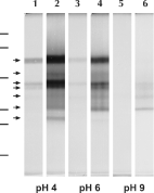
Fig. 1. Gelatin/SDS–PAGE analysis of protease activities in the Philasterides dicentrarchi crude extract (CE). The gel strips were incubated for 12 h at 37 °C at pH 4, 6 or 9. Lanes 1, 3 and 5: incubation in buffer without DTT. Lanes 2, 4 and 6: incubation in buffer containing 1 mM DTT. Relative molecular masses of standard markers (111, 73, 47·5, 33·9, 28·8 and 20·5 kDa from top to bottom) are shown to the left of the gel. The main proteolytic activities are labelled with arrows at the left of lane 1.
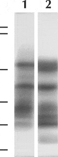
Fig. 2. Comparison of protease activities in crude extract (CE) and excretion/secretion (E/S) products of Philasterides dicentrarchi, by gelatin/SDS–PAGE analysis. The gel strips were incubated at pH 4 in the presence of 1 mM DTT. Lane 1: proteinases contained in CE. Lane 2: proteinases contained in E/S. Relative molecular masses of standard markers (113, 92, 52·3, 35, 28·9 and 21 kDa from top to bottom) are shown to the left of the gel.
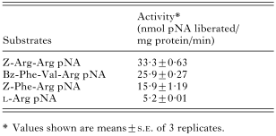
Table 1. Hydrolysis of p-nitroanilide substrates by proteases present in the crude extract (CE) of Philasterides dicentrarchi
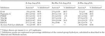
Table 2. Effects of protease inhibitors on the hydrolysis of Z-Arg-Arg pNA, Bz-Phe-Val-Arg pNA and Z-Phe-Arg pNA by the Philasterides dicentrarchi crude extract
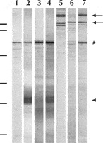
Fig. 3. Digestion of haemoglobin and type-I collagen by cysteine proteases present in the crude extract (CE) of Philasterides dicentrarchi. Lane 1: CE incubated without either substrates or E-64. Lanes 2 and 5: haemoglobin and type-I collagen, respectively, incubated in the absence of CE. The position of the haemoglobin dimer band (coinciding with that of an abundant unidentified parasite protein) is indicated with an asterisk. Lanes 3 and 6: haemoglobin and type-I collagen, respectively, incubated in the presence of CE. Note degradation of the haemoglobin monomer (arrow head) and the β- and α-chains of type-I collagen (uppermost and lowermost arrows to the right of the gel, respectively). Lanes 4 and 7: electrophoretic profiles of haemoglobin and type-I collagen, respectively, incubated in the presence of CE and E-64. Relative molecular masses of standard markers (111, 73, 47·5, 33·9, 28·8 and 20·5 kDa from top to bottom) are shown to the left of the gel.
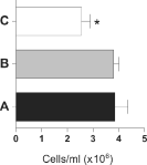
Fig. 4. Effect of the cysteine proteinase-specific inhibitor E-64 on the Philasterides dicentrarchi histolytic activity observed in ciliate–turbot fibroblast co-cultures. The turbot fibroblasts were cultivated without ciliates or inhibitors as negative control (A) or in the presence of ciliates with or without E-64 inhibitor (B and C respectively). Bars show mean number of cells (±S.D.) counted in each monolayer after 24 h of incubation (3 replicates per treatment). Asterisks indicate a significant difference (P<0·05) with respect to the control (A).
 You have
Access
- 48
- Cited by