Introduction
Nasal polyps are clinically diagnosed via rigid nasendoscopy on an out-patient basis. The appearance of a single unilateral mass on rigid nasendoscopy raises the possibility of neoplasia or intracranial extension. Current clinical practice is to perform urgent computed tomography (CT), followed by urgent surgical biopsy or resection. Some clinicians argue that if the mass is easily visualised in the out-patient clinic, then an in-office biopsy of the mass should be obtained. Considerations for this biopsy include the assurance that the lesion is not vascular or does not contain cerebrospinal fluid. This rationale varies significantly from the management of bilateral nasal polyps which is initially medical, with surgical polypectomy reserved for those cases unresponsive to maximal medical therapy.
This study aimed to assess the necessity for histopathological investigation as routine practice in every case of bilateral or unilateral nasal lesions by estimating the incidence of unexpected pathologies. We also investigated the value of CT scans in predicting histopathological diagnosis in patients with unilateral nasal lesions.
Materials and methods
This study was based on a retrospective analysis conducted in the University of Edinburgh Otolaryngology Department at St John's Hospital, a tertiary referral centre that covers otolaryngology services for the south-east of Scotland.
Ninety-eight adult patients underwent primary or revision elective endonasal polypectomy and sinus surgical procedures as in-patients during the 12-month period from January 2010 to December 2010, and had biopsies taken. The procedures were performed by ENT consultants (with or without a special interest in rhinology), specialty registrars and senior house officers. All operations were performed after failure of medical treatment, or if there was suspicion of a benign or malignant neoplasm. No pre-operative biopsies were performed in the clinic under local anaesthetic. Patients were reviewed in the out-patient department using rigid nasendoscopy. Those clinically diagnosed with bilateral polypoid disease were listed for routine polypectomy.
A total of 102 consecutive nasal specimens obtained during surgical polypectomy procedures were initially identified from the pathology database. Of these 102 specimens, 4 were taken from the same patients during revision surgery within the 12-month period. The total number of patients included in the study was therefore 98. The mean age of patients was 50.98 years (range, 16–89 years), and the male to female ratio was 2.5 to 1. 32 per cent of patients had undergone previous surgery, 28 per cent were asthmatic, and 9 per cent had asthma and aspirin sensitivity. Exclusion criteria included age younger than 16 years, and a known previous benign or malignant neoplastic sinonasal lesion. Patients for whom there was a high suspicion of neoplasia but who had undergone no previous biopsies were included in the study.
Patients clinically diagnosed with a unilateral nasal lesion were listed for an urgent polypectomy and underwent pre-operative CT. Other indications for scanning patients with clinically bilateral disease included revision surgery (simple polypectomy with or without sinus surgery) and pre-operative planning for primary sinus surgery with polypectomy. In the majority of cases (79 out of 98 cases), urgent or routine pre-operative CT scans were performed. In the remaining 19 cases, primary endoscopic polypectomy was performed without pre-operative CT scanning.
Clinical diagnoses made using rigid nasendoscopy were compared with histopathological diagnoses confirmed from biopsy samples obtained during surgical excision. The CT images were scored according to the Lund–Mackay staging system blindly (i.e. the scorer was not aware of the histopathological diagnosis or the clinical presentation of each case). In order to assess for asymmetry of disease on CT imaging, we compared the Lund–Mackay subscores for each side. The absolute difference between the two sides was recorded for all bilateral polyps patients. For patients with unilateral polyps or inverted papillomas, we used the side contralateral to that diagnosed in the out-patient setting as reference point (e.g. for a patient with a right unilateral lesion seen in clinic, the asymmetry score would be calculated as the score for the right side minus the score for the left side). Asymmetry scores could therefore range from 0 to 12.
All statistical tests were performed using SPSS for Windows software (version 11.5; SPSS, Chicago, Illinois, USA). Results were given as percentages, means, standard deviations (SDs) and 95 per cent confidence intervals. The unpaired t-test was used for testing statistical significance.
A previous study showed that the average total Lund–Mackay score in patients with chronic rhinosinusitis was 11, with an SD of 6.5.Reference Hopkins, Browne, Slack, Lund and Brown1 A sample size calculation was conducted to determine the minimum sample required to detect a significant change in the total Lund–Mackay score. Based on these data, it was determined that a sample size of 4 patients was needed to demonstrate a significant difference of 10 in the total scores in order to achieve the desired power of 80 per cent at an alpha value of 0.05 for a two-sided test. A sample size of 10 patients was needed to show a significant difference of 6 in the total scores.
Results
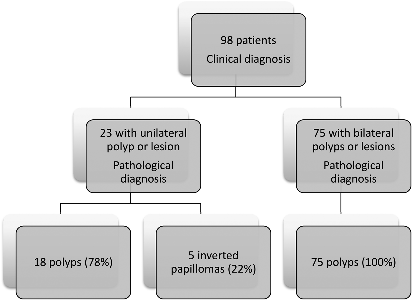
Of the 98 patients who underwent surgical polypectomy, 5 patients (5.1 per cent) had a histological diagnosis of inverted papilloma and 93 had simple inflammatory polyps (Figure 1). All five inverted papilloma patients were clinically diagnosed as having a unilateral lesion in the out-patient department. None of the 75 patients clinically diagnosed with bilateral lesions or polyps were found to have inverted papillomas or other neoplasms on histopathological examination.
Flow diagram of histopathological diagnosis of all patients.

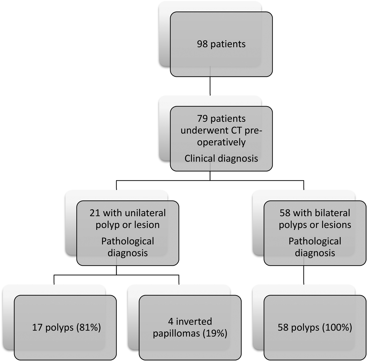
Of the 98 patients included in the study, 79 underwent pre-operative CT of their sinuses. Of the 23 patients with unilateral polyposis on clinical examination, 21 underwent CT prior to their operation. Of the two patients not scanned, one proved to have inverted papilloma. One was an elderly patient in whom no further surgery was planned because of significant co-morbidities. The other was a patient, for whom there was a low index of suspicion, who had a simple unilateral polyp confirmed on histopathology following simple polypectomy. Of the 75 patients with bilateral polyps on clinical examination, 58 underwent CT of their sinuses (Figure 2).
Flow diagram of histopathological diagnosis of patients who underwent computed tomography (CT) pre-operatively.
Total Lund–Mackay scores were calculated for all 79 patients who underwent CT (Figure 3). The mean score (± SD) for unilateral simple polyps patients was 9.29 ± 4.09, which was not quite statistically significant compared to a mean score of 4.75 ± 5.06 for inverted papilloma patients (p = 0.0702). There was, however, a strong statistical difference when the mean total scores for inverted papilloma patients were compared to those for bilateral polyps patients (4.75 ± 5.06 vs 15.33 ± 6.15; p = 0.0002). The mean scores for unilateral polyps patients were also statistically different to those for bilateral polyps patients (9.29 ± 4.09 vs 15.53 ± 6.15; p = 0.0011).
Mean total Lund–Mackay scores and 95 per cent confidence intervals for patients with unilateral and bilateral simple polyps and inverted papillomas.
Lund–Mackay asymmetry scores were calculated for all 79 patients who underwent CT, and statistical analysis was undertaken using the unpaired two-tailed t-test (Figure 4). The mean asymmetry scores for unilateral polyps patients were significantly lower than those for the inverted papilloma patients (0.75 ± 0.85 vs 2.75 ± 1.71; p = 0.0025), but not significantly different when compared to bilateral polyps patients (0.75 ± 0.85 vs 0.74 ± 0.85; p = 0.9661). Asymmetry scores were significantly higher for inverted papilloma patients when compared to those for bilateral polyps patients (2.75 ± 1.71 vs 0.74 ± 0.85; p = 0.0001).
Mean Lund–Mackay asymmetry scores and 95 per cent confidence intervals for patients with unilateral and bilateral simple polyps and inverted papillomas.
Discussion
There is controversy regarding whether all nasal polyp specimens removed during surgical excision should be sent for histopathological examination. Routine histopathological examination of all nasal polyps is performed by some surgeons merely because of the medicolegal consequences of missing an occult neoplasm. Inverted papillomas or other neoplastic lesions may also occur in clinically normal looking bilateral polyps.Reference Alun-Jones, Hill, Leighton and Morrissey2–Reference Kale, Mohite, Rowlands and Drake-Lee4 In this case, the consequences for the patient and the medicolegal implications of such an oversight argue in favour of processing every specimen.
None of our patients with clinically bilateral polyps had an unexpected histopathological diagnosis. A substantive proportion of our patients (22 per cent) clinically diagnosed with a unilateral lesion were found to have an inverted papilloma. This evidence suggests that only unilateral lesion specimens should be sent for histopathological diagnosis.
Patients with an inverted papilloma had lower total Lund–Mackay scores on their CT scans compared to patients with bilateral polyps. Those with proven inverted papilloma had significantly higher mean Lund–Mackay asymmetry scores than unilateral and bilateral polyps patients. Our findings also showed that there was no difference in the asymmetry scores between bilateral and unilateral polyps patients.
Our data sample was smaller compared to previous studies, but our study design and patient selection process reflects a more practicable approach. By including patients that underwent revision surgery and those with suspicious unilateral lesions on rigid nasendoscopy, our we are more likely to investigate a more representative sample of patients.
We refrained from using the arbitrary definition of unilateral maxillary sinus opacification to define unilateral disease, as unilateral polyps or inverted papillomas are associated with not just complete but also partial maxillary sinus opacification and isolated ethmoid opacification. Adopting this inclusion criterion probably misses out a substantive number of cases with suspicious underlying pathologies. We have instead devised an asymmetry score based on the Lund–Mackay staging system to assess this.
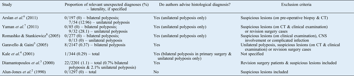
A review of the existing literature shows little consensus regarding when to send specimens in routine sinus surgery cases for histopathological examination. The majority of existing studies focus on occult pathology in nasal polyposis, but do not provide any established guidelines that can be used in routine cases of nasal polyposis. Seven major studies have been published on the matter, with variable advice given by the authors (Table I).Reference Alun-Jones, Hill, Leighton and Morrissey2–Reference Romashko and Stankiewicz8
Reported incidence of unexpected neoplasia following nasal polypectomy surgery
CT = computed tomography; CNS = central nervous system
In three recently published retrospective studies, the authors recommended that only unilateral lesion specimens should be sent for histopathological examination.Reference Arslan, Hidir, Durmaz, Karslioglu, Tosun and Gerek6–Reference Romashko and Stankiewicz8 Two of these studies reported 12.96 per centReference Arslan, Hidir, Durmaz, Karslioglu, Tosun and Gerek6 and 0 per centReference Romashko and Stankiewicz8 detection rates of neoplasms in unilateral lesions, which are lower compared to our results. However, these studies excluded patients with highly suspicious lesions based on radiological findings and pre-operative biopsy results taken in the out-patient clinic. This has therefore reduced the overall number of patients in their sample with a high possibility of neoplasm.
A large study by Garavello and Gaini reported a 0.37 per cent detection rate of neoplasms in patients with bilateral lesions.Reference Garavello and Gaini5 The authors of that study excluded patients with unilateral polyps, those with highly suspicious lesions based on pre-operative CT scans and patients who had undergone revision surgery. This latter exclusion criterion is in contrast to the high proportion of patients with revision surgery in our study (32 per cent). The former two exclusion criteria should theoretically reduce the detection rate of unexpected pathologies, but a small proportion of patients in their study still presented in such a manner.
Kale et al. demonstrated a detection rate of 0.29 per cent, suggesting that specimens from primary surgery patients with bilateral lesions and all cases of unilateral lesions should be sent for histology.Reference Kale, Mohite, Rowlands and Drake-Lee4 The exclusion criteria in that study were not clearly stated. In the only study in which a cost–benefit analysis was performed, an unexpected pathology detection ratio as low as 1.1 per cent did not deter the authors from recommending routine histopathological examination for all specimens.Reference Diamantopoulos, Jones and Lowe3 Highly suspicious lesions and revision surgery patients were excluded from the patient group under investigation. The former exclusion criterion would explain the low detection rate of neoplasia in unilateral lesions compared to our data. The first published study on the topic, conducted in 1990, showed a detection rate for unexpected pathologies of 0 per cent, and, therefore, the authors did not recommend routine histopathological examination of nasal polypoidal lesions.Reference Alun-Jones, Hill, Leighton and Morrissey2
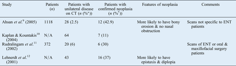
Our secondary aim in this project was to evaluate the use of CT scanning in predicting the risk of neoplasia in patients clinically diagnosed (using rigid nasendoscopy) with unilateral nasal polyposis. In four studies identified in a review of the current literature, the risk of malignancy in patients with radiological evidence of unilateral disease ranged from 11 to 42.9 per cent (Table II).Reference Ahsan, El-Hakim and Ah-See9–Reference Lehnerdt, Weber and Dost12 All of these studies used the term ‘unilateral maxillary sinus opacification’ to define unilateral disease. This diagnosis was made radiologically, rather than clinically as in our study. This important difference in the inclusion criterion makes our patient sample similar to that of a ‘real life scenario’. Patients present with findings on endoscopy rather than with radiological findings.
Unilateral opacification on computed tomography scans and histopathological diagnosis
*Percentage of all patients. †Percentage of patients with unilateral disease. CT = computed tomography; N/A = not applicable
To our knowledge, the Lund–Mackay score has only been used in one study to evaluate the significance of CT in determining diagnostic criteria for patients with unilateral sinus opacification.Reference Kaplan and Kountakis10 In that study, the overall Lund–Mackay scores were found to be higher in patients presenting with unilateral sinus opacification with a histopathological diagnosis of simple polyps compared to patients with inverted papillomas. However, it was not clarified whether patients with polyps had clinically bilateral or unilateral disease.
The study with the largest patient population was based on a retrospective review of all CT scans performed in a radiology department, not necessarily for ENT patients.Reference Ahsan, El-Hakim and Ah-See9 This means that a great number of incidental findings would have been picked up.
Current surgical practice in this area is very varied and depends on the individual surgeon's preferences. Our study findings suggest that only unilateral lesion specimens should be sent for histopathological diagnosis. Further studies are required to establish what the current practice is amongst ENT surgeons, particularly specialist rhinologists. In addition, a detailed cost–benefit analysis would be useful to investigate how much the current workload and cost could be reduced without compromising patient care. This has not been assessed in the last 10 years. During this period, medical litigation, patient expectations and costs of surgery have changed considerably. The cost of each nasal polyp specimen processed at the Royal Infirmary of Edinburgh pathology laboratories is approximately £70, which includes consumables, and the biomedical scientist's and consultant pathologist's time. The overall cost of sending the 75 bilateral nasal lesion specimens for histopathological analysis over this 12-month period was £5250.
The small foci of potential carcinoma in situ or neoplastic tissue within bilateral simple polypoid tissue are macroscopically undetectable by the operating surgeon. Nasal polypectomy tends to involve removal of all visible polypoidal tissue. Therefore, surgery for bilateral simple polyps harbouring potential pre-malignant or even malignant microscopic disease is a curative procedure for this group of patients. Hence, even if such a diagnosis is confirmed, arguably, the initial management plan will not change. One could argue though that more surveillance endoscopies and more frequent follow up might be necessary if such information is available.
• There is controversy regarding whether all nasal polyp specimens removed during surgical excision should be sent for histopathological examination
• Unilateral nasal lesions should be sent for routine histopathological diagnosis
• Computed tomography imaging may have a role in predicting histopathological diagnosis by demonstrating asymmetry and less overall sinus opacification in neoplasia patients
Finally, most clinicians are happy to manage bilateral polypoid disease medically without histological diagnosis if symptom control is adequate. One therefore has to question the logic of suddenly requiring a histological diagnosis if surgery is for simple bilateral polyposis for reasons of symptom control. An exception to this might be cases with eosinophilic infiltration of polypoidal mucosa, where the histological findings can be a predictive parameter of recurrent disease that therefore necessitate long-term follow up and treatment.Reference Matsuwaki, Ookushi, Asaka, Mori, Nakajima and Yoshida13
This study discusses a controversial area in ENT surgery in which no clear guidelines have been established. We propose that CT has a definite role in predicting histopathological diagnosis. Although patients with suspicious lesions still need to be scanned urgently, their operations do not have to be performed on an urgent basis if their CT findings are not highly suggestive of neoplasia. This will favour a less urgent approach towards surgical excision in this patient group, with a subsequent reduction in the waiting list workload.