Introduction
Acute rheumatic fever is a systemic inflammatory disease that develops 1 to 5 weeks after upper respiratory tract infections with group A beta-haemolytic streptococcus and primarily affects the heart, joints, brain, skin, and subcutaneous tissues. Although its incidence has markedly declined in developed countries, acute rheumatic fever remains the leading cause of acquired heart disease in children and young adults in many developing regions. Reference Karthikeyan and Guilherme1
In Turkey, acute rheumatic fever remains a clinically relevant condition and is classified as an intermediate- to high-risk population according to the Jones Criteria. Epidemiological data demonstrate regional and temporal variability; reported incidence rates include 21/100,000 in Ankara between 2000 and 2009 and 7.4/100,000 in Kayseri between 1998 and 2011. Reference Orün, Ceylan and Bilici2,Reference Narin, Mutlu and Argun3 More recent nationwide estimates suggest an overall incidence of approximately 8.84/100,000, with regional variation ranging from 3.3 to 14.4 per 100,000. Reference Gürses, Koçak and Tutar4 However, up-to-date comprehensive epidemiological data remain limited.
The diagnosis of acute rheumatic fever is based on the Jones Criteria, which require the presence of major and minor clinical findings in addition to evidence of preceding streptococcal infection. Reference Arı, Ertuğrul and Yoldaş5 Among these manifestations, carditis is the most important determinant of long-term morbidity and mortality. Clinically, carditis may present with murmurs, cardiomegaly, heart failure, pericardial effusion, or a pericardial friction rub, and echocardiography plays a central role in confirming valvular involvement. Reference Ralph, Noonan and Wade6 Importantly, some patients may lack overt clinical findings and instead demonstrate echocardiographic evidence of subclinical (silent) carditis. According to the revised Jones criteria, silent carditis is recognised as a major manifestation in both low- and high-risk populations. Reference Gewitz, Baltimore and Tani7
Antistreptolysin O is a widely used serological marker indicating recent streptococcal infection. Elevated antistreptolysin O titers are detected in approximately 80% of patients following infection, while up to 20% may have normal levels. Reference Gewitz, Baltimore and Tani7 In some settings, anti-DNase B is used in combination with antistreptolysin O to improve diagnostic sensitivity; however, its availability remains limited in certain regions. Reference Delice, Adaleti and Cevan8 Importantly, these serological markers provide evidence of preceding streptococcal exposure but do not establish the diagnosis of acute rheumatic fever in the absence of compatible clinical findings and should be interpreted within the framework of the Jones Criteria.
In clinical practice, antistreptolysin O testing is frequently requested by primary care or outpatient physicians to evaluate suspected streptococcal infections, including presentations such as recurrent pharyngitis, upper respiratory tract infections, or non-specific complaints such as fatigue or arthralgia. As a result, elevated antistreptolysin O titers may be incidentally detected in children who do not fulfil the Jones Criteria and have no clinical signs of acute rheumatic fever. In such cases, the clinical significance of isolated antistreptolysin O elevation and the need for further cardiac evaluation remain uncertain.
Therefore, this study aimed primarily to determine the prevalence of silent carditis in asymptomatic children with elevated antistreptolysin O titers and secondarily to describe their echocardiographic findings.
Material and methods
This descriptive cross-sectional study was conducted in the Paediatric Cardiology Department of a tertiary care hospital between September 2020 and September 2021. A total of 61 asymptomatic children aged 3–18 years with elevated antistreptolysin O titers were included.
Ethical approval was obtained from the Scientific Research Ethics Committee of the University of Health Sciences, Hamidiye Faculty of Medicine (approval date: October 23, 2020; protocol number: 20/376). The study was conducted in accordance with the principles of the Declaration of Helsinki. Written informed consent was obtained from the parents or legal guardians of all participants, and assent was obtained from children aged ≥8 years.
This study did not involve direct sampling from the general paediatric population. Instead, children with elevated antistreptolysin O titers identified in primary care or outpatient settings were referred to our tertiary paediatric cardiology centre for further evaluation. These patients were asymptomatic with respect to acute rheumatic fever and cardiac involvement, and antistreptolysin O testing had been performed during evaluation of upper respiratory tract infections or non-specific symptoms such as fatigue or arthralgia. Therefore, the total number of patients with normal antistreptolysin O levels during the study period was not available.
To ensure a homogeneous study cohort, predefined exclusion criteria were applied: a known history of rheumatic disease, clinical findings suggestive of acute rheumatic fever, or physical examination findings consistent with carditis. Among the referred patients during the study period, none met the exclusion criteria, and all eligible cases were consecutively included.
Demographic characteristics (age, sex), anthropometric measurements (height, weight), blood pressure values, electrocardiographic parameters (including PR interval), and timing of presentation were recorded.
An antistreptolysin O level greater than 150 IU/mL was considered elevated according to the institutional laboratory reference range.
Acute phase reactants, including C-reactive protein and erythrocyte sedimentation rate, were measured. Complete blood count parameters were analysed, including haemoglobin level, platelet count, total white blood cell count, neutrophil, lymphocyte, and monocyte counts, and derived inflammatory markers such as neutrophil-to-lymphocyte ratio, platelet-to-lymphocyte ratio, and lymphocyte-to-monocyte ratio.
All patients underwent screening transthoracic Doppler echocardiography performed by an experienced paediatric cardiologist. Pathological valvular regurgitation was defined according to the World Heart Federation 2012 criteria for the echocardiographic diagnosis of rheumatic heart disease. Reference Reményi, Wilson and Steer9 Silent carditis was defined as echocardiographic evidence of pathological valvular regurgitation in the absence of clinical signs of carditis on physical examination.
Patients diagnosed with silent carditis were initiated on benzathine penicillin G prophylaxis according to standard secondary prevention recommendations.
Statistical analysis
Statistical analyses were performed using SPSS version 25.0 (IBM Corp., Armonk, NY, USA). Continuous variables were expressed as mean ± standard deviation or median (interquartile range), depending on distribution. Group comparisons were performed using Student’s t-test or Mann–Whitney U test as appropriate. Correlations were assessed using Spearman’s rank correlation coefficient.
The strength of correlation was classified as follows: 0.00–0.25, very weak; 0.26–0.49, weak; 0.50–0.69, moderate; 0.70–0.89, strong; and 0.90–1.00, very strong. A p-value < 0.05 was considered statistically significant. All correlation analyses were exploratory and interpreted cautiously due to the limited sample size.
Sample size calculation
The sample size was calculated using OpenEpi software based on an estimated prevalence of subclinical rheumatic heart disease of 4%. This estimate is supported by epidemiological evidence from echocardiographic screening studies, including a systematic review and meta-analysis, as well as community-based studies reporting prevalence rates of 2–4%. Reference Rothenbühler, O’Sullivan and Stortecky10–Reference Beaton, Okello and Lwabi12 With a 95% confidence level and a 5% margin of error, the minimum required sample size was 56 participants. A total of 61 patients were included in the final analysis.
Results
The study included 61 children with elevated antistreptolysin O levels, of whom 37 (60.7%) were female, and 24 (39.3%) were male. The median age was 11 years (IQR: 7–14 years). Most admissions occurred during the winter season (41.0%). The demographic, clinical, and baseline echocardiographic characteristics of the study population are summarised in Table 1.
Demographic, clinical, and echocardiographic characteristics of the study group

Note: Data are presented as n (%) for categorical variables, mean ± standard deviation (SD) for normally distributed continuous variables, and median (interquartile range [IQR]) for non-normally distributed continuous variables. DBP = diastolic blood pressure; EF = ejection fraction; FS = fractional shortening; IVSd = interventricular septal thickness in diastole; LVIDd = left ventricular internal diameter in diastole; LVIDs = left ventricular internal diameter in systole; LVPWd = left ventricular posterior wall thickness in diastole; SBP = systolic blood pressure.
Silent carditis was identified in 8 patients (13.1%; 95% CI: 6.9–23.3), the majority of whom were female (75%), with a median age of 12 years (IQR: 9–14). Half of the cases were diagnosed during winter.
Echocardiographic evaluation of patients with silent carditis (n = 8) demonstrated pathological valvular regurgitation consistent with rheumatic involvement. Combined mitral regurgitation and aortic regurgitation were identified in four patients (three with mild mitral regurgitation and one with moderate mitral regurgitation, all with concomitant mild aortic regurgitation). Isolated mitral regurgitation was observed in four patients (all mild). No definite rheumatic morphological valve changes were identified according to the 2012 World Heart Federation criteria.
In patients without silent carditis (n = 53), echocardiographic findings were predominantly normal (n = 46, 86.8%). Trivial (physiological) grade 1 mitral regurgitation was observed in 3 patients (5.7%). Incidental congenital anomalies were rare and included a secundum atrial septal defect in 2 patients (3.8%) and a bicuspid aortic valve in 1 patient (1.9%).
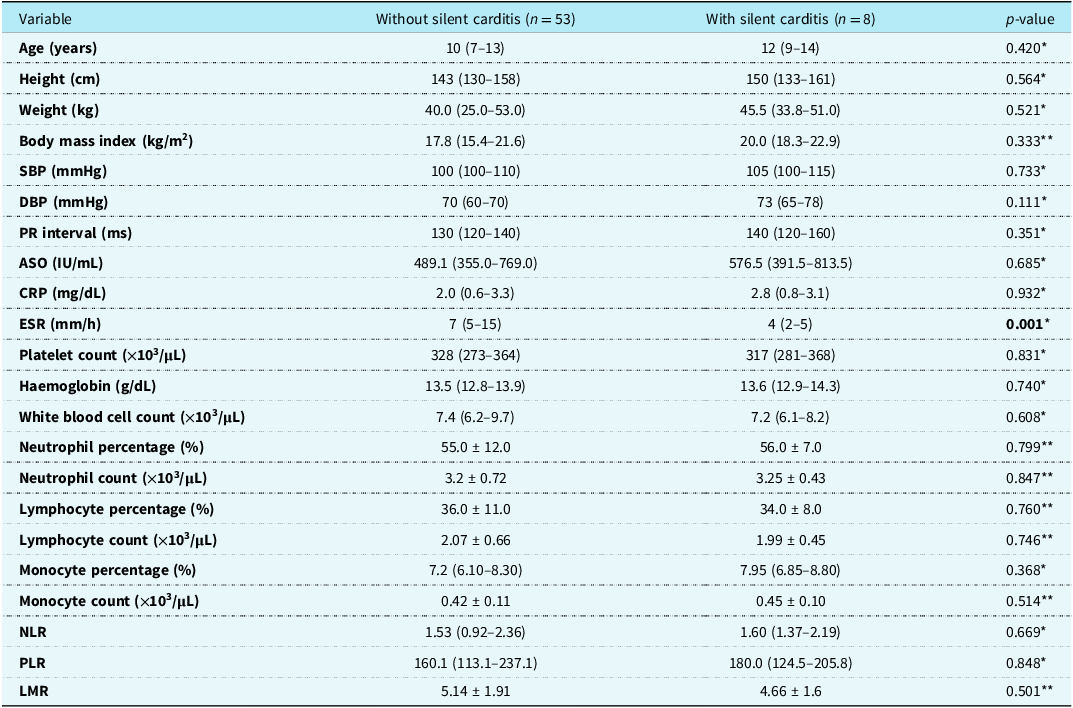
Comparison of clinical and laboratory parameters between children with and without silent carditis is presented in Table 2. A statistically significant difference was observed only in erythrocyte sedimentation rate (p = 0.001). In the group without silent carditis, elevated inflammatory markers were observed in a subset of patients: CRP in 13.2% (n = 7), ESR in 18.9% (n = 10), and leukocytosis in 13.2% (n = 7). Thrombocytosis and anaemia were observed in 9.4% (n = 5) and 7.5% (n = 4), respectively.
Comparison of clinical and laboratory parameters between children with and without silent carditis

Note: Data are presented as mean ± standard deviation (SD) for normally distributed continuous variables and median (interquartile range) for non-normally distributed variables. ASO = antistreptolysin O; CRP = C-reactive protein; DBP = diastolic blood pressure; ESR = erythrocyte sedimentation rate; LMR = lymphocyte-to-monocyte ratio; NLR = neutrophil-to-lymphocyte ratio; PLR = platelet-to-lymphocyte ratio; SBP = systolic blood pressure; WBC = white blood cell count. Bold values represents statistically significant differences between groups (p < 0.05).
*Compared using the Mann–Whitney U test.
**Compared using the independent samples t-test. A p-value < 0.05 was considered statistically significant.
By study design, all patients had elevated antistreptolysin O titers. Patients with silent carditis did not demonstrate anaemia or thrombocytosis, and acute phase reactants were within normal ranges in this subgroup.
Correlation analysis showed that antistreptolysin O levels had weak positive correlations with CRP (ρ = 0.361, p = 0.004) and ESR (ρ = 0.279, p = 0.030). Weak negative correlations were observed between antistreptolysin O titers and age (ρ = −0.325, p = 0.011), height (ρ = −0.334, p = 0.009), haemoglobin (ρ = −0.322, p = 0.011), mean arterial pressure (ρ = −0.263, p = 0.040), and left ventricular posterior wall diameter (ρ = −0.270, p = 0.036).
Discussion
Valvular pathologies detected exclusively by echocardiography in the absence of clinical signs of carditis are defined as silent or subclinical carditis and account for approximately 53% of all rheumatic carditis cases. Reference Gewitz, Baltimore and Tani7 In this study, we evaluated the prevalence and clinical characteristics of silent carditis in asymptomatic children with evidence of recent streptococcal infection and found a prevalence of 13.1% (n = 8).
Previous studies have reported variable rates of silent carditis. For instance, Güler et al. demonstrated an increase in the incidence of silent carditis over time, from 21.8 to 36%, likely reflecting improved awareness and echocardiographic detection. Reference Güler, Laloğlu and Olgun13 In contrast, Bülbül et al. reported a lower incidence of 10.8%. Reference Bulbul, Akyol and Civan14 These differences may be related to variations in study populations, endemic burden of streptococcal infection, and differences in diagnostic criteria and echocardiographic protocols.
Although acute rheumatic fever most commonly affects children aged 5–15 years, cases in late adolescence and adulthood have also been reported. Reference Zühlke, Beaton and Engel15 The median age of 12 years (IQR: 9–14) in our silent carditis group is consistent with previous reports, including a North Indian cohort (mean 11.27 ± 2.05 years) and the study by Özdemir et al. (mean 11.4 ± 2.5 years). Reference Mehrotra, Narain and Sethi16,Reference Özdemir, Işık and Abacı17 The increasing burden with age is thought to reflect cumulative exposure to group A streptococcal infections and repeated immune activation. Reference Carapetis, Hardy and Fakakovikaetau18
Although the overall incidence of acute rheumatic fever does not significantly differ between sexes, rheumatic heart disease is more frequently reported in females. Reference Carapetis, Beaton and Cunningham19 In our study, a female predominance was observed (female-to-male ratio 3.0), consistent with previous literature, although the underlying mechanisms remain unclear and may include biological and healthcare-related factors. Reference Saxena, Ramakrishnan and Roy20
Seasonal variation was also evident in our findings. Streptococcal infections are more prevalent during autumn and winter, paralleling the seasonal peak of acute rheumatic fever. Pirinçcioğlu et al. reported that 65.9% of acute rheumatic fever cases were diagnosed during winter. Reference Pirinççioğlu, Alyan and Kanğın21 Similarly, most cases in our cohort occurred in winter (41.0%) and autumn (31.1%), with half of silent carditis cases detected in winter, supporting a seasonal pattern in disease occurrence.
Echocardiography remains the cornerstone for detecting subclinical valvular involvement. In our study, to minimise overdiagnosis, pathological mitral and aortic regurgitation were defined according to the 2012 World Heart Federation Doppler criteria. Reference Reményi, Wilson and Steer9 The updated World Heart Federation 2023 recommendations emphasise stricter morphological and Doppler thresholds and highlight that valvular abnormalities should be interpreted along a continuum from physiological regurgitation to definite rheumatic heart disease. Reference Rwebembera, Marangou and Mwita22 In a post hoc re-analysis of the existing echocardiographic records using World Heart Federation 2023 criteria, case classification remained unchanged.
Mitral regurgitation was the most frequently observed valvular abnormality, either alone or in combination with aortic regurgitation, consistent with the known predilection of rheumatic carditis for the mitral valve. Importantly, no definite rheumatic morphological changes were identified, suggesting early functional valvular involvement rather than established rheumatic heart disease.
Our findings do not support universal echocardiographic screening. Instead, selective evaluation may be more appropriate in resource-limited settings. Echocardiography could be considered in selected patients with elevated antistreptolysin O titers and additional clinical or laboratory indicators; however, this remains hypothesis-generating and requires validation in larger prospective studies.
A weak negative correlation between antistreptococcal antibody levels and left ventricular posterior wall thickness was observed, without consistent evidence of ventricular dilation or global remodelling. In rheumatic carditis, ventricular remodelling typically manifests as progressive chamber dilation rather than isolated regional wall thickening. Reference Gaasch and Meyer23 Recent longitudinal data in children suggest that while cardiac function often remains preserved, those with severe rheumatic heart disease may exhibit increased end-systolic and end-diastolic volumes at diagnosis, with left ventricular dilation potentially worsening over time. Reference MacDonald, Tarca and Causer24 Given the cross-sectional design, these findings are likely incidental and should be interpreted cautiously.
This study has several limitations, including its single-centre design, relatively small sample size, and cross-sectional nature, which preclude assessment of disease progression and long-term outcomes. Additional limitations include the lack of age-adjusted antistreptolysin O reference ranges and the absence of anti–DNase B testing, which may have led to misclassification of streptococcal exposure. Socioeconomic and nutritional variables were not recorded, limiting the interpretation of haematological findings. Interobserver variability in echocardiographic assessment was not evaluated, and advanced imaging techniques such as strain imaging were not available. Therefore, subtle myocardial involvement may have been missed. Finally, the absence of elevated inflammatory markers in some patients does not clarify the temporal stage of disease, and prior unrecognised rheumatic episodes cannot be excluded.
This cohort represents a referred and pre-selected population rather than a true screening population, which may have led to an overestimation of prevalence. Therefore, the findings should not be generalised to the broader paediatric population and should be interpreted within a referral-based context.
Despite these limitations, the study has important strengths, including standardised echocardiographic evaluation and strict application of World Heart Federation criteria, which enhance the internal validity of the findings.
In conclusion, silent carditis may be detected in a subset of asymptomatic children with elevated antistreptolysin O titers. However, antistreptolysin O levels are not predictive of cardiac involvement, and echocardiography remains essential for diagnosis. These findings highlight a clinically relevant but underrecognised subgroup and underscore the need for larger, prospective studies to better define optimal evaluation strategies.
Acknowledgements
We would like to thank all participants.
Financial support
The authors declare that the study received no funding.
Competing interests
The authors declare that there are no conflicts of interest to disclose.
Ethical standard
This study was approved by the Scientific Research Ethics Committee of the University of Health Sciences, Hamidiye Faculty of Medicine (approval date: 23/10/2020, protocol number: 20/376), and was conducted in accordance with the principles of the Declaration of Helsinki.