Introduction
Until recently, only three diplozoid species were reported from Africa. Paradiplozoon ghanense (Thomas, Reference Thomas1957) was described from the gills of Brycinus macrolepidotus Valenciennes, 1850 in Ghana and Paradiplozoon aegyptense (Fischthal & Kuntz, Reference Fischthal and Kuntz1963) from the gills of Labeo forskalii Rüppell, 1835 in Egypt. These species have, respectively, been recorded from B. macrolepidotus and Alestes baremoze (Joannis, 1835) in Ghana (Thomas, Reference Thomas1957; Yamaguti, Reference Yamaguti1963; Paperna, Reference Paperna1969, Reference Paperna1979; Khotenovsky, Reference Khotenovsky1985) and L. forskalii, Labeo coubie Rüppell, 1832, Labeo victorianus Boulenger, 1901, Labeo cylindricus Peters, 1852, Raiamas senegalensis (Steindachner, 1870), Barbus paludinosus Peters, 1852 and B. macrolepidotus in Egypt, Ghana, Kenya, Tanzania and Uganda (Fischthal & Kuntz, Reference Fischthal and Kuntz1963; Yamaguti, Reference Yamaguti1963; Paperna, Reference Paperna1969, Reference Paperna1979; Petr & Paperna, Reference Petr and Paperna1979; Khotenovsky, Reference Khotenovsky1985). The third diplozoid, Afrodiplozoon polycotyleus (Paperna, Reference Paperna1973), was described from B. paludinosus, L. victorianus and Barbus cercops Whitehead, 1960 in Kenya and Barbus macrolepis Pfeffer, 1889 in Tanzania. This genus displays 8–10 pairs of clamps, in contrast to the two Paradiplozoon species which only display four. Afrodiplozoon polycotyleus has also been recorded from Barbus kerstenii Peters, 1868, Barbus neumayeri Fischer, 1884, Labeobarbus marequensis (Smith, 1841) Barbus trimaculatus Peters, 1852, Barbus neefi Greenwood, 1962 and A. baremoze in Nigeria, Uganda, Kenya, Tanzania and in the Limpopo province of South Africa (Paperna, Reference Paperna1973, Reference Paperna1979; Petr & Paperna, Reference Petr and Paperna1979; Khotenovsky, Reference Khotenovsky1985; Chapman et al., Reference Chapman, Lanciani and Chapman2000; Mashego, Reference Mashego2000; Echi & Ezenwaji, Reference Echi and Ezenwaji2009).
Paradiplozoon ichthyoxanthon Avenant-Oldewage, 2013 is the latest addition to the African diplozoids and was described from the gills of Labeobarbus aeneus (Burchell, 1822) in the Vaal River and Vaal Dam, South Africa (Avenant-Oldewage et al., Reference Avenant-Oldewage, Le Roux, Mashego and Jansen van Vuuren2013). The description of P. ichthyoxanthon was the first diplozoid description to include genetic confirmation of its distinctness and displayed 14.5–38.8% genetic distance from any other diplozoid taxon in terms of the second internal transcribed spacer (ITS2) fragment (Avenant-Oldewage et al., Reference Avenant-Oldewage, Le Roux, Mashego and Jansen van Vuuren2013). This species forms a completely isolated geographic clade when compared to sequences of other diplozoids (Avenant-Oldewage et al., Reference Avenant-Oldewage, Le Roux, Mashego and Jansen van Vuuren2013). In the same locality, another diplozoid was collected from the gills of moggel, Labeo umbratus (Smith, 1841), and it is described here as a new taxon: Paradiplozoon vaalense n. sp.
As for all monogeneans, the chitenoid structures of diplozoids are considered to be the most reliable features for morphological studies (Price, Reference Price1967; Khotenovsky, Reference Khotenovsky1985; Matějusová et al., Reference Matějusová, Koubková, Gelnar and Cunningham2002, Reference Matějusová, Koubková and Cunningham2004). Differences and sizes of sclerotized parts of the haptor, in particular the third pair of clamps and central hook, are used for diplozoid species discrimination (Gläser & Gläser, Reference Gläser and Gläser1964; Khotenovsky, Reference Khotenovsky1985; Matějusová et al., Reference Matějusová, Koubkova, D'Amelio and Cunningham2001). However, some authors state that use of morphological and biometrical characters in species discrimination is doubtful due to their variability (Bovet, Reference Bovet1967; Le Brun et al., Reference Le Brun, Renaud and Lambert1988). As such, morphological methods have been used in conjunction with molecular techniques for Diplozoidae (Le Brun et al., Reference Le Brun, Renaud and Lambert1988; Matějusová et al., Reference Matějusová, Koubkova, D'Amelio and Cunningham2001, Reference Matějusová, Koubková and Cunningham2004; Sicard et al., Reference Sicard, Desmarais and Lambert2001, Reference Sicard, Desmarais, Vigneux, Shimazu, Lambert, Combes and Jourdane2003; Gao et al., Reference Gao, Chen, Yao, Gao, Song and Wang2007). Several studies used molecular markers, especially the ITS2 marker, to study various diplozoid taxa (Matějusová et al., Reference Matějusová, Koubkova, D'Amelio and Cunningham2001, Reference Matějusová, Koubková and Cunningham2004; Sicard et al., Reference Sicard, Desmarais and Lambert2001, Reference Sicard, Desmarais, Vigneux, Shimazu, Lambert, Combes and Jourdane2003; Gao et al., Reference Gao, Chen, Yao, Gao, Song and Wang2007), but these studies focus on a select number of taxa and molecular data remain scarce, especially for African taxa. As such, morphology and relevant morphometric analyses, as well as comparative sequence data derived from the ITS2 region of rDNA, was used to confirm the distinctness of P. vaalense n. sp.
Materials and methods
Collection and examination of fish
A total of 101 moggel were collected between January 2012 and March 2012 in the Vaal Dam and Visgat area of the Vaal River using gill nets. Fish were euthanized by severing the spinal cord posterior to the skull with a single cut, after which the gills were removed. Gills were examined for the presence of parasites, which were preserved in either 70% or absolute ethanol for morphological and genetic analyses respectively. Some specimens were also fixed in a prostrate position in steaming hot aceto-formaldehyde alcohol and preserved in 70% ethanol.
During this survey, 67 adult pairs were collected. Some of these were studied using a TESCAN Vega 3 LMH scanning electron microscope (Brno, Czech Republic) at 5–10 kV acceleration voltages. Specimens were dehydrated through a graded ethanol series and then dried using hexamethyldisilazane (HMDS) (Merck, Germany) as per Nation (Reference Nation1983), but with gradually increased concentrations of HMDS for 15 min each. Graded series consisted of 40% (2 parts HMDS:3 parts absolute ethanol), 70% (7 parts HMDS:3 parts absolute ethanol) and two changes of 100% HMDS. Specimens were dried in a Sanpla dry keeper desiccator cabinet (Kita-Ku, Osaka, Japan) and coated with gold using an Emscope SC500 sputter coater (Quorum Technologies, Newhaven, UK).
For light miscopy most adult pairs were stained with Horen's trichrome and mounted in lactophenol, while the remainder were stained with Mayer's haematoxylin and mounted in Entellan® (Merck). Drawings of the parasite, sclerites and eggs were made from whole mounts using a drawing tube attachment, and measurements were made using a Zeiss Axioplan 2 imaging light microscope with AxioVision Release 4.7 and 4.8 software (Carl Zeiss, Jena, Switzerland). Mean measurements for relevant structures are given in the results section with number of measurements (n) and range of measurements provided in parentheses.
Genetic analysis
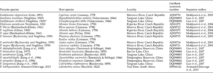
DNA was extracted from anterior sections of 20 adult pairs (i.e. from both individuals) using a DNeasy® tissue kit (QIAGEN Inc., Manchester, UK) following standard protocols suggested by the manufacturer. DNA was extracted from anterior sections of parasites to preserve haptors for morphological assessments as outlined above. Amplification was done using universal primers D and B1 (Bachellerie & Qu, Reference Bachellerie, Qu and Hyde1993). Polymerase chain reaction (PCR) amplification was done using a MultiGene Gradient system (Labnet International, Inc., Edison, New Jersey, USA) according to parameters set out by Matějusová et al. (Reference Matějusová, Koubkova, D'Amelio and Cunningham2001). Amplicons were sequenced following Avenant-Oldewage et al. (Reference Avenant-Oldewage, Le Roux, Mashego and Jansen van Vuuren2013). Sequences were aligned with those of several other diplozoid taxa (table 1) using MacClade 4 (Maddison & Maddison, Reference Maddison and Maddison2003). Eudiplozoon nipponicum was used to root resultant phylogenies, according to Gao et al. (Reference Gao, Chen, Yao, Gao, Song and Wang2007) and Avenant-Oldewage et al. (Reference Avenant-Oldewage, Le Roux, Mashego and Jansen van Vuuren2013). Parsimony (equally weighted characters) and distance (using uncorrected p-distances) approaches (as implemented in PAUP 4*; Swofford, Reference Swofford2002) were used to assess the genealogical relationships between taxa. Robustness of all resultant topologies was assessed by 1000 bootstrap replicates.
List of diplozoid species used for genetic comparison with Paradiplozoon vaalense n.sp., with their host species, locality, GenBank accession number and sequence author.
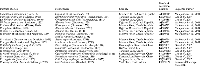
* GenBank record for Sindiplozoon ctenopharyngodoni (Ling, 1973), but according to Khotenovsky (Reference Khotenovsky1985) this is a synonym of S. strelkowi which is the valid name for this species.
Results
Morphology
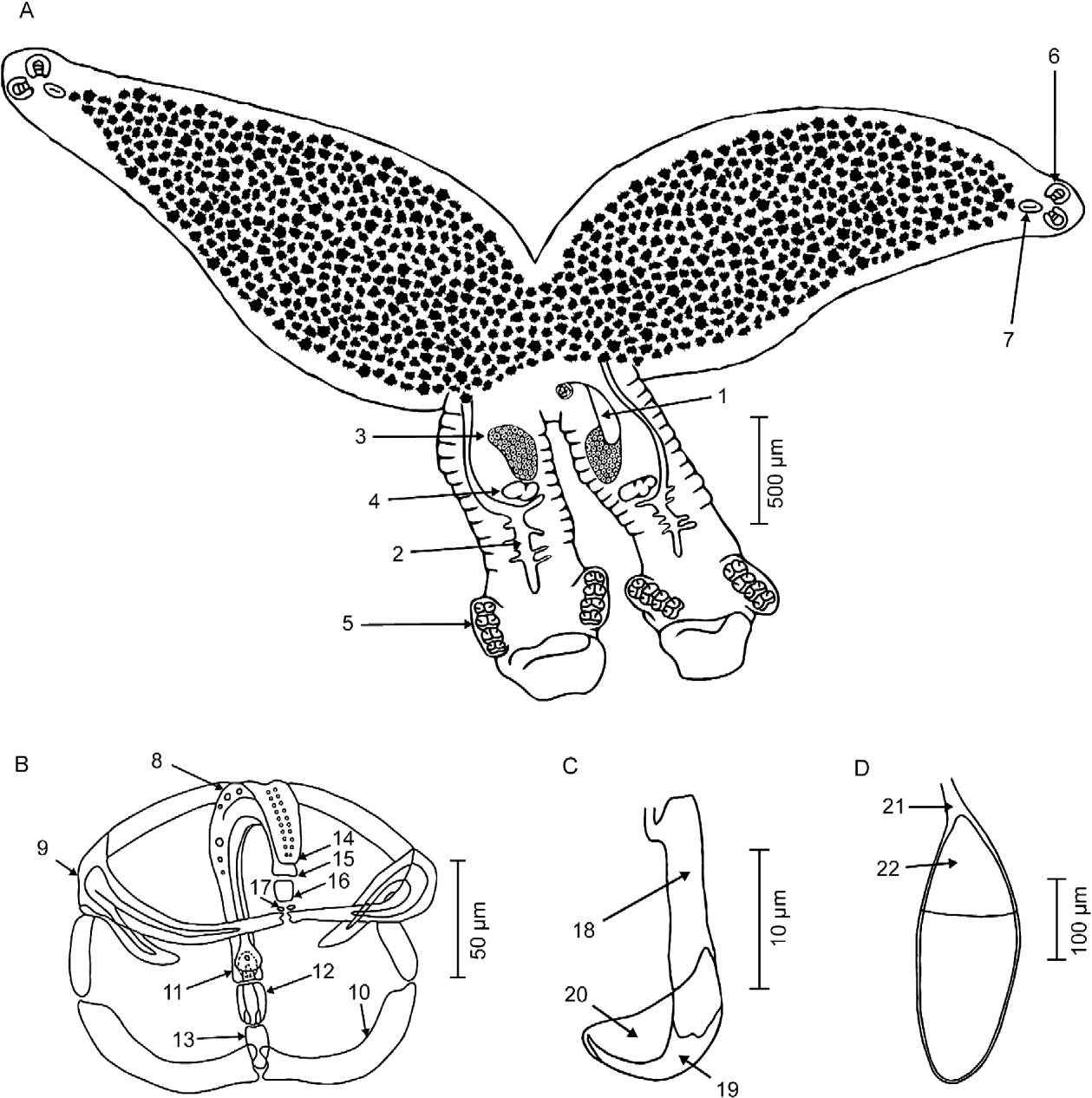
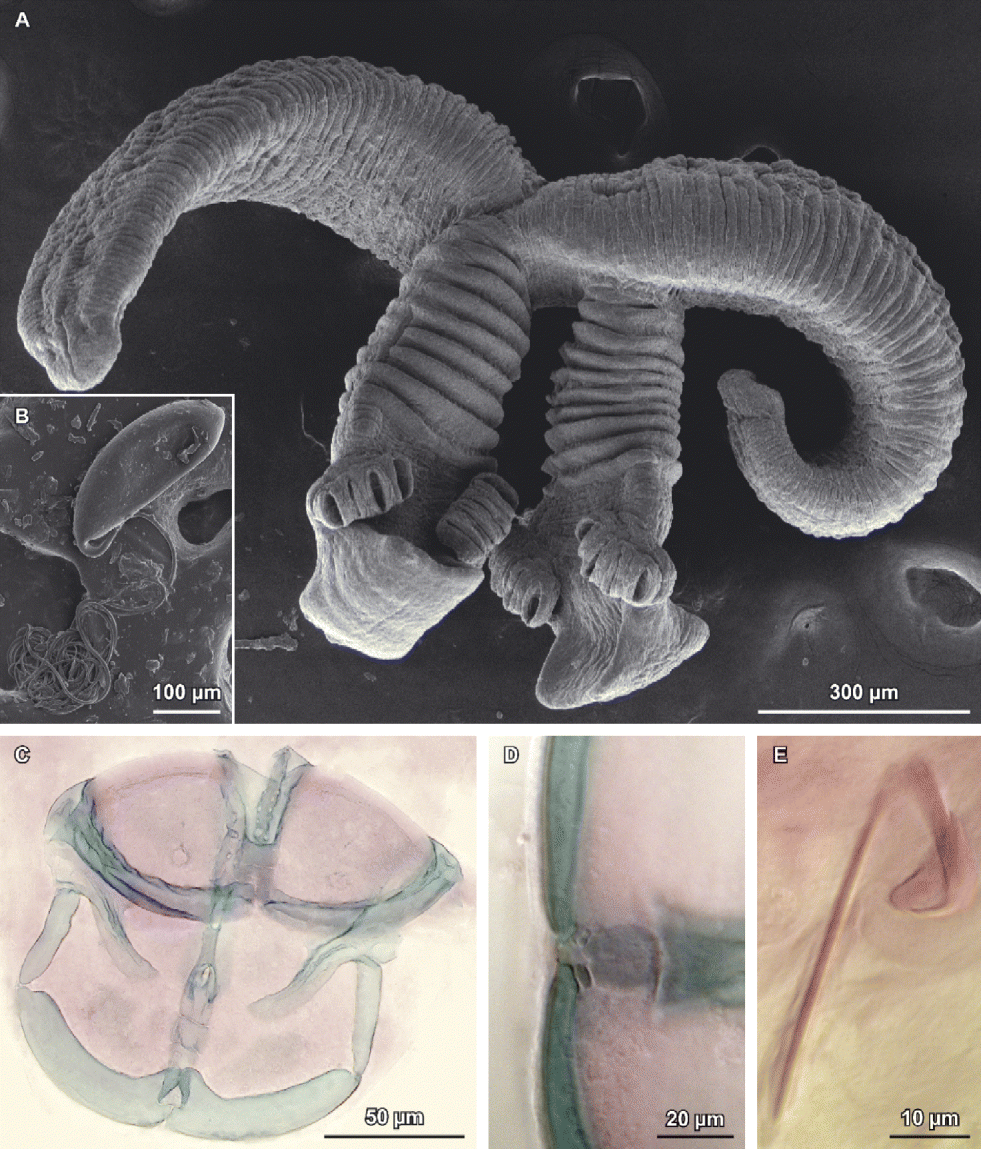
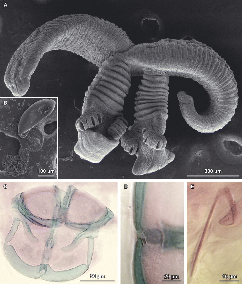
Adult specimens consist of two individuals in permanent ‘X’ copulation (figs 1A and 2A) with a total body length of 4.30 mm (n= 124, 2.89–7.10 mm). Anteriorly the tegument is relatively smooth with small inconspicuous annular transverse folds or plicae. In the region between the haptor and fusion area, or prehaptor, the plicae are significantly more pronounced, forming 8–12 deep transverse folds (fig. 2A).
Paradiplozoon vaalense n. sp. (A) Adult pair; (B) clamp; (C) hook and (D) egg. 1, Egg; 2, intestine in posterior region; 3, ovary; 4, testis; 5, clamps; 6, suckers; 7, pharynx; 8, median sclerite; 9, anterior clamp jaw; 10, posterior clamp jaw; 11, posterior end of median sclerite; 12, anterior additional sclerite; 13, posterior additional sclerite; 14, anterior end of median sclerite; 15, trapezoid projection of anterior end of median sclerite; 16, sclerites connecting anterior end of median sclerite to clamp jaws; 17, additional anterior sclerites connecting the median sclerite to the clamp jaws; 18, shaft; 19, point; 20, wing; 21, egg filament; 22, operculum.

Paradiplozoon vaalense n. sp. Scanning electron micrographs of (A) adult pair and (B) egg with filament. Light micrographs of (C) isolated third clamp; (D) anterior end of the median sclerite with connection to the clamp jaws; and (E) central hook showing handle (stained with Horen's trichrome).
Anterior region is 2.85 mm long (n= 124, 1.79–4.77 mm). A large semi-circular, subterminal mouth is present on antero-ventral surface. Mouth opens into a buccal cavity containing two large buccal suckers, 130 μm long (n= 124, 96–195 μm) and 112 μm wide (n= 124, 74–156 μm). Buccal cavity leads to an oval muscular pharynx which is 109 μm long (n= 62, 85–131 μm) and 75 μm wide (n= 62, 54–99 μm). Pharynx opens into a highly branched intestine. From just posterior to pharynx, to onset of fusion region, the anterior region displays a large number of vitellaria.
Posterior region is 1.41 mm long (n= 124, 0.90–2.33 mm). Prehaptor houses reproductive organs of parasite, comprising a large ovary and a smaller, lobed testis which appears almost diffuse in the posterior (fig. 1A). Intestine extends from fusion area into prehaptor, traversing reproductive organs, before terminating close to haptor. Intestine is highly diverticulated in prehaptor and often protrudes into haptor when engorged. Haptor is disc-shaped with a pronounced cucullate terminal protrusion (fig. 2A). This structure carries primary attachment organs of this parasite, which consist of two rows of four clamps each arranged ventro-laterally. First clamp is 134 μm long (n= 212, 100–174 μm) and 90 μm wide (n= 212, 55–144 μm); second clamp is 145 μm long (n= 214, 105–194 μm) and 88 μm wide (n= 214, 46–149 μm); third clamp is 145 μm long (n= 210, 108–192 μm) and 89 μm wide (n= 210, 52–154 μm) and fourth clamp is 143 μm long (n= 204, 97–184 μm) and 93 μm wide (n= 204, 50–159 μm).
Internal structures of clamps of P. vaalense n. sp. consist of a U-shaped median sclerite riddled with fenestrations (figs 1B and 2C). Anterior end of median sclerite is round with a trapezoid anterior projection (figs 1B and 2D). This end of median sclerite articulates to anterior jaw clamp via a single sclerite, but two small additional sclerites are sometimes seen between this sclerite and clamp jaws. Posterior end of median sclerite bears a claw-shaped extension ventrally and a fish-tail-shaped extension dorsally. Posterior end of median sclerite articulates to posterior clamp jaws via an anterior additional sclerite and a posterior additional sclerite. Anterior clamp jaw consists of two curved sclerites approaching each other centrally with projections posteriorly. Posterior clamp jaws are thickened medially extending towards posterior where they meet centrally.
A pair of central hooks is located between terminal protrusion of haptor and first pair of clamps (figs 1C and 2E). These central hooks consist of a shaft and a point, articulating via a small joint to a long handle. A wing is present and envelopes the point of the hook, terminating just below the suture indicating the border of the point and shaft. Hooks are 19 μm long (n= 136, 18–20 μm) and handles are 43 μm long (n= 124, 39–49 μm).
Each egg exhibits an extremely tightly coiled filament attached to the operculum in utero which unravels once released (figs 1D and 2B). Eggs are 384 μm long (n= 20, 341–495 μm) and 170 μm wide (n= 20, 135–301 μm).
Molecular analysis
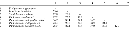
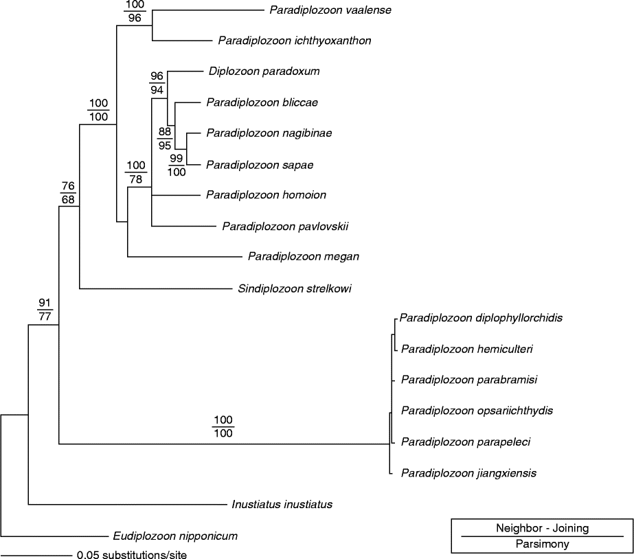
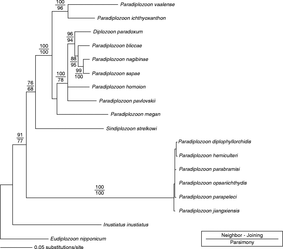
The ITS2 region of rDNA was successfully amplified and sequenced for all 40 samples (two individuals from 20 adult pairs). All 40 sequences of P. vaalense n. sp. included here had identical sequences across 700 bp of the ITS2 region and collapsed into a single haplotype (i.e. unique sequence). Uncorrected sequence divergences separating distinct taxa are given in table 2. Paradiplozoon vaalense n. sp. is separated from other recognized species by 12–37% uncorrected sequence distance. Similar topologies were retrieved irrespective of the method used to analyse data (parsimony or distance). For ease of representation, the neighbour-joining tree is shown in fig. 3. Paradiplozoon vaalense n. sp. forms a distinct clade with P. ichthyoxanthon, receiving more than 95% statistical support.
Average uncorrected (p-distance) sequence divergence separating the different diplozoid taxa. Values are given as a percentage (%).
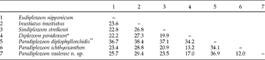
* Representative of clade including D. paradoxum, P. bliccae, P. nagibinae, P. sapae, P. homoion, P. pavlovskii and P. megan. Average distances were used for this comparison.
** Representative of clade including P. diplophyllorchidis, P. hemiculteri, P. parabramisi, P. opsariichthydis, P. parapeleci and P. jaingxiensis. Average distances were used for this comparison.
Distance-based phylogenetic tree, rooted with Eudiplozoon nipponicum, indicating the evolutionary relationships among different diplozoid taxa. Parsimony and neighbour-joining bootstrap support for nodes are indicated as fractions on the branches preceding the nodes.
Discussion
Species delimitation
Species delimitation was done using dichotomic diagnostic keys produced by Khotenovsky (Reference Khotenovsky1985). According to the key to subfamilies of Diplozoidae, the two subfamilies (Diplozoinae and Neodiplozoinae) are separated on the basis of number of clamps displayed by adult parasites (Khotenovsky, Reference Khotenovsky1985). Paradiplozoon vaalense n. sp. displays four pairs of clamps on each haptor and therefore belongs to the Diplozoinae. According to the key regarding genera of the subfamily Diplozoinae, of which Diplozoon Nordmann, 1832, Paradiplozoon Akhmerov, 1974, Sindiplozoon Khotenovsky, 1981, Eudiplozoon Khotenovsky, Reference Khotenovsky1985 and Inustiatus Khotenovsky, 1978 are members, the species was placed into the genus Paradiplozoon on account of the absence of a pronounced dilatation in the posterior of the prehaptor.
The key to Paradiplozoon was divided into two geographical regions by Khotenovsky (Reference Khotenovsky1985), namely those species which occur throughout South-East Asia and Europe (13 species) and those occurring in Europe, the Far East and West and Middle Asia (25 species). Combined, these two keys contain 34 species. Considering the key for species in South-East Asia and Europe, only Paradiplozoon kashmirense (Kaw, 1950) displays a plicated posterior and was included in the comparison. On account of the absence of posterior plicae in P. ghanense and P. aegyptense, both were excluded by this key. However, they are included in this comparison on the grounds of their geographical proximity to the current study. Using the key for species occurring in Europe, the Far East and West and Middle Asia, which was later amended by Pugachev et al. (Reference Pugachev, Galli and Kristsky2010), Paradiplozoon capoetobrame (Gavrilova, 1964) possesses a single sclerite connecting the anterior end of the median sclerite to the clamp jaws and was included into the comparison.
Paradiplozoon kashmirense differs from P. vaalense n. sp. in several morphometric aspects, of which the egg size is most notable. In P. kashmirense, eggs range from 270 to 290 μm in length and 70–90 μm in width (Khotenovsky, Reference Khotenovsky1985), while those in P. vaalense n. sp. are much larger. This trend was seen in the comparison of most measurements between these two species, with P. vaalense n. sp. being larger in size in total, anterior and posterior body lengths, as well as in the size of suckers, pharynx and clamps. These measurements overlapped to some degree, but in terms of mean measurements, a clear difference was seen. The intestine in the posterior of the body is also without diverticula in P. kashmirense while many diverticula can be seen in P. vaalense n. sp. (Khotenovsky, Reference Khotenovsky1985). No detailed drawing or description of the sclerites of P. kashmirense could be found and, as such, the comparison of these structures with P. vaalense n. sp. was not possible. Paradiplozoon kashmirense has previously been recorded from members of the genus Schizothorax Heckel, 1838 in India (Khotenovsky, Reference Khotenovsky1985; Fotedar & Parveen, Reference Fotedar and Parveen1987; Khan & Rasheed, Reference Khan and Rasheed1993) while P. vaalense n. sp. was only found on L. umbratus during this study. Species of the genus Schizothorax do not occur in southern Africa (Skelton, Reference Skelton2001).
Paradiplozoon capoetobrame also differs from P. vaalense n. sp. in several morphometric aspects, of which the size of the central hook and egg are most notable. The central hook, which was the most morphometrically stable characteristic, only ranges between 18 and 20 μm in P. vaalense n. sp. while in P. capoetobrame it is 20–23 μm in length (Khotenovsky, Reference Khotenovsky1985). In P. capoetobrame the eggs range from 227 to 260 μm in length and 83–107 μm in width (Khotenovsky, Reference Khotenovsky1985), while those in P. vaalense n. sp. are, once again, much larger. This trend was also seen when comparing measurements between these two species, with P. vaalense n. sp. being larger in total, anterior and posterior body lengths, as well as size of suckers, pharynx and clamps. The large plicae on the posterior of the body also differ, with P. vaalense n. sp. displaying 8–12 while P. capoetobrame displays 11–18 (Khotenovsky, Reference Khotenovsky1985). When considering the ultrastructure of sclerites, joining of the anterior end of the median sclerite in P. capoetobrame is similar to that of P. vaalense n. sp., as both are rounded and connect to the anterior clamp jaws by a single broad sclerite. However, P. capoetobrame lacks the trapezoid projection seen in P. vaalense n. sp. Paradiplozoon capoetobrame has only been recorded from Capoetobrama kuschakewitschi kuschakewitschi (Kessler, 1872) in Tajikistan (Khotenovsky, Reference Khotenovsky1985; Pugachev et al., Reference Pugachev, Galli and Kristsky2010) which does not occur in southern Africa (Skelton, Reference Skelton2001).
Both P. ghanense and P. aegyptense lack plicae on the posterior of the body, which are a prominent feature of P. vaalense n. sp. All three of these parasites have similar body sizes, with that of P. ghanense and P. aegyptense falling within the range of measurements obtained for P. vaalense n. sp. The only exception was the posterior of P. ghanense which is significantly smaller than that of the other two species. In terms of clamps, those of P. ghanense are similar to those of P. vaalense n. sp. while those of P. aegyptense are shorter. The drawing and description of ultrastructure of sclerites accompanying the description of P. ghanense do not show much detail, while no such information accompanied the description of P. aegyptense. The central hooks of P. aegyptense measure 17–18 μm, thus smaller than those in P. vaalense n. sp., while measurements for hooks of P. ghanense are not available. The suckers of P. aegyptense and P. vaalense n. sp. overlap in measurement, while those of P. ghanense are smaller. The pharynx of P. aegyptense is also smaller than that of P. vaalense n. sp. Unlike the situation in most diplozoids, including P. vaalense n. sp., reproductive organs of P. ghanense occur in the fusion area of the body. The only hosts known to harbour P. ghanense and P. aegyptense with distribution ranges in southern Africa are L. cylindricus and B. paludinosis, suggesting the possible existence of these parasites in southern Africa. These hosts have not been recorded in the localities of this study. In addition to this, the Vaal River forms part of the Orange River system which is separated from water bodies in which other Paradiplozoon species have been recorded by a number of large river systems, such as the Congo and Zambezi River systems.
Similar to the case presented for P. ghanense and P. aegyptense, P. ichthyoxanthon was also included in this delimitation. This species has only recently been described from South Africa in the same system as P. vaalense n. sp., and they occur in close proximity to one another. Paradiplozoon ichthyoxanthon differs notably from P. vaalense n. sp. in both morphology and genetics. The most striking differences occur in the morphology of eggs, the posterior and sclerites. The eggs in P. ichthyoxanthon lack a polar filament which is present in P. vaalense n. sp. In terms of the posterior region, P. vaalense n. sp. displays a prominent cucullate terminal protrusion from the haptor as well as distinct folds in the posterior body, which are both absent in P. ichthyoxanthon. The clamps of P. ichthyoxanthon are similar in size to those of P. vaalense n. sp. and range from 50 to 150 μm in length and 50–200 μm in width. However, the clamps of P. ichthyoxanthon increase in size towards the anterior region, unlike the clamps of P. vaalense n. sp. which do not display any significant trend in their sizes relative to one another. In terms of the ultrastructure of the clamps, the anterior end of the median sclerite in P. ichthyoxanthon is rounded while that of P. vaalense n. sp. displays a trapezoid projection. The central hooks also differ as in P. ichthyoxanthon they are 25 μm and thus larger than those of P. vaalense n. sp. The posterior portion of the intestine in P. ichthyoxanthon appears to be simple, without any diverticula, in contrast to that of P. vaalense n. sp. which displayed numerous diverticulations.
Furthermore, genetic distances obtained relinquish any doubt of their independent taxonomic validity. These two species are separated by a genetic distance of 12.0% and, as such, fall within the range of distances observed for the separation of known species (Matějusová et al., Reference Matějusová, Koubkova, D'Amelio and Cunningham2001; Gao et al., Reference Gao, Chen, Yao, Gao, Song and Wang2007; Avenant-Oldewage et al., Reference Avenant-Oldewage, Le Roux, Mashego and Jansen van Vuuren2013).
For the sake of performing a comprehensive delimitation of diplozoids collected from moggel, this parasite was also compared to all available morphological descriptions of diplozoids. Species not included in Khotenovsky (Reference Khotenovsky1985) were also included. Thus, Paradiplozoon species excluded by the keys set out by Khotenovsky (Reference Khotenovsky1985), members of the other four genera of Diplozoinae and the species not recorded in Khotenovsky (Reference Khotenovsky1985) were compared to P. vaalense n. sp. Several of the morphometric and morphological characteristics of other diplozoids overlapped with one another and with those of P. vaalense n. sp., but when comparing clamp ultrastructure as well as other morphological and morphometric characters of the parasites, it was concluded that none of these diplozoids were likely to represent species infecting moggel. Thus, in terms of morphology, P. vaalense n. sp. is distinct from all other diplozoid taxa.
Molecular findings
Both DNA and morphological data support P. vaalense n. sp. as a distinct taxonomic entity. All specimens of P. vaalense n. sp. formed a monophyletic group (in fact, all were genetically identical) which is distinct from all other taxa included here. If distances between species nested in the European clade of Paradiplozoon, including Diplozoon paradoxum Nordmann, 1832 and P. ichthyoxanthon, are used as the standard to discriminate species (uncorrected distances of 1.9–16.1%) there can be no doubt that P. vaalense n. sp. is a distinct entity.
Identical phylogenetic topologies were retrieved with both distance and parsimony approaches which, when taken with bootstrap support for nodes, provide some confidence in relationships. When looking at the clade which stems from Sindiplozoon strelkowi as its basal taxon, three clades emerge. Paradiplozoon megan (Bychowsky & Nagibina, 1959) is separate from a group including the European Paradiplozoon and D. paradoxum, with P. ichthyoxanthon and the study species forming a third group. It is interesting to note that both species from South Africa (P. ichthyoxanthon and P. vaalense n. sp.) form a distinct clade based on their DNA profiles as well as geographic origin. When taking the distance of P. megan from the European species and the fact that it separates from these diplozoids at almost the same node as the African diplozoids, the idea set forward by Sicard et al. (Reference Sicard, Desmarais, Vigneux, Shimazu, Lambert, Combes and Jourdane2003) that P. megan may represent a separate genus becomes more plausible. If it is then accepted that the distinctness of Eudiplozoon, Inustiatus and Sindiplozoon signify the standard to discriminate genera, the sequence divergence based on the ITS2 marker is in the range of 19–38% between genera. As such, a similar dilemma arises to the one noted by Sicard et al. (Reference Sicard, Desmarais, Vigneux, Shimazu, Lambert, Combes and Jourdane2003) in that P. vaalense n. sp. shows a distance of 16.2–18.7% from the European Paradiplozoon (including P. megan and D. paradoxum) and thus is in the ‘grey area’ between the upper limit of differentiating species and the lower limit for constituting a distinct genus. But, as also observed by Sicard et al. (Reference Sicard, Desmarais, Vigneux, Shimazu, Lambert, Combes and Jourdane2003), there is no morphological criterion which is robust enough to support recognition of three separate genera. Thus the present study makes a significant contribution to molecular data currently available for diplozoids, especially from Africa.
Species details: Paradiplozoon vaalense n. sp
Type host. Moggel (Labeo umbratus (Smith, 1841)).
Type locality. Vaal Dam, Gauteng, South Africa (26°52′S; 28°10′E). Other locality: Vaal River (26°49′S; 28°03′E).
Etymology. The name of the species refers to the Vaal River System in which the hosts were collected.
Type material. Type material in Iziko South African Museum, Cape Town (SAM A61817–A61820). Additional paratype specimens stored in the collection of the Natural History Museum, London (NHMUK 2013.8.5.1–2, NHMUK 2013.8.5.3–4, NHMUK 2013.8.5.5–6).
Genetic material. The unique ITS2 sequence was deposited in the European Molecular Biology Laboratory (EMBL) (accession number HG423142).
Site. Gills, interhemibranchial septum of gill arches.
Prevalence. 28.7% at Vaal Dam; 28.6% in Vaal River (January to March 2012).
Species diagnostics. Paradiplozoon vaalense n. sp. differs from other diplozoids in the size of the clamps, shape of the haptor, size of the eggs and number of folds in the prehaptor. In particular, the size of the central hook (18–20 μm) and the internal structure of the clamps are diagnostic features of this species. Furthermore, the genetic distance of the 700 bp sequence of the ITS2 region is unique and distinct.
Acknowledgements
The authors would like to thank the National Research Foundation and the University of Johannesburg Faculty Research Commission for funding to A.A.-O., Professor Hank Swanepoel (UJ) for translation from Russian texts and Professor Bill Henderson (UJ) for the species name.
Financial support
This work was supported by the National Research Foundation and the University of Johannesburg Faculty Research Commission.
Conflict of interest
None.
Ethical standards
The authors assert that all procedures contributing to this work comply with the ethical standards of the relevant national and institutional guides on the care and use of laboratory animals and the study was approved by the Ethics Committee of the University of Johannesburg.