





Non-technical Summary
There are many hooved, plant-eating mammals in the fossil record of the John Day region in Oregon. One especially common set of these mammals is the oreodonts, a now-extinct group related to camels. Past paleontologists have named 34 different oreodont species using skulls that come from this area. But it’s possible that what they thought was an important difference between species is normal variation, like the difference between two people’s noses. This is especially problematic because we don’t know how much variation should be present in the bones of similar modern species. In this paper we measure and report variation of modern camels, llamas, vicuñas, pigs, muntjaks, and duiker antelope, and then apply that to oreodonts from the John Day region. We found that many of the characters used to identify different species were probably just different individuals of the same species. There are two groups of oreodonts (eporeodontine and promerycochoerine) that had especially large numbers of species assigned but our analysis shows that for each group there probably was only one species. We call these Eporeodon occidentalis and Promerycochoerus superbus, and don’t recognize other species of these genera in the John Day region.
Introduction
Within the John Day region of eastern and central Oregon the oreodonts (superfamily Merycoidodontoidea) have received considerable taxonomic attention and subsequent taxonomic disagreement. Over the past century and a half, 34 different holotypes have been named from the abundant fossiliferous rocks of the Clarno, John Day, and Mascall formations (Cope, Reference Cope1884; Thorpe, Reference Thorpe1921, Reference Thorpe1937; Schultz and Falkenbach, Reference Schultz and Falkenbach1968; Lander, Reference Lander, Janis, Scott and Jacobs1998; Stevens and Stevens, Reference Stevens, Stevens, Prothero and Foss2007; Ludtke, Reference Ludtke2008; Stevens et al., Reference Stevens, Prothero, Cleaveland, Welsh, Marriott, Htun, Balassa, Olson, Watmore and Wheeler2023). Far fewer are still recognized as separate species today, although the exact number varies by researcher.
Of the two families of oreodonts, species of Merycoidodontidae have received more taxonomic adjustments. Schultz and Falkenbach (Reference Schultz and Falkenbach1949, Reference Schultz and Falkenbach1950, Reference Schultz and Falkenbach1954, Reference Schultz and Falkenbach1968) recognized 16 species of merycoidodontid oreodonts from 7 genera (up to 10 when subgenera are included). Lander (Reference Lander, Janis, Scott and Jacobs1998) recognized 11 species and 9 genera of Merycoidodontidae, while Stevens et al. (Reference Stevens, Prothero, Cleaveland, Welsh, Marriott, Htun, Balassa, Olson, Watmore and Wheeler2023) recognized 14 species and 11 genera.
Agriochoeridae suffers from similarly varying taxonomy, although to a lesser extent. Cope (Reference Cope1884) recognized 5 species from 2 genera in the John Day Formation, while Lander (Reference Lander, Janis, Scott and Jacobs1998) considered those specimens to represent one species, with a second species in the Clarno Formation. Thus, the recent overall estimates for oreodont diversity vary from 13 species on the low end to 21 species on the high end.
The issues surrounding fossil systematics are compounded by the relative paucity of modern species identified primarily by their osteology. Few if any modern taxonomic works link the biological species concept with a corresponding osteological species concept. As a result, what paleontologists refer to as a species is not always comparable to a modern standard (Alroy, Reference Alroy2002; Emery-Wetherell and Davis, Reference Emery-Wetherell and Davis2018). Larger sample sizes of fossil taxa often capture a broader spectrum of possible morphological variation than is considered typical for extant species. Indeed, work describing osteological characters for modern species has suggested that artiodactyls are broadly more variable than previously expected (e.g. Onuk et al., Reference Onuk, Kabak and Atalar2013; Emery-Wetherell and Davis, Reference Emery-Wetherell and Davis2018; Martini et al., Reference Martini, Schmid and Costeur2018).
The issue is not simply a somewhat semantic dispute over what constitutes a species, but of cross-identification as well. For example, Hypsiops johndayensis Shultz and Falkenbach, Reference Schultz and Falkenbach1950, was initially established as an endemic species. Lander (Reference Lander, Janis, Scott and Jacobs1998) considered it a valid representation of the genus but part of a more broadly ranging species, Hypsiops breviceps Douglass, Reference Douglass1907. Stevens et al. (Reference Stevens, Prothero, Cleaveland, Welsh, Marriott, Htun, Balassa, Olson, Watmore and Wheeler2023) disagreed completely and referred the holotype specimen to Eporeodon pacificus Cope, Reference Cope1884, a completely different subfamily of oreodont.
In this paper we set out to analyze the diagnostic power of key qualitative and quantitative characters used to identify John Day region oreodonts, specifically focusing on the species named explicitly from local holotypes. This includes taxa from the Clarno (Eocene), John Day (late Eocene to Early Miocene), and Mascall (Middle Miocene) formations, which date from 42.7 ± 0.3 Ma (Bestland et al., Reference Bestland, Hammond, Blackwell, Kays, Retallack and Stimac1999) to 13.564 ± 0.018 Ma (Maguire et al., Reference Maguire, Samuels and Schmitz2018). The John Day Formation includes the Big Basin (Eocene to Oligocene), Turtle Cove (Oligocene), Kimberly (Oligocene), Haystack Valley (Oligocene), Balm Creek (Early Miocene), Johnson Canyon (Early Miocene), and Rose Creek (Early Miocene) members (Bestland and Retallack, Reference Bestland and Retallack1994; Hunt and Stepleton, Reference Hunt and Stepleton2004; Albright et al., Reference Albright, Woodburne, Fremd, Swisher, MacFadden and Scott2008; Mohr et al., Reference Mohr, Famoso, Samuels, Laib and Schmitz2025). Additional geologic context is available in Supplementary Data Set 2. We compared these characters to modern analog taxa (Muntiacus, Cephalophus, Camelus, Vicugna, Lama, Hylochoerus) and to oreodonts from across North America. By using large local sample sizes and comparative specimens, we set out to evaluate local oreodont taxonomy for signs of taxonomic inflation specifically deriving from intraspecific variation, sexual dimorphism, geography, or ontogeny.
Our goal was to reject single-species hypotheses for the general clades within the John Day area. The characteristics that would cause us to reject a single-species hypothesis are as follows: (1) high coefficients of variation (CVs) that are not related to statistical uncertainty or recognizable forms of intraspecific variation in modern analogues; (2) CVs that increase when taxa are pooled and decrease when they are split according to other likely diagnostic characteristics; (3) qualitative character states that do not represent intraspecific variation in modern analogues; (4) clusters of correlated character states within a genus, quantitative and/or qualitative, that cannot be explained by intraspecific variation; and (5) statistically significant, non-gradational quantitative character changes over time or between geographic divisions.
Methods
Terminology
We follow Bärmann and Rössner (Reference Bärmann and Rössner2011) for dental terminology. Certain terms used in oreodont cranial and dental anatomy were not defined in that paper or may not have common use and so are defined here. (1) Fossettes are the wearable surface of each cone on a premolar or molar. (2) Molar ribs refer to linear ridges on the fossette’s enamel; outer ribs are those on the most labial fossette, inner ribs are those on the most lingual fossette. (3) What Lander (Reference Lander, Janis, Scott and Jacobs1998) referred to as the lacrimal fossa we call the pre-orbital fossa, because the equivalent fossa is not confined to the lacrimal bone in all oreodonts or in all modern artiodactyls. (4) The paraconule refers to an additional cusp situated between the paracone and protocone of the upper molars. (5) We use the term jugal to refer to what is commonly called the malar bone in oreodont literature, because jugal is more commonly used across Artiodactyla.
Data collection and measurements
All data were collected by MMW. Our data consisted of a combination of physical specimens, casts, and digital models. For physical specimens and casts, we used digital calipers (Mitutoyo CD-s6”C and CD-12”C) for measurements. Specimen digital models were built using photogrammetry in Agisoft Metashape, versions between 1.0.0 and 2.2.11 (Agisoft, Reference Agisoft2013). Digital model measurements were taken predominantly in Agisoft Metashape, with some additional measurements made using the free software Blender (Roosendaal and Siddi, Reference Roosendaal and Siddi2025). In Blender version 4.4, models were scaled and measured using the Measure and Scale add on (Belaid-ZIANE, 2025). Repeated measurements of specimens across software packages showed that measurements were directly comparable between software. All measurements used were selected according to best practices to ensure comparability between digital and physical models (Emery, Reference Emery2016). The lead author conducted extensive repeat measurements as part of the data cleaning process and remeasured on 3D models where necessary. Measurements are available in Supplementary Data Set 1. A selection of 3D models are available as supplementary information on Morphosource.
Our 3D models were made and measured as separate dorsal and ventral halves, because this was an easier way to quickly photograph specimens on brief museum visits. Lacking combined models did necessitate some adjustments for measurements that evaluated angles (e.g., the angle of the basioccipital bone compared to the palate) or measured across dorsal and ventral models (e.g., true muzzle height). At the time of initial data collection, Agisoft Metashape did not have an easy way to measure angles, so these adjustments were made by using trigonometry and the law of cosines (Hazewinkel, Reference Hazewinkel2013). Later measurements of angles were taken using Blender; these were also confirmed by the lead author to be directly comparable to trigonometry measurements.
To measure pre-orbital fossa depth, we used the law of cosines on the length across the fossa and the length from the deepest part of the fossa to the anterior of the orbit. For cranial flexion (also called basicranial tilt), we measured from the end of the palate to the beginning of the occipital condyles and from the palate to the midpoint of the foramen ovale. Selecting the foramen ovale as the midpoint reduced the extent of cranial flexion measured but maximized the available sample given that many specimens were not prepared between the pterygoid processes and would have been excluded if the midpoint for the flexion measurement had been any farther anterior. Finally, we used trigonometry to calculate muzzle height by measuring the palate width at P2 on the ventral model of the skull, and from P2 to the dorsal surface of the muzzle then used the Pythagorean theorem to solve for the true height (Hazewinkel, Reference Hazewinkel2013). Simply measuring from P2 to the height of the muzzle would have included transverse variation and not been representative of the character as reported in the literature.
We included several qualitative characters that were specific to Camelus dromedarius Linnaeus, Reference Linnaeus1758, and Camelus bactrianus Linnaeus, Reference Linnaeus1758, to demonstrate how qualitative characters can help separate species. These characters were coded following Martini et al. (Reference Martini, Schmid and Costeur2018) along with an additional character regarding the shape of the occipital condyles (flexion stops).
The exact measurements and character states used are recorded and described in more detail in Supplementary Data Set 1.
Systematics and taxonomic scope
Although there are 34 holotypes named from the John Day region, our initial assessment found that most of the still-disputed taxa were within two groups from the family Merycoidodontidae. We concentrated the bulk of our character analysis on these taxa. The larger taxa with prominent zygomatic arches were treated as a potentially multi-species sample of Promerycochoerus (Megoreodon, Merycochoerus, Promerycochoerus, and some Desmatochoerus) and the smaller taxa as a potentially multi-species sample of Eporeodon (Eporeodon, Eucrotaphus, and some Desmatochoerus). The rest of this paper refers to them grouped by these genus names.
Coefficient of variation and multimodality
Most of the characters used to identify species of Eporeodon, Promerycochoerus, and related oreodont taxa have not been previously tested for their diagnostic capacity or for their intraspecific variation. Those characters that have been tested previously have often been found to be influenced by more than species delineations. For example, the “dolichocephaly” and “mesocephaly” reported in several species by Schultz and Falkenbach (Reference Schultz and Falkenbach1968) represent a byproduct of deformation of the skulls during fossilization (Stevens and Stevens, Reference Stevens, Stevens, Prothero and Foss2007).
As part of our taxonomic assessment, we collected both qualitative and quantitative characters from the original descriptions and recent treatments of these taxa. Ostensibly, single-species samples should have normal distributions of quantitative characters, with variation of about 10% (Simpson and Roe, Reference Simpson and Roe1939; Cope and Lacy, Reference Cope and Lacy1992). Previous work in artiodactyls has found that particularly with large artiodactyls or small measurements, that percentage may be much higher, easily between 10% and 20% for dentition (Emery-Wetherell and Davis, Reference Emery-Wetherell and Davis2018). Samples that exceed or fall into the high end of these percentages may contain multiple species or display variation resulting from factors such as ontogeny or sexual dimorphism. Similarly, species with much lower variation may be over split.
We calculated the scaled coefficient of variation (Sokal and Braumann, Reference Sokal and Braumann1980) for quantitative characters in different subgroups: John Day Promerycochoerus, John Day Eporeodon, all measured Promerycochoerus, and all measured Eporeodon. CVs with high variation within a subgroup were evaluated for signs of multimodality using Hartigan’s dip test using the ‘diptest’ package in R (Hartigan and Hartigan, Reference Hartigan and Hartigan1985; Maechler, Reference Maechler2015).
We also tested these characters where possible in comparative modern taxa. Camelus dromedarius (19 adults, 8 juveniles), C. bactrianus (12 adults, 4 juveniles), Lama guanicoe (Müller, Reference Müller1776) (13 adults, 2 juveniles), and Vicugna vicugna Molina, Reference Molina1782 (9 adults, 12 juveniles) were chosen as the closest phylogenetic relatives of oreodonts. Hylochoerus meinertzhageni Thomas, Reference Thomas1904 (15 adults, 7 juveniles) has a similar inflation of the zygomatic arches as is found in Promerycochoerus, so it was chosen as an analogue for that set of characters. Cephalophus weynsi Thomas, Reference Thomas1901 (13 adults, no juveniles) and C. leucogaster Gray, Reference Gray1873 (25 adults, no juveniles) are small sympatric artiodactyls that have enlarged pre-orbital fossae and corresponding scent glands similar to those found in Eporeodon. Muntiacus muntjak (Zimmerman, Reference Zimmerman1780) (16 adults, no juveniles) and M. reevesi (Ogilby, Reference Ogilby1838) (8 adults, 2 juveniles) were also chosen for their preorbital fossae.
Qualitative character notes
We coded for two forms of potentially diagnostic premolar patterns in our Eporeodon sample: enamel wells (an additional crest that surrounds a small circle of dentine on the lingual cone) and enamel ridges on the labial cone of the P4 (Fig. 1).
Variations in the shape of P4 demonstrated in (1) JODA 6315 and (2) UOMNH F-76529. Abbreviations: LaC, labial cone; LiC, lingual cone; EW, enamel well; R, ripples. Scale bar corresponds to photographs, not illustrations.

Figure 1. Long description
Two different examples of enamel irregularities in the upper fourth premolar of Eporeodon. In the first panel the specimen has a bifurcation of the crest on the labial cone which forms a circle, known as an enamel well. In the second panel, the premolar both has an enamel well and has a split lingual cone where the crest does not meet in the middle.
We used Hylochoerus as an analogue for several of the characteristics of Promerycochoerus and its zygomatic arch. One of these was the presence of a ‘rugose texture’ on the arches. The term ‘rugose’ in terms of texture is perhaps not as informative as it could be. While the literal definition means ‘wrinkled’ or ‘corrugated’, that could easily describe textures ranging from sun-related damage to rodent chew marks. For our study we defined rugosity as specifically referring to only raised cortical bone surface rather than eroded bone surfaces. We also limited our definition to cases where there were small lumps, valleys, and ridges that were on the order of 2 mm or smaller in spacing. Even within this definition of rugosity we noticed variation (Fig. 2).
Different ‘rugose’ textures observed on the zygomatic arches of Promerycochoerus and Hylochoerus. All images are approximately 2 cm across. (1) CM 109 showing pits, grooves, and raised texture on the left side of the image. (2, 3) JODA 7795: (2) grooves and ridges departing from a white sediment-filled seam on the underside of the zygomatic arch, accompanied by small pits; (3) intertwining ridges forming a ‘filigree’ texture on the posterior raised component of the opposite zygomatic arch from that in (2). (4) MCZ 27851, a male Hylochoerus specimen with regular raised sections of periosteum that are pointed at the tips. All images of MCZ specimens remain © President and Fellows of Harvard College.

Our 3D models were taken as ventral and dorsal photogrammetry models; areas that were along the seam of these two models had high measurement uncertainty, such as the thickness of the zygomatic arch. We chose to code this as a character instead of a quantitative variable.
Many modern ruminants possess an additional articulation surface on their occipital condyles sometimes referred to as flexion stops, ventral ‘saddles’, or as unnamed ventral extensions of the condyles themselves (Webb, Reference Webb1981; Geisler, Reference Geisler2001; O’Leary, Reference O’Leary2016). These had not been coded previously in oreodonts to our knowledge.
Paroreodon, Hypsiops, and Ticholeptus are all described in part by the presence of a lacrimal vacuity, although little work has been done to establish the ranges of modern variation in this character (Schultz and Falkenbach, Reference Schultz and Falkenbach1941, Reference Schultz and Falkenbach1947, Reference Schultz and Falkenbach1950; Stevens et al., Reference Stevens, Prothero, Cleaveland, Welsh, Marriott, Htun, Balassa, Olson, Watmore and Wheeler2023). We measured qualitative characteristics of the size, shape, and symmetry of this character in modern camelids.
Qualitative variation
To measure the degree of character state variation we calculated the deviation from the mode (DM) using the ‘qualvar’ package in R (Wilcox, Reference Wilcox1973; Gombin, Reference Gombin2018). DM is a measurement of category spread, where a 0 indicates all data are in a single category and a 1 indicates all data are spread evenly between categories. Where applicable, we measured these in all oreodont subgroups and in modern analogues.
Principal Coordinate Analysis and Gower’s Distance Matrix
To assess whether combinations of qualitative and quantitative characteristics were capable of diagnosing distinct subgroups within Promerycochoerus or Eporeodon, we evaluated them using a Principal Coordinate Analysis (PCoA) with a Gower’s Distance Matrix using the packages ‘cluster’, ‘ape’ and ‘gowerdist’ in R (Gower, Reference Gower1966; Paradis and Schliep, Reference Paradis and Schliep2019; van der Loo and Turner, Reference van der Loo and Turner2024; Maechler et al., Reference Maechler, Rousseeuw, Struyf, Hubert, Hornik, Studer, Roudier, Gonzalez and Kozlowski2025). Unlike Principal Component Analysis (PCA), the combination of PCoA and a Gower’s Distance Matrix does not assume linear relationships among variables and can incorporate both quantitative and qualitative data (Gower, Reference Gower1966, Reference Gower1971; Tschopp et al., Reference Tschopp, Napoli, Wencker, Delfino and Upchurch2022). Because the distance matrix is run on pairwise comparisons, it can also handle data with missing values (Gower, Reference Gower1966). There are some disadvantages to this method, with previous studies demonstrating the method may be overly reliant on qualitative data (D’Orazio, Reference D’Orazio2021). Thus, we also re-ran results where necessary without qualitative characters to determine if the subgroups identified were still stable.
Additionally, although the process of calculating the PCoA can handle missing data, we found that it often over-weighted the importance of variables with abundant missing data. To avoid this, for each subgroup tested we removed characters with >50% missing values, or where the character was uniform across individual specimens (e.g., zygomatic arch shape was uniformly 1 in the John Day Promerycochoerus so was dropped for this subgroup).
The purpose of the PCoA and Gower’s Distance Matrix was to detect subgroups, but it was unlikely all detected groups would represent separate species. We assumed that a specimen’s relative age would also influence certain characteristics. We measured the height of the upper M1 in Promerycochoerus and used a generalized wear stage as a proxy for age of modern taxa, Eporeodon and Promerycochoerus. Definitions of these wear stages are in Supplementary Data Set 1. We also subsampled the oreodont PCoA analyses and ran them with and without dental wear characters.
Unlike PCA, PCoA does not return factor loadings or rotation scores (the estimates of influence from each original qualitative or quantitative variable). Instead, it returns a list of eigenvectors that correlate to the relationships between each specimen. Similar to PCA, the first two axes contain the most variation in the groups (Wold et al., Reference Wold, Esbensen and Geladi1987). To determine which characters held the greatest influence on each subgroup of PCoA, we calculated the correlation (r) between the first two PCoA axes and all input variables.
Follow-up character analyses
Some characters appeared to meet one of the hypothesis-rejecting conditions noted in the Introduction (e.g., high coefficients of variation). We ran additional follow-up analyses to see if these were caused by variation factors such as sexual dimorphism, ontogenetic change, disease, or malnutrition. These included using: (1) multiple linear regression to evaluate whether skull length and the dimensions of the preorbital fossa were related in modern taxa; (2) correlation to determine if zygomatic injuries and rugosity were related to one another or other quantitative characters in Promerycochoerus and Hylochoerus to determine if rugosity could therefore be linked to disease; and (3) two-sample, two-tailed t-tests to assess whether quantitative measurements differed between males and females in modern taxa, or between John Day and non-John Day specimens, or between individuals with certain character states.
Repositories and institutional abbreviations
Specimens examined in this study are deposited in the following institutions: Frick Collection at the American Museum of Natural History (AMNH F:AM), New York, USA; Fossil Mammal Collection at the American Museum of Natural History (AMNH FM), New York, USA; Modern Mammal Collection at the American Museum of Natural History (AMNH M-), New York, USA; Field Museum of Natural History (FMNH) Chicago, USA; University of Oregon Museum of Natural and Cultural History (UOMNH F-), Eugene, USA; John Day Fossil Beds National Monument (JODA), Kimberly, USA; Carnegie Museum of Natural History (CM), Pittsburgh, USA; the Princeton University collections (PU) which are now stored at the Yale Peabody Museum (YPM VPPU), New Haven, USA; other collections at the Yale Peabody Museum (YPM VP), New Haven, USA; California Institute of Technology collection (CIT) now stored at the Natural History Museum of Los Angeles County, now at the LACM (LACM-CIT), Los Angeles, USA; University of California Museum of Paleontology (UCMP), Berkley, CA; Museum of Vertebrate Zoology at th University of California, Berkley (MVZ); Museum of Comparative Zoology, Museum of Comparative Zoology at Harvard (MCZ), Cambridge, USA; Texas Memorial Museum (TMM), Austin, USA; University of Washington’s Burke Museum (UWBM VP), Seattle, USA.
Results
Discarded characters
Several characters were discarded early on. In the course of character scoring, we noticed that the high variation in overall skull profile reported by Cope (Reference Cope1884) for Eporeodon directly related to whether or not the sagittal and occipital crests were preserved: if incomplete or absent, the skull appeared convex rather than flat. For the purposes of this study, we discarded the overall outline of the skull as a character. Although skull silhouette may be valid for differentiating other taxa, in the studied sample of Eporeodon it was a clear byproduct of preservation.
Similarly, the thin (<1mm) laminae of bone separating the stylomastoid foramen from the tympanohyal canal (Cope, Reference Cope1884; Douglass, Reference Douglass1906) is easily broken off or removed during fossil preparation, and its presence or absence was therefore not a reliable generic character.
Finally, the lacrimal (or lachrymal) tubercle was mentioned by Thorpe (Reference Thorpe1937) in a species description. However, this tubercle is an attachment point for the muscles of the eyelid and was likely present in all oreodonts (as in all mammals). It seems unlikely that larger eyelids were a reliable species indicator, especially considering that the lacrimal tubercle grows through ontogeny (Silver, Reference Silver, Brothwell and Higgs1963).
Coefficients of variation
Coefficients of variation ranged from 1% to 43%, with the highest coefficients of variation found in the dental characteristics of modern single species taxa, especially in the caniniform teeth (Figure 3). Modern animals typically had higher or similar variation of dental characteristics, but fossil taxa showed higher degrees of variation in several cranial characters, including pre-orbital fossa length and jugal height (Fig. 3). Most single-species measurements within modern taxa fell below 15%, thus this was chosen as our cutoff for unusually high variation (Fig. 3).
Coefficients of variation for cranial and dental characters at the genus or subgroup level (species for modern taxa, John Day only for fossil taxa). Modern points are calculated by genus but colored by family for simplicity. Abbreviations: AP, anterior–posterior (length); T, transverse (width); H, ventral–dorsal (height); D, depth. Full list of character explanations available in Supplementary Data Set 1. Gray line indicates 15% cutoff.

Figure 3. Long description
Scatterplot of different coefficients of variation by character and taxon, including organisms pooled at the genus level and separated out by species. On the y axis are the characters, on the x axis is the coefficient of variation ranging between 0 and 45. A vertical line marks 15% variation,a bove which is considered unusua for a species (though in many cases, modern species also show this amount of variation). The canines have the highest degree of variation both in their width and length, ranging up to 45% variation in some species. Most moderm measurements for a single pspecies are below the 15% cutoff, though camels often range above that. Specific instances of high variation are described in table 1.
When CVs were evaluated within John Day Eporeodon and Promerycochoerus versus our full pools of each genus, the differences were minimal (0–4% change). Modern taxa showed a larger range of CVs, with some characters showing higher variation within single species than in pooled genus groups and vice versa (Fig. 4). Camelus CVs increased by as much as 9% in some characters when grouped by genus, while genus-grouped Muntiacus CVs increased up to 20%. Not all characters increased in CV when grouped by genus. Camelus had 36 characters that increased, however 20 other measurements decreased when the genus was pooled (Fig. 4). Table 1 shows some of the higher CV characters in Eporeodon and Promerycochoerus and their corresponding CVs in modern taxa.
Q–Q plots of the coefficient of variation for pooled genera versus single species. Modern points are calculated by genus but colored by family for simplicity. The x axis shows the CV for animals grouped by their genus while the y axis shows the CV of that character in each subgroup (species for modern taxa, or John Day only for oreodonts). Data above the gray line have higher variation within the subgroup, data below the gray line have higher variation when pooled. Characters that show higher intraspecific variation should fall below the line.

Figure 4. Long description
Q–Q plots of the coefficient of variation for pooled genera versus single species. Modern points are calculated by genus but colored by family for simplicity’s sake. The x axis shows the CV for animals grouped by their genus while the y axis shows the CV of that character in each subgroup (species for modern taxa, or John Day only for oreodonts). Anything above the grey line has higher variation within the subgroup, anything below the grey line has higher variation when pooled. Characters that show higher intraspecific variation should fall below the line. Both Eporeodon and Promerycochoerus stick close to the line, suggesting that there are few changes in coefficients of variation when split into John Day versus non-John Day oreodonts. Most of the artiodactyls show a great deal of deviation from the line when they are pooled by their genus.
High coefficients of variation (≥ 15%) for the John Day subsamples of Eporeodon and Promerycochoerus and their corresponding values in other taxa. Anatomy abbreviations: AB, auditory bullae; P. Fossa, Pre-orbital fossa; PO, width at postorbital bar; TRL, toothrow length; Zyg, width at zygomatic arches. Measurement abbreviations: H, dorsal–ventral (height); AP anteroposterior (length); T transverse (width); D, depth. Taxa abbreviations: Ca., Camelus; Ce., Cephalophus; H., Hylochoerus; L., Lama; M., Muntiacus; V.; Vicugna.

Table 1. Long description
High coefficients of variation (equal to or greater than 15%) for the John Day subsamples of Eporeodon and Promerycochoerus and their corresponding values in other taxa.
This includes, for Promerycochoerus, the anteroposterior length of the canine, first premolar, second molar, and third molar; the transverse width of the first premolar; the total toothrow length; the skull width at the zygomatic arches, third molar, canine, orbits, and post-orbital bar; the height of the muzzle. Eporeodon coefficients of variation do not exceed 14% for any of these measurements. All modern relatives have similarly high or higher coefficients of variation for the canine. In all of these measurements save the total tooth row length and dimensions of the first premolar (as other modern artiodactyls do not have this tooth) at least one species has similar variation.
Both Eporeodon and Promerycochoerus had high variation in the height of the auditory bullae, though not as high as the variation in vicunas. Both oreodonts had high variation in the height of the jugal; the height in Promerycochoerus was higher than any modern relative, but both camels had higher variation than Eporeodon.
Eporeodon had high variation in the pre-orbital fossa length and depth. Fossae were measured in Cephalophus and in Muntiacus, and while they were variable in both modern genera, that variation was half that found in Eporeodon.
For Eporeodon, we measured 80 quantitative variables (Supplementary Data Set 1). Of these, 77 had more than 5 samples. Ten variables had CVs of 15% or higher in our John Day region subset. For Promerycochoerus, we measured 64 quantitative variables. Of these, 50 had more than 5 samples. Eighteen variables had CVs of 15% or higher in our John Day subset.
We also evaluated CVs within our identified sample of John Day oreodonts other than Eporeodon and Promerycochoerus. The highest CVs were often the result of low sample size or small measurements, which are prone to higher amounts of measurement uncertainty (Polly, Reference Polly1998; Emery-Wetherell and Davis, Reference Emery-Wetherell and Davis2018). Only the width of the third premolar of Diplobunops kardoula Emery, Davis, and Hopkins, Reference Emery, Davis and Hopkins2016, had both a high CV (17%) and more than five samples. All other measurements with higher CVs had five or fewer samples and were typically of features less than 2 cm in length (Fig. 5).
Comparison of average measurement, coefficient of variation, and sample size for the John Day oreodonts other than Eporeodon and Promerycochoerus. Horizontal bar indicates unusually high variation (15%). The largest sample size was 12.

Figure 5. Long description
Comparison of average measurement, coefficient of variation, and sample size for the John Day oreodonts other than Eporeodon and Promerycochoerus. Horizontal bar indicates unusually high variation (15%). The largest sample size was 12. Most measurements fall below the 15% cutoff, and of those that do not only one has more than 5 samples. Most measurements are also small, with only one reaching 150 mm.
In Eporeodon, we found high CVs for both the length (21%) and depth (30%) of the pre-orbital fossa. These were much larger than those measured in modern taxa. When only John Day specimens were considered, the length variation barely changed (22%) but the depth variation decreased (27%). In Muntiacus muntjak the pre-orbital fossa showed higher variation (12% length, 14% depth) than any other cranial character.
Multimodality
We performed Hartigan’s dip test on any pooled-genus modern measurements or full genus oreodont measurements that had a CV higher than 15% and a sample size of seven or higher. No univariate measurement within oreodonts was statistically significant. Only one modern measurement was statistically significant—the ratio of the preorbital fossa depth divided by length in Muntiacus muntjak (p = 0.017).
Qualitative variation
Many of the qualitative characters used to diagnose oreodonts showed gradational character states, both in oreodonts and in modern specimens. Table 2 shows the character states and general degree of variation in different taxa. Specific cases are discussed here and within the discussion section.
Qualitative variation in oreodonts and modern analogues. DM is the deviation from the mode where 1 means perfectly even distributions between all measured character states and 0 indicates a single character state. Character states show all states found in that species (character definitions in the Supplementary Data Set 1). Asterisk indicates single-state character when juveniles are removed. Taxa abbreviations: Ca., Camelus; Ce., Cephalophus; H., Hylochoerus; L., Lama; M., Muntiacus; V.; Vicugna.

Table 2 Long description
Table 2 summarizes qualitative morphological characters that exhibit within-taxon polymorphism in both modern and fossil taxa. For each character, the table lists the taxa exhibiting multiple character states, the deviation from the mode (DM), and the character states observed. DM measures the evenness of the distribution of observed character states, where a value of 1 indicates that character states are represented equally and a value of 0 indicates that only a single character state is present. Thus, higher DM values indicate a more even distribution among multiple states rather than a greater amount of variation.
For the auditory bullae shape, the modern species Ce. weynsi has three observed character states with a DM of 0.69, indicating that multiple states occur with a relatively even distribution. The fossil taxon Eporeodon exhibits 2 different character states with DM values of 0.77 and 0.83, likewise indicating that both states are well represented.
For the auditory meatus shape, several living species are polymorphic. Ce. leucogaster (DM = 0.45), Ce. weynsi (0.72), and M. muntjak and M. reevesi (both 0.67) each exhibit multiple character states with differing degrees of evenness, whereas H. meinertzhageni is invariant (DM = 0). Eporeodon is also polymorphic, spanning 4 different character states with DM values of 0.71 and 0.67, indicating that these states are relatively evenly represented.
The basioccipital ridge character is invariant in H. meinertzhageni (DM = 0) but polymorphic in M. muntjak (DM = 0.67). Eporeodon also exhibits more than one character state, although the lower DM values (0.25–0.32) indicate that one state predominates.
The dental characters P4 enamel ridge and P4 enamel well were evaluated only in Eporeodon. Both characters are polymorphic. The enamel ridge has DM values of 0.55–0.57, indicating a moderately even distribution of character states, whereas the enamel well has lower DM values (0.29–0.36), reflecting a stronger predominance of one state.
For the presence of flexion stops on the occipital condyles, most taxa are invariant. Several living species and both fossil genera have DM values of 0, while a few camelids have low DM values (0.08–0.13), indicating that an alternate state is present but uncommon.
The position of the infraorbital foramen is polymorphic in both modern and fossil taxa. H. meinertzhageni has a DM of 0.83, while Eporeodon has DM values of 0.86 and 0.68 and spans 3 different character states. These values indicate that multiple states are present and are relatively evenly represented.
The lacrimal vacuity character is polymorphic among modern camelids. DM values range from 0.41 to 0.87, reflecting varying degrees of evenness among the observed character states. No fossil taxa were scored for this character.
The position of the palate terminus is polymorphic in several living taxa, particularly M. muntjak (DM = 0.83). Eporeodon exhibits lower DM values (0.19 and 0), indicating that the full fossil dataset contains a rare alternate state that disappears when only JODA specimens are considered.
The shape of the palate terminus is polymorphic in most living species, with DM values between 0.59 and 0.83, indicating relatively balanced representation of multiple states. Eporeodon has lower DM values (0.08 and 0), showing that one state predominates strongly and that in JODA, only a single state is present.
The orientation of the paroccipital process is polymorphic across several living species, with DM values ranging from 0.22 to 0.92. Eporeodon also exhibits multiple character states, with intermediate DM values (0.50 and 0.48), indicating that the observed states occur in moderately similar frequencies.
The presence or absence of zygomatic arch rugosity is polymorphic in both H. meinertzhageni (DM = 0.50) and the fossil Promerycochoerus (DM = 0.58 and 0.35). In each case, more than one character state is present, although the balance between states differs.
For zygomatic curvature, Eporeodon exhibits two different character states with DM values of 0.32–0.38, indicating that one state is more common than the other. Promerycochoerus has three observed character states and higher DM values (0.80–0.88), indicating a more even distribution among those states.
Finally, zygomatic depth is polymorphic in H. meinertzhageni (DM = 0.70), Eporeodon (DM = 0.88–0.96), and Promerycochoerus (DM = 0.78–0.84). In each case, multiple character states are present, with the higher DM values indicating that those states occur in relatively similar proportions.
Auditory meatus and bullae
The shape of the auditory meatus was highly variable in most modern taxa except for Hylochoerus, which only showed one character state. Other taxa showed three or four different character states with uneven representation among states (DM <1).
Auditory bullae showed gradational and multiple character states in many taxa. We noticed a high degree of variation in modern Cephalophus weynsi regarding the shape and constrictions of the overall profile. Shapes in Cephalophus weynsi varied from oval to ‘kidney bean’ to almost ‘figure eight’ in shape, with gradation in between (Fig. 6).
Structure of the auditory meatus and bullae of modern specimens of Cephalophus weynsi: (1) general anatomy overview; (2) AMNH M-53066; (3) AMNH M-53055; (4) AMNH M-53037. Outlines on (2–4) show general shape of auditory meatus. Abbreviations: AM, auditory meatus; AB, auditory bullae; PGF, post-glenoid foramen; PG, post-glenoid process; FO, foramen ovale; BO, basioccipital; OC, occipital condyle; P, paraoccipital process; SF, stylomastoid foramen; FM, foramen magnum.

Figure 6. Long description
Structure of the auditory meatus and bullae of modern specimens of Cephalophus weynsi. The first panel is an overview of the relevant anatomy, which includes the auditory meatus, auditory bullae, post-glenoid foramien, post-glenoid process, foramen ovale, basioccipital, occipital condyle, paraoccipital process, stylomastoid foramen, and foramen magnum. The other three panels are examples of the species. The auditory bullae in panel 4 is smooth and round, while it is skinny in panel 2 and figure-8 shaped in panel 3. The auditory meatus is trumpet-shaped in panels 3 & 4, but straight in panel 2.
Flexion stops
Flexion stops were a very stable character in both modern and fossil taxa (Table 2, Supplementary Data Set 3). In camelids we found three juveniles (one each of Camelus dromedarius, Lama guanicoe, and Vicugna vicugna) without flexion stops; these individuals were so young their skulls had not finished fusing, and the presence of erupting deciduous premolars made them all less than 6 months old (Silver, Reference Silver, Brothwell and Higgs1963; Wheeler, Reference Wheeler1982).
Infraorbital foramen
Multiple individuals of Camelus had more than one infraorbital foramen on each side of their maxilla, and this was not always symmetrical (see Supplementary Data Set 3 for images). Within Hylochoerus, we found two character states for the exact placement of the infraorbital foramen (Table 2), with relatively equal distributions (DM closer to 1). The Eporeodon sample had three different character states, with similarly even distributions.
Lacrimal vacuity
The lacrimal vacuity showed significant variation in our sample of modern camelids, ranging from quite large to nonexistent, and often were asymmetric in presentation (see Supplementary Data Set 3 for images). All Lama had both vacuities, but 33% of Vicugna, 12% of Camelus dromedarius, and 35% of C. bactrianus were missing a vacuity on one or both sides. There were also many different expressions of the lacrimal vacuity shape, from rounded as in L. guanicoe to triangular, square, rectangular, or teardrop shaped in Camelus and Vicugna. Some individuals of Camelus had multiple vacuities separated by thin fragments of bone (e.g., AMNH M-139842 had four vacuities on the right side, and two on the left).
Variation in the lacrimal vacuity initially appeared to relate potentially to ontogeny. However, we did not find any statistically significant relationships between the lacrimal vacuity and wear stage, M1 height, or skull length for any species of camelid (p > 0.1).
Palate terminus
The terminus of the palate where it meets the pterygoid processes in our Eporeodon sample was either rounded (U-shaped) or pointed (V-shaped); in other oreodont and modern taxa it could have a small, pointed extension of the palate along the suture (W-shaped). Most of Eporeodon (96%) showed the U-shaped condition. Of the six modern taxa we evaluated, three showed more than one character state for this character, with DMs from 0.59 (moderately distributed) to 0.83 (nearly even between categories) (Table 2). The exact placement of the terminus of the palate was also variable. For example, in Cephalophus leucogaster, the palate terminates at M2 or M3 and in all three shape varieties (Table 2).
Paraoccipital process
Although the paraoccipital process only had two morphologies in Eporeodon, there was an additional character in modern animals where the lateral edge of the process was folded towards the posterior of the skull rather than towards the anterior. Although most species showed more than one character state, the distribution between character states varied. For example, in Cephalophus weynsi the two states had relatively even representation (Table 2). However, in Hylochoerus, almost all specimens showed character state 0 (Table 2). Vicugna showed two character states only when juveniles were included (Table 2). We also noticed other highly variable qualities of the paraoccipital processes (e.g., their exact positioning; see Fig. 6). While we coded these as parallel or perpendicular, we did find signs of gradation between the states and this character may not be as clearly delineated as expected.
Premolar characteristics
Two specimens among our sample of Eporeodon had both P4 ridges and enamel wells (UCMP 76529, UOMNH F-29689). We also found these characteristics were not always symmetrical within an individual: two specimens had ridges on one P4 and not on the other (UWBM VP 51725, AMNH FM 7567). In AMNH FM 7695, both P4s had enamel wells but the right P4 had a prominent, unusual labial style that almost acted as an additional cusp, which the left P4 lacked.
Zygomatic arches
The range of variation in zygomatic arch thickness was much higher in Promerycochoerus than in Hylochoerus, ranging from far thinner than the post-orbital bar to much thicker than the orbit in Promerycochoerus, while the minimum in Hylochoerus, even within juveniles, was as thick as the orbit. Sixteen specimens of Promerycochoerus had rugosity on at least one zygomatic arch. This was less common in John Day Promerycochoerus, where 17% of the specimens had rugosity compared to 34% of Promerycochoerus from elsewhere.
In Hylochoerus, rugosity was bilaterally symmetrical in all specimens. Of the 14 specimens of Promerycochoerus with rugosity that had both zygomatic arches present, only four showed symmetry in the pattern and extent of that rugosity.
Principal Coordinates Analysis
We used distance matrices in multiple subgroup-based PCoAs. The family Camelidae was used as a control, to determine if this technique worked on known multi-species samples (Fig. 7). Some separation is visible in both examples, regardless of whether qualitative data is included alongside quantitative data or not. Both analyses show some separation, with llamas and vicuñas more easily distinguished from each other than the two species of Camelus.
PCoA biplot of the dental characteristics of Camelus bactrianus, Camelus dromedarius, Vicugna vicugna, and Lama guanicoe. (1) Combinations of qualitative and quantitative characters; (2) results when only quantitative characters are included.

Figure 7. Long description
PCoA biplot of the dental characteristics of Camelus bactrianus, Camelus dromedarius, Vicugna vicugna, and Lama guanicoe. Panel 1 shows combinations of qualitative and quantitative characters; Panel 2 shows results when only quantitative characters are included. The x axis is Axis 1 and the y axis is Axis two for both. In both panels the Camelus species show separation from Vicugna and Lama. In only quantitative, there is less mixing between vicunas and guanicoes, but the camels show greater mixing.
For Promerycochoerus, correlation analyses showed that the first two axes of the PCoA were most strongly influenced by qualitative zygomatic arch characters (e.g., shape, whether they were textured and ‘rugose’) and the width of the zygomatic arches. John Day specimens clustered together regardless of whether wear characteristics were included or not, although they show overlap with other taxa (Fig. 8). These clusters were driven primarily by the evenness of the zygomatic arch (zygomatic shape) and the degree of hooking (zygomatic curvature) (Fig. 9). When all qualitative variables were dropped, the apparent clustering of John Day specimens disappeared, although the width of the zygomatic arches continued to be an important variable (Table 3). When John Day specimens were evaluated independently, the sizes of the canines became important characters, and rugosity and injury became more influential to the algorithm results (Fig. 8).
PCoA biplots of Promerycochoerus subgroups; (1, 3) include wear stage and M1 crown height in the analyses, while (2, 4) do not. All Promerycochoerus specimens are present in (1, 2); only John Day specimens are present in (3, 4).

Figure 8. Long description
Figure 8. PCoA biplots of Promerycochoerus subgroups. Panels 1 and 3 include wear stage and M1 crown height in the analyses, while panels 2 and 4 do not. The analyses in panels 1 and 2 have all Promerycochoerus in the analysis, while panels 3 and 4 are just John Day specimens. In panels 1 and 2, shapes indicate the four different shape categories of the zygomatic arches as that appears to be the primary form of separation. While all the John Day groups fall together, there are two clusters of non-john day Promerycochoerus. In Panels 3 and 4, there is no apparent relationship between the wear characters and John Day Promerycochoerus characters.
Two Promerycochoerus specimens in profile showing some of the difference in zygomatic arch shape. (1) AMNH FM 7430, from John Day Formation, showing even zygomatic arches with rugose edges. (2) CM 109 from Monroe Creek Formation, showing the crescent-shaped morphology, also with rugose edges. Both specimens have extreme curvature, with the arches curling forward more than 90 degrees. The ventral side of this zygomatic arch of CM 109 is shown in Figure 2.1.

Figure 9. Long description
PCoA biplots of Eporeodon subgroups. Panels 1 and 3 include wear stage in the analyses, while panels 2 and 4 do not. The analyses in panels 1 and 2 have all Eporeodon in the analysis, while panels 3 and 4 are just John Day specimens. In panels 1 and 2 there is a faint relationship between skull width at the orbits and axis 1, but none of the four panels show any signs of clustering. All Eporeodon specimens appear to be mixed across analyses.
Top contributing variables to the PCoA analysis of Promerycochoerus and Eporeodon by subgroup. Those subgroups are whether wear characters were included, and whether the sample represents all specimens or only those from the John Day region. Asterisk indicates qualitative characters.

Table 3. Long description
Top contributing variables to the PCoA analysis of Promerycochoerus and Eporeodon by subgroup. Those subgroups are whether wear characters were included, and whether the sample represents all specimens or only those from the John Day region.
For Eporeodon, the skull length was the most important contributing factor across all subgroups. No qualitative character fell into the top 5 characters in any analysis.
For Promerycochoerus, the most important characters were almost always related to the zygomatic arches including their shape, thickness, injury state, or the skull width at that point. The canine dimensions were also regularly very important.
Unlike in Promerycochoerus, PCoA did not show any geographically separate clusters in Eporeodon. The quantitative characters were most important for all subgroup analyses (Table 3), but these still did not reliably create visible clusters (Fig. 10). Removing qualitative variables showed no improvement to the analysis, and John Day and non-John Day specimens still grouped together.
PCoA biplots of Eporeodon subgroups; (1, 3) include wear stage in the analyses, while (2, 4) do not. All Eporeodon specimens are present in (1, 2); only John Day specimens are present in (3, 4). Color gradient corresponds to all parts.

Follow-up analyses in Promerycochoerus
Certain characters exceeded the expected degree of variation found in modern taxa. These were evaluated for their relationships with other characters to test specific hypotheses of intraspecific variation sources and are described below and summarized in Table 4.
Results from various follow-up analyses. Injury relates to zygomatic arch injuries other than rugosity (e.g., bone callus, puncture, etc.). If no subset is indicated, all specimens in that category are considered. Statistical significance is indicated by * (<0.05), ** (<0.01), and *** (<0.001). Ratios are the average of groups with that character divided by the average of the group without (With divided by Without rugosity, Male divided by Female, JODA divided by non-JODA). Taxa abbreviations are Promerycochoerus (Prom.), Eporeodon (Epor.), Hylochoerus (Hylo.), Muntiacus (Munt). Anatomy abbreviations: Fossa, Pre-orbital fossa; IF, infraorbital foramina; SKW, skull width; Zyg, zygomatic arches.

Table 4. Long description
Table 4 summarizes follow-up statistical analyses conducted to investigate potential causes of unusually variable qualitative and quantitative characters identified in earlier analyses. For each biological question, the table lists the variables compared, the statistical test performed, significance level, p-value, and effect size.
The first group of analyses examined whether injuries observed in Promerycochoerus were related to age or the presence of rugosity on the zygomatic arches. Neither age metric (M1 Height or Dental Wear Stage) showed significant correlations with rugosity or with injuries. However, the presence of zygomatic injuries was significantly associated with rugosity when all specimens were analyzed together (p = 0.002, correlation coefficient = 0.41). This relationship became stronger when specimens from outside of JODA were excluded (p < 0.001, r = 0.54), whereas no relationship was detected within the JODA subset alone.
The second group of analyses investigated whether rugosity on zygomatic arches, zygomatic arch dimensions, and jugal depth reflected sexual dimorphism in Promerycochoerus and Hylochoerus. In Promerycochoerus, neither zygomatic width nor overall skull length differed significantly between specimens with and without rugosity, and neither measurement was associated with injury. Zygomatic width was also unrelated to first molar height, indicating that relative arch width does not simply scale with tooth size. Dental wear, however, showed a significant positive correlation with zygomatic width (p = 0.023, r = 0.32), suggesting that zygomatic arches become wider with increasing age. Injury was associated with wider zygomatic arches (p = 0.006), but rugosity itself was not associated with increased width.
In Hylochoerus, both zygomatic width (p = 0.016) and jugal height (p = 0.008) differed significantly between sexes, indicating measurable sexual dimorphism in cranial proportions. Rugosity was also more common in one sex (p = 0.049), although the direct correlation between rugosity and sex did not reach statistical significance (p = 0.111). Together, these analyses suggest that cranial dimensions exhibit sexual dimorphism in the living comparative species, while rugosity alone is only weakly associated with sex.
The third analysis examined factors influencing the orientation of the paraoccipital process in Eporeodon. Skull length differed significantly between paraoccipital character states (p = 0.011). Dental wear showed no relationship with paraoccipital morphology (p = 0.815), suggesting that the observed variation is related to size rather than age.
The final group of analyses investigated whether dimensions of the pre-orbital fossa were associated with sexual dimorphism. JODA Eporeodon had deeper pre-orbital fossae (p = 0.018, ratio = 1.36), sightly longer skulls (p = 0.045, ratio 1.08), and narrower skulls at the infraorbital foramen (p = 0.006, ratio = 0.87). There were no statistically significant differences in pre-orbital fossa length or skull width at the orbits.
In the extant comparison taxon Muntiacus muntjak, males had deeper pre-orbital fossae (p = 0.027, ratio = 1.16) and wider skulls at the orbits (p = 0.033, ratio = 1.15). Fossa length and skull width at the infraorbital foramen approached but did not reach statistical significance, and skull length showed no significant difference between sexes.
The high coefficients of variation in the width of the zygomatic arches (19%) and the height of the jugal (29%) in Promerycochoerus slightly exceeded variation within Hylochoerus (15% zygomatic width, 23% jugal height). We suspected these were related to sexual dimorphism, which we tested for in Hylochoerus. A t-test confirmed that both the jugal height (p = 0.008) and zygomatic arch width (p = 0.016) were different between male and female Hylochoerus (Table 4).
Rugosity showed a strong, if not statistically significant, correlation with sex in Hylochoerus (Table 4). A t-test demonstrated that individuals with rugose textures also had wider zygomatic arches in Hylochoerus (Table 4). This was not mirrored in Promerycochoerus, where no subdivision showed a statistically significant relationship between rugosity and zygomatic width (Table 4).
Unlike Hylochoerus, the oreodont sample showed both rugosity and the presence of other forms of injuries to the zygomatic arches. These were positively correlated within our full sample of Promerycochoerus (Table 4). However, the relationship was weak and not significant for John Day specimens, indicating the relationship was driven by non-John Day specimens (Table 4).
Follow-up analyses in Eporeodon
Given the role of the pre-orbital fossa and corresponding gland in scent marking in this species, we suspected some of the variation might be the result of sexual dimorphism. A t-test of pre-orbital fossa depth between males and females was statistically significant (Table 4). Pre-orbital fossa length was approaching statistical significance (Table 4). In Muntiacus muntjak, t-tests showed that while skull length was not related to sex, skull width was, with width at the orbits being significant and width at the infraorbital foramina approaching significance (Table 4).
We suspected the depth of the pre-orbital fossa may be additionally affected by variation from other traits. We conducted a multiple linear regression evaluating the relationship between pre-orbital fossa depth, skull length, and the ratio of skull width at the orbit versus the infraorbital foramina. This was statistically significant in both Eporeodon and Muntiacus muntjak (Table 5), although not for non-John Day specimens of Eporeodon alone.
Results from a multiple linear regression predicting pre-orbital fossa depth using skull length, orbital width, infraorbital width, and their interaction terms. Columns show adjusted R 2, significance of the overall model, and significance of two-way interactions (Skull Length vs. Orbit Width) and three-way interactions (Skull Length vs. Orbit Width vs. Infraorbital Foramen Width).

Table 5 Long description
Results from a multiple linear regression predicting pre-orbital fossa depth using skull length, orbital width, infraorbital width, and their interaction terms. Columns show adjusted R2, significance of the overall model, and significance of two-way interactions (Skull Length vs. Orbit Width) and three-way interactions (Skull Length vs Orbit Width vs Infraorbital Foramen Width).
Within Muntiacus muntjak, the model was significant with a strong correlation (R2 = 0.75) and both two-way and three-way interaction terms were significant. For the three subgroups of Eporeodon (All, JODA only, Non-JODA), the non-JODA specimens were the only subgroup that did not have a significant relationship. This relationship was stronger within JODA (R2 = 0.71) than in the full group (R2 = 0.52), but both models were significant overall yet without any significant interaction terms.
The shape of the paraoccipital process was highly variable within both eporeodontine oreodonts and modern taxa. We suspected that in eporeodontine oreodonts, this character was related to muscle reworking and therefore potentially influenced by the length of the skull. A t-test of skull length versus the paraoccipital process shape was statistically significant for all eporeodontine oreodonts (Table 4), which was mirrored when only John Day specimens were considered.
Discussion
Coefficients of variation
Several of the measurements within the John Day sample of Eporeodon and Promerycochoerus showed high variation on larger measurements with reasonably large sample sizes. Most were very clearly linked to sexual dimorphism, such as the size of the canine, various width measurements in the skull, and muzzle and jugal height. The length and depth of the preorbital fossa in Eporeodon was more variable than expected even considering sexual dimorphism; this is explained in more detail below.
No John Day oreodont CV increased more than a few percent when combined with other species and formations, unlike the much larger increases seen in modern analogues (Fig. 4). That is not conclusive evidence that there were single species within these genera, but it does confirm that whatever is causing high variation in these characters was not the geographic divisions of John Day versus other regions.
Auditory bullae and meatus
Shape and size of the auditory bullae and auditory meatus have historically been prominent generic and species characters in oreodonts, particularly in eporeodontine oreodonts (Osborn and Wortman, Reference Osborn and Wortman1894; Schultz and Falkenbach, Reference Schultz and Falkenbach1968; Lander, Reference Lander, Janis, Scott and Jacobs1998). Bullae height variation in our Eporeodon sample was not greater than the intraspecific variation shown in modern examples (Fig. 6), and the extent of shape variation in both the bullae and the meatus was not greater than that seen in modern duikers (Fig. 6). The CV for auditory bullae height in Promerycochoerus was larger than in Eporeodon, but smaller than that of modern Vicugna.
This only means that fine-scale variation does not seem diagnostic in artiodactyls with inflated, rounded bullae, not that the size and shape should be disregarded completely. Camelids had completely different auditory bullae shapes from oreodonts or duikers, as did Hylochoerus. Auditory bullae shapes and sizes are therefore still a useful characteristic, with appropriate understanding of variation kept in mind.
Basioccipital ridge
Although past workers have suggested it as a species-level characteristic of Eporeodon (e.g., Cope, Reference Cope1884), the presence or absence of a ridge on the basioccipital bone showed multiple character states in some modern taxa (Table 2). This may relate to its role as a muscle attachment. The basioccipital ridge is formed by the meeting point of the two sides of longus capitis, a weak head flexor (Ramirez et al., Reference Ramirez, Jorgensen and Thrall1998). In modern dogs, ridge shape of the basioccipital is sexually dimorphic (Trouth et al., Reference Trouth, Winter, Gupta, Millis and Holloway1977). We found tentative evidence for sexual dimorphism in Muntiacus muntjak where all specimens with ridges were male, but not all males had this ridge. The variability of the basioccipital ridge in modern artiodactyls and caniniform teeth suggests that while there may be interesting sexual dimorphism at play, it should not be considered a diagnostic character for a species.
Paraoccipital process
The shape of the lateral edge of the paraoccipital process was strongly emphasized by previous workers as diagnostic for Eucrotaphus versus Eporeodon (Lander, Reference Lander, Janis, Scott and Jacobs1998). The two primary morphotypes of paraoccipital process in the Eporeodon sample reflect changes in the external edge of the process near the auditory meatus: in some, the external edge points perpendicular to the line of the skull with little to no curvature, and in others the external edge is folded anteriorly, becoming parallel with the skull (See Supplementary Data Set 3 for images). The paraoccipital process serves as the posterior attachment point for the digastric muscle, which attaches to the bottom of the anterior of the mandible and helps open the jaw (Cuccia et al., Reference Cuccia, Caradonna, Bruschetta, Vaccarino and Milardi2014). As such, longer faces would potentially change the exact direction of force on this muscle attachment point, causing it to face greater anterior tension. We found longer skulls in specimens with anterior-projecting paraoccipital processes (Table 4). Skull length showed variation within what we would expect for a single species, and multiple character states were found in other single-species modern taxa, therefore we suggest this character is related to muscle-driven bone remodeling. There may also be an ontogenetic component, because this character also showed different morphologies in juvenile Vicugna than in adults (Table 2).
The influence of dental variation on character states
Several qualitative characters are coded by examining their proximity to certain teeth (e.g., the nasals are retracted ‘posterior to P1’). This is the case with the exact placement of the maxillary notch, the infraorbital foramen, and the zygomatic root. However, the variation in artiodactyls is typically highest in the anterior of the toothrow or the diastema (Emery-Wetherell and Davis, Reference Emery-Wetherell and Davis2018), which may add some non-diagnostic variation to this character. High quantitative variation in teeth and diastemata may affect other characters such as retraction of the maxillary notch if measured compared to precise tooth position. While nasal retraction is an important evolutionary process in Merycoidodontidae, fine-scale differences in species without significant nasal retraction are likely to vary in relation to the already large intraspecific variation in diastema and premolar row length.
Similarly, the placement of the infraorbital foramen is classified in relation to teeth. In Eporeodon, this is in relation to specifically P3 and P4. Foramina form when bone tissue ossifies around nerves, and the placement of the nerve itself dictates the formation of the foramen (Canan et al., Reference Canan, Asim, Okan, Ozek and Alper1999), which this makes the placement of foramina highly variable (Leo et al., Reference Leo, Cassell and Bergman1995; Canan et al., Reference Canan, Asim, Okan, Ozek and Alper1999; Agthong et al., Reference Agthong, Huanmanop and Chentanez2005; Macedo et al., Reference Macedo, Cabrini and Faig-Leite2009). Given the variation inherent in tooth placement as well as the variation in nerve placement, this is more likely to be intraspecific variation than a diagnostic criterion.
Premolar crest patterns
Size, shape, and crest pattern of premolars have all been used as characters in oreodont systematics within and outside of the John Day region oreodonts (Douglass, Reference Douglass1906; Schultz and Falkenbach, Reference Schultz and Falkenbach1968; Lander, Reference Lander, Janis, Scott and Jacobs1998). These do often prove useful, particularly in identifying agriochoerid oreodonts (Theodor, Reference Theodor1999; Ludtke, Reference Ludtke2008; Emery et al., Reference Emery2016), although studies of other artiodactyls suggest caution in their use may be advised, particularly when looking at crest pattern (Heaton and Emry, Reference Heaton, Emry, Prothero and Emry1996). We coded two potentially diagnostic characters of the P4: enamel ridges, as used to diagnose Eucrotaphus (Lander, Reference Lander, Janis, Scott and Jacobs1998), and an enamel well, used to diagnose Mesoreodon (Douglass, Reference Douglass1906). Some John Day specimens showed both characters, or had asymmetric representations of one (Table 2). As such, these characteristics in eporeodontine oreodonts seem to represent individual variation, not species- or genus-level differences.
Flexion stops
This additional articulation surface on the occipital condyles is a derived character in Artiodactyla (O’Leary, Reference O’Leary2016). These may assist in locking the head in place during combat or provide additional support to avoid fatigue while grazing (Mead, Reference Mead1906; Webb, Reference Webb1965, Reference Webb1981). We found flexion stops were present in Agriochoerus and Promerycochoerus but absent in Eporeodon. Eporeodon have historically been confused for Agriochoerus when missing their dentition, and we found that large Eporeodon and small Promerycochoerus were often cross identified within the collection, consequently this characteristic may aid in their separation.
Injury to the zygomatic arches
The species of Promerycochoerus, Desmatochoerus, and Megoreodon have been identified in large part by their general size and the nature of their zygomatic arches, whether by their thickness (e.g. Schultz and Falkenbach, Reference Schultz and Falkenbach1949), degree of curvature (e.g. (Thorpe, Reference Thorpe1921), relative shape along the lateral border or ‘hooking’ (e.g. Schultz and Falkenbach, Reference Schultz and Falkenbach1949), or how rugose the texture on the bone may be (e.g. Thorpe, Reference Thorpe1937). Previous authors (e.g. Schultz and Falkenbach, Reference Schultz and Falkenbach1949; Stevens et al., Reference Stevens, Prothero, Cleaveland, Welsh, Marriott, Htun, Balassa, Olson, Watmore and Wheeler2023) have commented that some or all of these characters may be influenced by sexual dimorphism, similar to the patterns seen in the anterior portion of the zygomatic arch in peccaries and pigs (Herring, Reference Herring1972; Harris and White, Reference Harris and White1979).
Both Hylochoerus and Promerycochoerus have rugosity on the zygomatic arches of some individuals, but the patterns of placement and textures were dissimilar. In Hylochoerus, these rugose patches are used to support protective cartilaginous pads (Cooke and Wilkinson, Reference Cooke, Wilkinson, Maglio and Cooke2013). The pads may assist in protecting the bone during the violent head-on clashes between conspecifics (Frädrich, Reference Frädrich, Geist and Walther1974; Reyna-Hurtado et al., Reference Reyna-Hurtado, d’Huart, Turkalo, Melletti and Meijaard2017; Mekonnen et al., Reference Mekonnen, Bekele and Balakrishnan2018). Males also have enlarged pre-orbital glands that leak exudate onto and stain the cheekbones (Reyna-Hurtado et al., Reference Reyna-Hurtado, d’Huart, Turkalo, Melletti and Meijaard2017), presumably as a component of display. Only the largest Hylochoerus males in our sample had this rugose texture, suggesting there is both sexual dimorphism and individual variation at play in this character.
As an important side note, Hylochoerus is often used as an analogue in research on headbutting in dinosaurs (e.g., Hieronymus et al., Reference Hieronymus, Witmer, Tanke and Currie2009). We would caution researchers to keep the sexual dimorphism of this character in mind when interpreting soft tissue from bone textures.
Unlike Hylochoerus, the rugose texture of Promerycochoerus was rarely symmetrical and displayed many different textures (Fig. 2). In addition to generalized lumps and ridges, some Promerycochoerus specimens showed textures made of complex, intersecting raised ridges that looked lace-like in their layout. These textures, also known as ‘applique’ or ‘filigree’ textures, are characteristic of pathologies such as bone infection (Rothschild and Jellema, Reference Rothschild and Jellema2020; Rothschild et al., Reference Rothschild, Surmik and Bertozzo2023). Many were associated with small (<1 mm) pits in the bone, or larger (>5 mm) holes. Rugosity in Promerycochoerus was also positively correlated with other bone injuries and deformities such as bone spurs or calluses.
Given the strong asymmetry of the rugosity and its correlation to other injuries we suggest that rugosity in Promerycochoerus is primarily a symptom of disease or other damage. It is not likely to directly reflect species-level differences but may do so indirectly because John Day Promerycochoerus were half as likely to have rugosity as Promerycochoerus from other areas. This suggests there may have been behavioral or inflammatory response differences to consider in future evaluations of the genus as a whole, but does not support rejecting a single-species hypothesis within the John Day region.
Pre-orbital fossa
The pre-orbital fossa depth and length in Eporeodon had variation far higher than predicted by any of the modern species (Table 1). Possible causes of high pre-orbital fossa variation include postmortem deformation, sexual dimorphism, ontogenetic change, and multiple species in this sample. The bone of the pre-orbital fossa is very thin in modern and fossil artiodactyls. It creates accommodation space for a gland or sac (Rehorek et al., Reference Rehorek, Hillenius, Kennaugh, Chapman, Mason, LeMaster and Müller-Schwarze2005) and in many modern specimens, the bone was either transparent or broken. While we excluded fossil specimens that had broken fossa, it is possible we may not have appropriately accounted for plastic deformation.
The specific measurement of the pre-orbital fossa depth is related to the size of the skull itself, showing the same relationships to length and width as that in Muntiacus muntjak (Table 5). These interactions suggest that skulls with wider-set eyes and a thinner muzzle are more likely to have a deeper pre-orbital fossa, a relationship that also changes with the length of the skull itself. In muntjaks this character and the related width measurements are sexually dimorphic and seems likely to be the same in Eporeodon.
Even with this relationship considered, the variation of the pre-orbital fossa in Eporeodon was still much higher than that in modern muntjaks suggesting not all of this variation results from sexual dimorphism. The nuances of pre-orbital fossa morphology are also why we advocate against subsuming other eporeodontine species outside of John Day into this definition for the moment. John Day specimens have a fossa depth 1.4 times deeper than our non-John Day sample (Table 4). Within John Day, neither the length nor the depth of the fossa meet our specified criteria for rejecting a single-species hypothesis. The length and depth of the pre-orbital fossa do not show bimodality or clear, non-gradational groupings and do not appear to cluster with any other characters in our principal coordinates analyses (Fig. 10).
Failure to reject the null hypothesis
We found no combination of characters that we could use to reject the null hypothesis of a single species within either the eporeodontine or the promerycochoerine groups of John Day region oreodonts. For quantitative characters, several did show high coefficients of variation (Fig. 3, Table 1). Except for dimensions of the pre-orbital fossa, all of these characters were directly linked to sexual dimorphism or were within the range of variation expected from modern analogues.
Qualitative characters often showed patterns of sexual dimorphism or ontogeny within modern taxa. For example, both character states of the basioccipital ridge are found in modern Muntiacus muntjak where they were likely related to sexual dimorphism. In some cases, these characteristics were explainable by bone remodeling, such as the shape of the lateral edge of the paraoccipital process. In the case of the rugosity of the zygomatic arches in promerycochoerine oreodonts, this is most likely linked to injuries and disease rather than species difference.
Variation in the pre-orbital fossa depth and length in Eporeodon is partially explainable with sexual dimorphism and the width of the face, but not entirely. However, no clear grouping appeared within this character, only gradational character states. Additionally, when compared with other characters in a principal coordinates analysis, no distinct clusters appeared, which is far different from the distinct geographic groupings found in Promerycochoerus. Without clear division points or relationships to other character sets, we cannot use the pre-orbital fossa depth and length to reject a single-species hypothesis either.
We thus recognize only a single species for each within the John Day region: Eporeodon occidentalis Marsh, Reference Marsh1875, and Promerycochoerus superbus Leidy, Reference Leidy1870. A full systematic paleontology section for these and the other oreodonts in John Day is available in Emery-Wetherell et al. (Reference Emery-Wetherell, Famoso and Samuels2026).
Conclusions
Many of the characters used to diagnose new oreodont species from the John Day region represent intraspecific variation. When considered separately or together, no characters are sufficient to reject a single-species hypothesis for the 14 holotypes of eporeodontine oreodonts or the 9 holotypes of promerycochoerine oreodonts. Thus, we consider there to be a single species of each: Eporeodon occidentalis and Promerycochoerus superbus. Many of the characters we have evaluated are applied to holotypes outside of the John Day region, and further applications of our findings may reduce other over-split oreodont taxa.
Acknowledgments
The initial work on the project in 2014 was conducted during an internship though the Geological Society of America’s GeoCorps Program, which was funded by the National Park Service. Travel was funded by the Field Museum Research Grant from the Field Museum of Natural History in Chicago, the Theodore Roosevelt Grant from the American Museum of Natural History, the Ernst Mayr Grant from Harvard University, and the Thomas Condon Scholarship and Smith Scholarship from the University of Oregon. Access to and assistance in collections was provided by J. Chupasko (MCZ), R. Banasiak (FMNH), M. Brown (TMM), A. Atwater (Montana State University), E. Westwig (AMNH), C. Sagabiel (Vertebrate Paleontology Lab at the University of Texas, Austin), P. Holroyd (UCMP), G. Wilson (UWBM), and C. Conroy (MVZ). B. McHorse provided housing and assistance with photographs. K. Burns, T. and M. Sorenson, and D., B., L. and I. Houts provided much-needed housing during museum trips. B. Lander and J. Ludtke provided invaluable taxonomic advice and clarification. A.E. Kort, M. Ruga, M. Bouwens, Y. Patel, S. Masser, S. Lundblad, E. Baker, and M. McKnight assisted with geologic mapping and digitization of analog specimen localities. K. Walters (UO), K. Grande (CWU), S. Canada (CWU) and E. Welsh (NPS) helped collect some initial data. E. Davis (UO), S. Hopkins (UO), S. Frost (UO), D. Warrick (OSU), and J. Roering (UO) contributed invaluable advice to early versions of this research project. Feedback from S-V. Robson (U Calgary), E. Welsh (National Park Service) and one anonymous reviewer improved the quality of this manuscript. V. Rhue (YPM), E. Sargis (YPM), D. Brinkman (YPM), S. Davies (Carnegie Museum of Natural History) and M. Lamanna (Carnegie Museum of Natural History) gave permission to upload 3D models for download and study by others.
Competing interests
The authors declare no competing interests.
Data availability statement
Supplementary Data Set 1, Supplementary Data Set 2, Supplementary Data Set 3, and the Supplementary R Code used for analyses are available at https://www.doi.org/10.6084/m9.figshare.29374427
A selection of 3D models representing John Day oreodont species have been uploaded and made available for download with permission from the Yale Peabody Museum, John Day Fossil Beds National Monument, and Carnegie Natural History Museum. These are available at https://www.morphosource.org/projects/000786750.


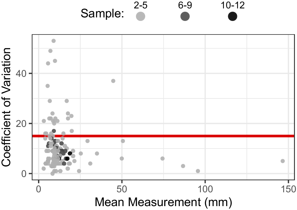


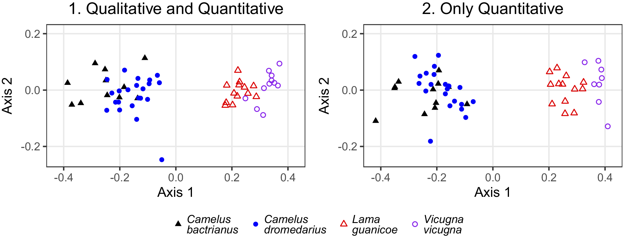




 Open access

