Introduction
Patients with repaired tetralogy of Fallot or Fontan circulation are exposed to chronic haemodynamic alterations and transient hypoxaemia that may predispose them to hepatic congestion and progressive liver injury. Reference Yamamura, Sakamoto and Morihana1,Reference Innocenzi, Rangel and Póvoa-Corrêa2 One concern in following these patients is the risk of liver fibrosis development as a result of haemodynamic alterations; such fibrosis may progress silently and even advance to cirrhosis. Reference Goldberg, Surrey and Glatz3
Metabolic dysfunction–associated steatotic liver disease has become the leading cause of chronic liver disease in young people. Reference Serbis, Polyzos, Paschou, Siomou and Kiortsis4,Reference Goyal, Xanthakos and Schwimmer5 Additionally, recent studies have revealed increased risks of obesity and metabolic syndrome in adults with CHD. Reference Deen, Krieger and Slee6–Reference García-Cruz, Manzur-Sandoval and Gopar-Nieto8 The coexistence of metabolic dysfunction–associated steatotic liver disease and cardiac-related hepatic injury may have synergistic effects, accelerating the progression of liver fibrosis. Reference Terzi, Camargo and Parente9,Reference Katz, Peck and Lubert10
Magnetic resonance imaging (MRI) is a valuable noninvasive tool for the assessment of liver fibrosis, enabling tissue characterisation and assessment of fibrosis. Reference Kupczyk, Mesropyan and Isaak11 T1 mapping and extracellular volume quantification provide insights into early and diffuse fibrotic changes, enhancing the role of MRI in comprehensive liver evaluation. Reference de Oliveira Terzi, Parente and Camargo12,Reference Hoffman, Ayoola, Nickel, Han, Chandarana and Shanbhogue13
The aim of this study was to compare the hepatic extracellular volumes and native T1 values of patients with cyanotic CHD and those with metabolic dysfunction–associated steatotic liver disease, to estimate the burden of liver involvement in these potentially synergistic conditions.
Materials and methods
Study population
This cross-sectional observational study was performed with five groups of participants: one group of healthy subjects, two groups of patients with metabolic dysfunction–associated steatotic liver disease, and two groups of patients with cyanotic CHD. Patients with metabolic dysfunction–associated steatotic liver disease were divided according to magnetic resonance elastography findings into F0–F1 (no or mild fibrosis; liver stiffness <3.5 kPa) and F2–F4 (significant fibrosis; liver stiffness ≥ 3.5 kPa) groups. The cyanotic CHD groups consisted of patients with tetralogy of Fallot and those with univentricular physiology who had undergone the Fontan procedure. Patients with tetralogy of Fallot were also compared according to the status of the pulmonary outflow tract.
Consecutive outpatients with tetralogy of Fallot or Fontan circulation were enrolled prospectively at cardiology clinics. Patients with steatotic liver disease were identified in hepatology outpatient clinics based on ultrasonographic diagnoses of liver steatosis and cardiometabolic criteria. Control subjects with fewer than two cardiovascular disease risk factors and no symptoms or MRI evidence of liver or heart disease were also enrolled prospectively. Patients with conditions that precluded MRI examination were not included. The institutional Research Ethics Committee approved the study protocols. All patients/guardians gave written informed consent to participate.
Study protocol
Clinical data were collected directly from patients/parents during clinical evaluation before MRI examination. All participants underwent liver assessment by cardiac or combined cardiac-abdominal MRI examination using a 3.0-T system (Magnetom Prisma; Siemens Healthcare, Erlangen, Germany). Examinations were conducted after 2–4-h fasts and lasted approximately 50 min. Peripheral blood samples for haematocrit analysis were obtained immediately before MRI examination. A gadolinium-DOTA contrast agent (Dotarem, 0.2–0.3 mmol/kg; Guerbet) was administered, and contrast-enhanced T1-weighted images were acquired 15 min post-infusion. MRI protocol is described in the Supplementary material. Image analysis and post-processing were performed using OsiriX MD (Pixmeo) for general imaging and Circle CVI42 (Circle Cardiovascular Imaging) for liver and cardiac evaluation. Specialists with>10 years of experience in cardiovascular or hepatic imaging conducted all analyses.
Statistical analysis
Continuous variables are reported as means ± standard deviations, and categorical variables are reported as counts and percentages. The distribution of continuous variables was assessed using the Shapiro–Wilk test. Between-group differences in hepatic extracellular volumes and native T1 values were evaluated using linear regression models. Unadjusted models were first constructed to estimate crude mean differences across groups. Multivariable linear regression was then performed with adjustment for potential confounders [body mass index and sex]. Age was not included in the model due to the lack of overlap among groups. Regression coefficients (β values), 95% confidence intervals (CIs), and p-values are reported. Pairwise comparisons were conducted to explore differences between clinical subgroups. Model diagnostics, including the assessment of residual plots and interaction terms, were performed to verify adherence to regression assumptions. Two-sided p values<0.05 were considered significant. All analyses were conducted using Stata (version 17.0; StataCorp, College Station, TX, USA).
Results
Participant characteristics
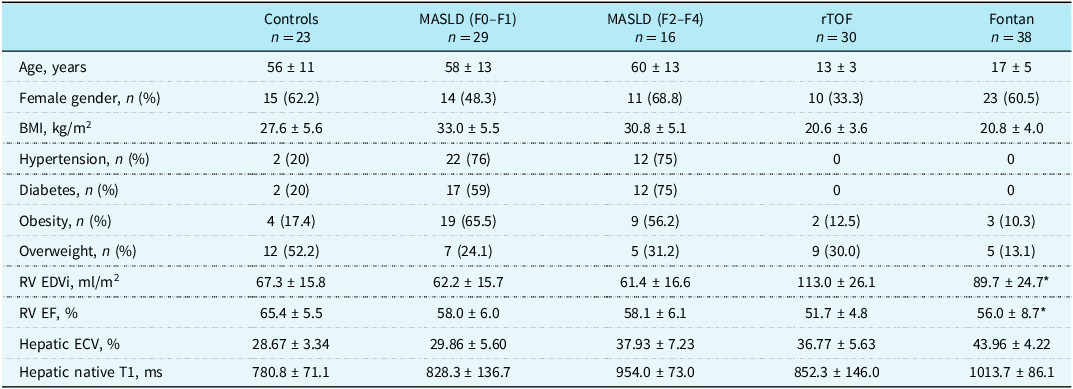
The study sample comprised 23 healthy controls, 45 patients with metabolic dysfunction–associated steatotic liver disease, 30 patients with tetralogy of Fallot, and 38 patients with Fontan circulation. Patients with tetralogy of Fallot and Fontan were younger (13 ± 3 and 17 ± 5 years, respectively) than controls (56 ± 11 years) and patients with steatotic liver disease (58.5 ± 12.6 years). Male sex was predominant only in the Fallot group. In the steatotic liver disease groups, the mean body mass index was >30 kg/m2, and the prevalence of obesity was >50%. Among controls and patients with CHD, the mean body mass index was <30 kg/m2. Overweight and obesity were more prevalent in the Fallot group (30.0% and 13.1%, respectively) than in the Fontan group (12.5% and 10.3%, respectively). The indexed right-ventricular end-diastolic volumes were larger in the Fallot group than in subjects with biventricular hearts. The mean right-ventricular and univentricular ejection fractions were normal. All patients were NYHA functional class I. The subjects’ baseline characteristics are presented in Table 1.
Clinical and MRI characteristics of patients

* For univentricular hearts, values were obtained for the systemic ventricle.
Values are described as n (%) ou mean ± standard deviation.
MRI = magnetic resonance imaging; MASLD = metabolic dysfunction–associated steatotic liver disease; rTOF = repaired tetralogy of Fallot; BMI = body mass index; RV = right-ventricular; EDVi = indexed end-diastolic volume; EF = ejection fraction; ECV = extracellular volume.
Hepatic extracellular volumes
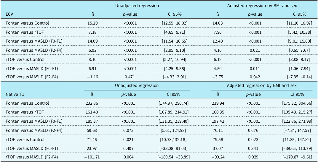
The hepatic extracellular volume was significantly larger in the Fontan circulation group (43.96% ± 4.22%) than in the other groups. The extracellular volume of patients with Fallot was smaller than that of patients with significant metabolic dysfunction–associated steatotic liver disease (36.77% ± 5.63% and 37.93% ± 7.23%, respectively), but significantly larger than those of patients with mild steatotic liver disease (29.86% ± 5.60%) and controls (28.67% ± 3.34%; Figures 1( a ) and 2). The unadjusted regression analysis indicated that the hepatic extracellular volume was significantly larger in patients with Fontan than in the other groups, with mean increases of 15.29% relative to controls and 7.18% relative to patients with Fallot (both p < 0.001; Table 2). The extracellular volume of patients with Fallot was 8.10% larger than that of controls (p < 0.001). The extracellular volumes of patients with mild and significant metabolic dysfunction–associated steatotic liver disease were 14.09% and 6.02% smaller, respectively, than that of patients with Fontan circulation (both p < 0.001). The extracellular volume in the mild steatotic liver disease group was 6.91% smaller than that of patients with Fallot (p < 0.001); the difference in the extracellular volume between the advanced steatotic liver disease and Fallot groups was not significant.
Hepatic extracellular volumes ( a ) and hepatic native T1 values ( b ) in patients with repaired tetralogy of Fallot, Fontan circulation, controls, and patients with mild (F0–F1) and significant (F2–F4) metabolic dysfunction–associated steatotic liver disease. ECV = extracellular volume; rTOF = repaired tetralogy of Fallot.

Sagittal native hepatic T1 parametric maps generated to provide quantitative assessment of hepatic tissue characterisation and to derive extracellular volume in three patients: ( a ) Fontan: native T1 = 1026 ms; ECV = 48.9%; ( b ) Tetralogy of Fallot: native T1 = 991 ms; ECV = 39.7%; ( c ) Control: Native T1 = 850 ms; ECV = 26.6%. ECV, extracellular volume.

Differences in the hepatic ECV and hepatic native T1 value from unadjusted and adjusted (for BMI and sex) linear regression models

ECV = extracellular volume; BMI = body mass index; CI = confidence interval; rTOF = repaired tetralogy of Fallot; MASLD = metabolic dysfunction–associated steatotic liver disease.
In the regression model adjusted for body mass index and sex, the hepatic extracellular volume of patients with Fontan circulation remained significantly larger than that in all other groups, with mean increases of 14.03% relative to controls and 7.90% relative to patients with Fallot (both p < 0.001; Table 2). Relative to that of patients with Fontan circulation, the extracellular volumes of patients in the mild and significant steatotic liver disease groups were 12.40% (p < 0.001) and 4.16% (p = 0.021) smaller, respectively. The extracellular volume of patients with Fallot was significantly larger than that of controls and mild metabolic dysfunction–associated steatotic liver disease (β = 6.12%; p<0.001 and β = 4.50%; p = 0.011, respectively). Finally, relative to patients with Fallot, the extracellular volumes of patients in the significant steatotic liver disease group was 3.75% larger (p = 0.042).
Among patients with tetralogy of Fallot, none had significant (moderate or severe) pulmonary stenosis. Seventeen patients (56.7%) presented with moderate or severe pulmonary regurgitation. Hepatic extracellular volume was not significantly higher in patients with significant pulmonary regurgitation compared with those with no/mild regurgitation (38.1% vs. 35.0%; p = 0.09).
Hepatic native T1 values
The mean hepatic native T1 value was longest in patients with Fontan circulation (1013.7 ± 86.1 ms), followed by those with significant metabolic dysfunction–associated steatotic liver disease (954.0 ± 73.0 ms), Fallot (852.3 ± 146.0 ms), mild steatotic liver disease (828.3 ± 136.7 ms) and controls (780.8 ± 71.1 ms; Figures 1( b ) and 2). The unadjusted regression model indicated that the T1 value of patients with Fontan circulation was significantly longer than those of all other subjects, by averages of 232.86 ms (p < 0.001) relative to controls, 161.40 ms (p < 0.001) relative to patients with Fallot, 185.37 (p < 0.001) relative to mild metabolic dysfunction–associated steatotic liver disease group, and 59.68 (p = 0.073) relative to significant steatotic liver disease. The native T1 value of patients with Fallot was 71.46 ms (p = 0.021) longer than that of controls. The T1 value was shorter in patients with Fallot than in those with significant metabolic dysfunction–associated steatotic liver disease (β = –101.71; p = 0.004), with no significant difference observed between the mild steatotic liver disease and Fallot groups (Table 2).
In the model adjusted for body mass index and sex, the hepatic native T1 value remained longest in patients with Fontan circulation, with mean differences of 239.94 ms from controls and 160.35 ms from patients with Fallot (both p < 0.001). The native T1 value remained significantly longer in patients with Fallot than in controls (β = 79.58 ms; p = 0.023]. This value also remained longer in the Fontan circulation group than in the mild metabolic dysfunction–associated steatotic liver disease group (β = 197.42; p < 0.001), with no significant difference between the significant steatotic liver disease and Fontan circulation groups. The T1 value remained significantly shorter in the Fallot group than in the significant metabolic dysfunction–associated steatotic liver disease group (β = –90.24 ms; p = 0.029; Table 2), with no difference observed between the mild steatotic liver disease and Fallot groups.
Discussion
The hepatic extracellular volume, a noninvasive marker of fibrosis, varied significantly across patient groups in our cohort. It was significantly larger among patients with Fontan circulation than in Fallot, mild and significant metabolic dysfunction–associated steatotic liver disease, and healthy control groups. Even after adjustment for sex and body mass index, Fontan circulation remained independently associated with markedly elevated hepatic extracellular volumes. Patients with Fallot had significantly larger extracellular volumes than did controls. Patients with steatotic liver disease with mild fibrosis had significantly smaller extracellular volumes than did patients with Fallot, whereas values for those with significant fibrosis were larger than those of patients with Fallot, after adjustment for sex and body mass index. These trends were corroborated by the native T1 values, which were highest in patients with Fontan circulation, although not significantly higher in this group than those observed in patients with steatotic liver disease with significant fibrosis. Adjusted regression analyses confirmed that Fontan physiology and Fallot were associated independently with increased native T1 values relative to healthy controls, reinforcing the occurrence of diffuse liver tissue remodelling, presumably in response to chronic haemodynamic stress and transient hypoxaemia.
Liver fibrosis is highly prevalent among patients with Fontan circulation. Reference Acosta Izquierdo, Rai and Saprungruang14–Reference Munsterman, Duijnhouwer and Kendall20 Recently, advanced imaging techniques, including magnetic resonance elastography, have demonstrated the elevation of hepatic extracellular volumes and native T1 values in patients with Fontan circulation. Reference Innocenzi, Rangel and Póvoa-Corrêa2,Reference Serai, Tsitsiou and Wilkins21 In the present study, we showed that the hepatic extracellular volumes and native T1 values of patients with Fontan circulation were higher not only than those of controls, but also than those of patients with steatotic liver disease.
Residual haemodynamic disturbances such as pulmonary regurgitation and right-ventricular volume overload can lead to chronic hepatic congestion and progressive parenchymal injury in patients with tetralogy of Fallot. Despite this potential risk, liver abnormalities in these patients remain unrecognized and poorly characterised. Reference Ravndal, Borgwardt and Juul22 Yamamura et al. Reference Yamamura, Sakamoto and Morihana1 reported that circulating markers of liver fibrosis were elevated in adults with Fallot, and Ozkok et al. Reference Ozkok, Sorkun and Erdemli23 suggested that hepatic microvascular perfusion abnormalities precede the development of fibrosis in this population. Previous studies of the hepatic extracellular volume in patients with Fallot have been performed with small samples and have revealed no significant difference from healthy controls. Reference Kazour, Serai, Xanthakos and Fleck24 In the present study, however, we observed that the hepatic extracellular volume was significantly larger in patients with Fallot than in healthy controls and also exceeded values seen in patients with metabolic dysfunction–associated steatotic liver disease with mild fibrosis. Notably, extracellular volumes in the Fallot group were similar than those observed in patients with steatotic liver disease with significant fibrosis, underscoring the potential risk of clinically significant liver disease in this population and reinforcing the need for ongoing hepatic surveillance. Although we found higher hepatic extracellular volume in patients with Fallot and significant pulmonary regurgitation compared with those with no/mild regurgitation, this difference did not reach statistical significance, possibly due to the small sample size available for this stratification.
Hepatic extracellular volume calculation and native T1 mapping are new imaging biomarkers for the assessment of liver fibrosis. In the present study, the hepatic extracellular volume effectively distinguished patients with Fallot from those with metabolic dysfunction–associated steatotic liver disease with mild fibrosis, whereas native T1 mapping did not. Similarly, hepatic extracellular volumes were significantly larger in patients with Fontan circulation than in those with steatotic liver disease and significant fibrosis, whereas native T1 mapping did not distinguish these groups. These findings align with recent literature indicating that the diagnostic performance of native T1 values is generally inferior to that of extracellular volumes for fibrosis staging. Reference Mesropyan, Kupczyk and Dold25 The extracellular volume is considered to be a more robust parameter than native T1 values due to its reduced sensitivity to variations in magnetic field strength and acquisition protocols. Reference Luetkens, Klein and Traeber26 The expansion of the extracellular matrix in chronic liver injury leads to an increase in extracellular space, which is captured by elevated extracellular volumes. Reference Luetkens, Klein and Träber27 Mesropyan et al. Reference Mesropyan, Kupczyk and Dold25 demonstrated that the hepatic extracellular volume reliably differentiated Child–Pugh classes, supporting its utility as a noninvasive marker of cirrhosis severity. Despite the inferior diagnostic accuracy of native T1 mapping for fibrosis staging, native T1 values were significantly elevated in the tetralogy of Fallot and Fontan circulation groups relative to healthy controls in this study.
The present comparison of hepatic native T1 values and extracellular volumes among patients with cyanotic CHDs and those with metabolic dysfunction–associated steatotic liver disease showed that Fontan circulation was associated with higher values and that values in the Fallot and steatotic liver disease groups were similar. The risks of metabolic syndrome and diabetes mellitus development are greater among patients with CHD than in the general population. Reference Umapathi, Thavamani, Bosco, Dhanpalreddy and Nguyen7,Reference Björk, Mandalenakis, Giang, Rosengren, Eriksson and Dellborg28
García-Cruz et al. Reference García-Cruz, Manzur-Sandoval and Gopar-Nieto8 identified high rates of cardiometabolic risk factors in a large cohort of Mexican adults with CHD. Additionally, recent studies have shown that the prevalence of obesity is increased among patients with CHD. Reference Umapathi, Thavamani, Bosco, Dhanpalreddy and Nguyen7,Reference Vangedal, Dahl, Thuraiaiyah, Lim, Jørgensen and Jensen29 These patients are likely to lead sedentary lifestyles, which is associated strongly with increased risks of obesity, atherosclerosis, and metabolic syndrome. Reference Siaplaouras, Niessner and Helm30,Reference Pelosi, Kauling and Cuypers31 Their reduced physical activity stems from multiple factors, including parental fear, anxiety, and an unexpectedly high frequency of physician-recommended activity restrictions, as highlighted by a recent German nationwide survey. Reference Siaplaouras, Niessner and Helm30,Reference Venna, d’Udekem and Figueiredo32 Taken together, these cardiometabolic risk factors may contribute to an increased risk of metabolic dysfunction–associated steatotic liver disease development in patients with CHD, particularly those with cyanotic heart defects or Fontan physiology. These findings were corroborated in our cohort of patients with CHD, as 23.4% of individuals in the Fontan group and 43.1% of those in the Fallot group were classified as overweight or obese. They underscore the importance of a comprehensive understanding of the overlapping factors contributing to liver disease in this population. In summary, extracellular volume calculation and native T1 mapping offer complementary insights into liver alterations in distinct populations of patients with CHD, highlighting the need not only for the careful screening and monitoring of metabolic risk factors in these groups, but also for the active promotion of healthier lifestyles to reduce the risks of liver disease, atherosclerotic cardiovascular disease, and other complications associated with metabolic disorders.
This study has certain limitations, including the lack of age matching among groups. Given that liver fibrosis increases with age, Reference Tong, Xue, Wang and Chen33,Reference Roy, Slimani and de Meester34 the older ages of the control and metabolic dysfunction–associated steatotic liver disease groups could have attenuated the differences observed. Still, we were able to demonstrate greater degrees of liver fibrosis in the younger groups of patients with cyanotic CHDs. If the patients in the control and steatotic liver disease groups were younger, they would likely have had even smaller extracellular volumes, further reinforcing the magnitude of the observed differences. In addition, due to ethical concerns, we were unable to perform liver biopsies for the histological confirmation of our findings; however, the extracellular volume has been validated as a noninvasive surrogate marker of liver fibrosis, Reference Luetkens, Klein and Träber27,Reference Lyu, Liu and Rui35,Reference Obmann, Ardoino and Klaus36 supporting the reliability of our results. Moreover, T1 values and extracellular volumes may reflect not only liver fibrosis, but also congestion, and these processes cannot always be distinguished based solely on these parameters. Reference de Lange, Reichert and Pagano37,Reference Bogaert, Dresselaers and Imazio38
In this study, extracellular volume assessment revealed hepatic involvement in patients with Fontan circulation that exceeded in patients with metabolic dysfunction–associated steatotic liver disease and significant fibrosis, while patients with Fallot showed intermediate involvement between those with steatotic liver disease and significant or mild fibrosis. The study findings highlight the potential of the extracellular volume as a sensitive, noninvasive biomarker capable of detecting early interstitial fibrosis before clinical symptoms emerge. Individuals with cyanotic CHD are increasingly recognised as being at elevated risk of metabolic syndrome development, which contributes significantly to hepatic steatosis and fibrosis progression. This synergistic burden underscores the need for the application of comprehensive clinical strategies that not only incorporate regular screening and the monitoring of metabolic risk factors but also emphasise proactive lifestyle modifications to mitigate liver disease progression in patients with CHD.
Supplementary material
The supplementary material for this article can be found at https://doi.org/10.1017/S1047951125110792.
Acknowledgements
We are very grateful to the staff and research assistants at the D’Or Institute for Research and Education, and who dedicated their time to support this study.
Financial support
This work was supported by the D’Or Institute for Research and Education, Rio de Janeiro (IDOR) and Fundação Carlos Chagas Filho de Amparo à Pesquisa do Estado do Rio de Janeiro (FAPERJ#E-26/010.002144/2019;#E-26/010.001806/2019).
Competing interests
None declared.
Ethics standard
The study was approved by the Ethics Committee of the D’Or Institute for Research and Education (CAAE:#70699417.7.0000.5249 & CAAE#34035120.1.0000.5249/2018). All participants provided informed written consent.