




No CrossRef data available.
Article contents
Dissociation of remote and anterograde memory impairment and neural correlates in alcoholic Korsakoff syndrome
Published online by Cambridge University Press: 01 May 2004
Abstract
Alcoholic Korsakoff's syndrome (KS) is marked by remote memory impairment together with characteristic profound anterograde memory deficits. Despite previous studies of memory processes in KS, questions remain regarding the nature and severity of these impairments and identification of brain systems that underlie these different memory impairments. This study examined remote and anterograde memory function in 5 KS patients in comparison with 8 patients with Alzheimer's disease (AD) and 24 normal control subjects (NC). In addition, relationships between memory performance and regional brain volumes were examined in the KS group. Overall, the KS group showed severe impairment on both remote and anterograde memory measures, performing at the level of the AD group on most measures. Differences were observed on the pattern of temporal gradient for verbal recognition, with KS exhibiting a more steeply graded rate of decline over the most recent period examined. Severity of the remote memory deficit in KS was not associated with severity of anterograde memory deficit. Examination of brain structure–function relationships in the KS subjects revealed that photo naming of remote historical information was related to posterior cortical white matter volumes but not hippocampal volumes; sequencing was related to prefrontal but not hippocampal volumes. By contrast, a measure of anterograde memory for nonverbal visual material showed a relationship to hippocampal but not regional cortical white matter volumes. This set of dissociations, which parallels that observed in our earlier study of AD, is now documented in KS and provides further evidence that these separate cortical and limbic brain systems are principal neural substrates of the remote and anterograde memory and sequencing deficits in KS. (JINS, 2004, 10, 427–441.)
Information
- Type
- Research Article
- Information
- Journal of the International Neuropsychological Society , Volume 10 , Issue 3 , May 2004 , pp. 427 - 441
- Copyright
- © 2004 The International Neuropsychological Society
INTRODUCTION
Korsakoff's syndrome (KS), a neurological disorder commonly arising from alcoholism-related thiamine deficiency, is characterized by a profound anterograde amnesia, and the inability to learn new information (Cermak et al., 1971; Corkin et al., 1985; Milner, 1958; Squire, 1982; Victor et al., 1989). In addition to anterograde memory deficits, impairment in remote memory, evidenced as a disability in recall of past public and autobiographical events, occurs in alcoholic KS (Albert et al., 1979, 1981; Butters & Albert, 1982; Kopelman, 1989, 1995; Kopelman et al., 1999; Parkin et al., 1990; Sanders & Warrington, 1971; Squire et al., 1989). Remote memory in KS is typically characterized as a temporally extensive and graded deficit, being relatively greater for more recent than remote time periods. Despite the co-occurrence of remote and anterograde memory deficits in KS, the severity of each has not been shown to be consistently related and the underlying neural substrates compromised that result in these dissociable deficits are still under investigation.
KS is generally referred to as an example of a diencephalic amnesia because the anterograde memory deficit observed in KS has been associated with lesions of diencephalic structures, including the anterior and dorsomedial nucleus of the thalamus and the mammillary bodies (Aggleton & Brown, 1999; Butters & Stuss, 1989; Harding et al., 2000; Shimamura, 1989; Squire et al., 1990). Recent reports, however, have indicated that profound memory deficits can be observed in KS individuals with relatively intact mammillary bodies (Shear et al., 1996; Sullivan et al., 1999) and that alcoholic individuals with severely damaged mammillary bodies do not always suffer from global amnesia (Davila et al., 1994; Sullivan et al., 1999). Additionally, structures outside of the diencephalon, most notably the hippocampus, show volume deficits in alcoholics with (Jernigan et al., 1991; Sullivan et al., 2001) and without amnesia (Agartz et al., 1999; Sullivan et al., 1995b), suggesting that either limbic or diencephalic or both loci contributes to KS amnesia. Importantly, however, alcoholics with KS do show greater volume deficits in the mammillary bodies (Sullivan et al., 1999) and hippocampi (Sullivan & Marsh, 2003) than nonamnesic alcoholics, raising the possibility that a threshold of tissue compromise must be crossed in order for impairment to be manifest. Furthermore, postmortem findings highlight white matter pathology as especially prominent in chronic alcoholism (Harper & Kril, 1993) and imply that such tissue damage contributes significantly to concomitant cognitive and motor deficits observed in alcoholic individuals (cf. Pfefferbaum & Sullivan, 2002; Pfefferbaum et al., 2000; Sullivan et al., 2000).
Similar to individuals with KS, patients with Alzheimer's disease (AD), exhibit remote and anterograde memory deficits (Beatty et al., 1988; Corkin, 1982; Fama et al., 2000; Kopelman, 1989; Sagar et al., 1988). Remote memory deficits in AD are extensive, spanning decades (Hodges et al., 1993; Wilson et al., 1981), although the deficits observed are less temporally graded than in KS (Beatty et al., 1988; Kopelman, 1989; Sagar et al., 1988). AD is a neurodegenerative process, a dementia, characterized by deficits in a number of cognitive domains, most notably, anterograde memory. Studies examining the principal brain areas underlying the progressive anterograde memory deficits in AD have found relationships between severity of anterograde memory impairments and medial temporal structures, particularly the hippocampus (Deweer et al., 1995; Fama et al., 1997; Jack et al., 1998; Killiany et al., 1993, 2000; Laakso et al., 1995).
Aggleton and colleagues, however, have questioned the classical distinction made between diencephalic and medial temporal amnesia (Aggleton & Brown, 1999; Aggleton & Saunders, 1997). These authors proposed that the anterograde memory deficits associated with both diencephalic and medial temporal amnesia are a result of dysfunction within an “extended hippocampal system,” which includes the hippocampus, fornix, mammillary bodies, and anterior thalamic nuclei. Differences observed in the pattern of anterograde memory impairment in diencephalic amnesia and medial temporal lobe amnesia are hypothesized to be a result of the influence of extra-hippocampal damage associated with these different neurological conditions.
In contrast to anterograde memory deficits attributable to diencephalic, thalamic, and hippocampal lesions, remote memory impairments are related to cortical abnormalities (Damasio & Damasio, 1993; Squire et al., 1993). It has been hypothesized that remote memories are distributed throughout the cortex and in particular may rely on the integrity of cortical association areas (Ungerleider, 1995). Although medial temporal structures are thought to have a time-limited role in the consolidation process, recent studies have reported the importance of the hippocampus in retrieval of remote autobiographical (episodic) and spatial memory (Maguire et al., 2001; Nadel et al., 2000; Ryan et al., 2001). Temporal neocortical lesions, without medial temporal or diencephalic involvement, have been associated with remote memory deficits (Hodges, 1995; Kapur et al., 1994). Posterior cortical areas, parieto–occipital and occipital lobes, have been implicated in focal deficits of autobiographical information (Hunkin et al., 1995). In a recent study (Fama et al., 2001), we reported relationships between recognition and sequencing of remote public information and posterior cortical volumes in AD, and these deficits and relationships were dissociable from those observed between new learning and hippocampal volumes. Thus, it appears that remote memory, at least for past public and semantic information, and in contrast to anterograde memory, is mediated by non-hippocampal systems.
The goals of this study were (1) to establish the severity and pattern of remote memory performance for past public historical information in KS, (2) to identify brain correlates of component processes of remote memory in KS, and (3) to compare the pattern of neuropsychological performance and brain correlates of remote and anterograde memory in KS with those documented in AD. Similarities between KS and AD in memory performance and brain–behavior relationships would provide support for the notion that observed impairments may be due to dysfunction of the same brain system in both conditions and support the extended hippocampal hypothesis of Aggleton and colleagues for anterograde memory deficits in KS and AD. Differences between these groups may be attributed to differences in neural systems disrupted. We hypothesized that the KS group would be impaired on all remote memory measures, as would the AD group, and that the deficit would be most pronounced for the recent past in both groups. Similar to AD (Fama et al., 2001), we expected that the remote and anterograde deficits would not be strongly related in the KS group. We hypothesized that the frontal executive dysfunction observed in KS would be reflected in their remote memory performance, such that the KS group would perform particularly poorly on remote memory tasks requiring temporal ordering and frontal executive abilities (e.g., free recall and ability to pair, date, and sequence presidential candidates) compared with tasks without temporal processing demands (e.g., recognition of presidential candidate names, photo naming). Finally, we hypothesized that selective remote memory deficits, particularly recognition of public figures, would be related to volume deficits in cortical brain regions quantified from structural MRI (cf. Fama et al., 2001). In light of postmortem findings, we anticipated that regional cortical white matter volume would be associated with selective cognitive performance in the KS group.
METHODS
Research Participants
Study participants included 5 KS patients, 8 patients with probable AD, and 24 normal control (NC) subjects. The KS subjects were recruited from inpatient units of a Veterans Administration Medical Center. Each KS subject had an extensive history of alcoholism, which was verified through medical charts or family reports, and met criteria for DSM–IV Alcohol-Induced Persisting Amnestic Disorder (American Psychiatric Association, 1994). Because of their amnesia, however, the KS patients were unable to provide a credible account of their drinking quantities. The AD subjects were recruited from the Geriatric Psychiatry Rehabilitation Unit and the National Institute of Mental Health Aging Clinical Research Center, both housed at the VA Medical Center. All AD patients met the National Institute of Neurological and Communicative Diseases and Stroke-Alzheimer's Disease and Related Disorders Association criteria for probable Alzheimer's disease (Khachaturian, 1985; McKhann et al., 1984). These AD subjects, a subset of a larger AD group used in earlier reports of remote memory functioning (Fama et al., 2000, 2001), were selected to match the KS in age range. The NC subjects were recruited from the community. Potential control subjects who scored below 25 on the Mini-Mental State Examination (MMSE; Folstein et al., 1975) were excluded from the study. Screening for all participants (KS, AD, and NC) included a structured psychiatric diagnostic interview and medical examination. Subjects were excluded for significant history of psychiatric or neurological disorder not related to their primary diagnosis, past or present alcohol or drug abuse or dependence in the AD and NC groups, or serious medical condition. Written informed consent was obtained from all participants and, where relevant, from conservators. Demographics for all subject groups are in Table 1.
Subject demographics

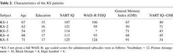
The subject groups were not significantly different in age (Kruskal Wallis H = 4.45, p = .11) or years of formal education (H = 4.52, p = .80). Groups were significantly different on measures of premorbid intelligence, which was estimated with the National Adult Reading Test (Nelson, 1982; H = 5.85, p = .05), and current global functioning, which was assessed with the Mini-Mental State Exam (Folstein et al., 1975; H = 22.93, p = .0001). Follow-up group comparison indicated that the KS group did not differ from the NC group on the NART (Mann-Whitney z = .12, p = .91) but scored significantly lower than the NC group on the MMSE (z = 3.37, p = .0008). The difference between the KS group and AD group on the NART approached significance (z = 1.68, p = .09), with the KS group performing better than the AD group. The difference between the KS and AD group on the MMSE was significant (z = 2.11, p = .04), with the KS group scoring higher than the AD group. Additional information including individual demographic scores for each of the KS patients is provided in Table 2.
Characteristics of the KS patients

Neuropsychological Measures
Although the neuropsychological measures used in this study likely rely on several component processes for successful completion, we grouped measures that have been shown to share similar task demands and labeled them accordingly. Not all subjects completed all tests.
Anterograde memory
The following measures were used to assess episodic memory:
- Recognition Memory Test (Warrington, 1984) assessed recognition of words and faces separately. Each subtest consists of 50 items.
- Wechsler Memory Test–Revised (Wechsler, 1987) provided indices of immediate memory (verbal and visual) and an assessment of memory after a short delay.
Semantic memory
The following measures assess the ability to retrieve semantically based information that is well learned throughout one's lifetime:
- Modified Boston Naming Test (Huff et al., 1986) used 42 line drawings of common objects, plants, and animals taken from the original 85 item BNT (Kaplan et al., 1976).
- Vocabulary subtest of the WAIS–R (Wechsler, 1981) assessed ability to define increasingly difficult words.
Executive function
We used the following measures to assess abilities to plan and sequence:
- Wisconsin Card Sorting Test (Milner, 1963) required subjects to sort 128 cards into predetermined categories. Scores used in this study were number of categories correctly sorted (all subjects completed 128 cards) and number of perseverative responses committed (Heaton, 1981). Studies have shown these test parameters to be sensitive to frontal lobe dysfunction (cf. Milner, 1963; Sullivan et al., 1993).
- Picture Arrangement subtest of the WAIS–R (Wechsler, 1981) assessed the ability to sequence the events of an action or a story which was visually presented. This subtest has been reported to be a sensitive measure of frontal lobe/executive function integrity (cf. McFie & Thompson, 1972; Sullivan et al., 1989).
Remote memory
- Presidential Candidates Test, modified from Hamsher and Roberts (1985), assessed subjects ability to name and recognize all the Democratic and Republican presidential candidates (elected and defeated) dating back to 1920.
- Candidate Recall, in which subjects were given a sheet of paper divided into columns and were asked to write down, within a 5-min time limit, the names of all the Democratic and Republican presidential candidates since 1920, identify which political party they were affiliated with, and the year(s) those candidates ran for office. Last names of the candidates were deemed sufficient for credit.
- Candidate and Election Year Recognition, in which each item consisted of six names (two of which were presidential candidates who ran against each other in a particular election) and 3 years (one of which was the correct election year). The remaining four names were those of high profile individuals who had been politically or socially active in the same era as the presidential candidates. The remaining two years were balanced across items such that one of the incorrect years was ± 4 years and the other incorrect year was ± 12 years from the correct election year. Subjects were to identify which two of the six candidates ran against each other in a particular election and choose the year that election occurred.
- Photo Naming, in which subjects were shown black-and-white photographs (9 × 11 cm) of all presidential candidates from the elections of 1920 to 1980 and asked to name each of them. Participants received credit for naming as long as the correct last name of the candidate was given.
- Candidate Sequencing, in which subjects sequenced the names of presidential candidates within a single political party. Three sets of index cards, each card containing the name of a candidate, were presented in a random order (fixed across subjects) for each political party (Set 1 = 1920–1940; Set 2 = 1944–1964; Set 3 = 1968–1988). Subjects were asked to place the candidates in chronological order from the earliest to the most recent. Final sequence scores were based on the number of cards placed in the correct serial position, with a range of zero to 6 points for each of the six sets of cards. Because there are presidential candidates who ran for office more than one time, trials contain cards that are scored correct in more than one position.
MRI Scanning and Quantification
MRI was conducted with 1.5 T General Electric Signa scanners. Image acquisition and quantification procedures used herein have been previously described (axial: Pfefferbaum et al., 1994; coronal: Sullivan et al., 1995a, 1995b; see figure legends for MRI acquisition parameters). For the axial series, the cortical rim was automatically segmented into gray matter, white matter, and CSF compartments and was also divided geometrically into six cortical regions: prefrontal, frontal, anterior superior temporal, posterior superior temporal, anterior parietal, and posterior parietal–occipital (see Figure 1a). White matter subjacent to the gray matter in these designated regions are referred to as cortical volumes, albeit white matter (as opposed to gray matter) cortical volumes, throughout this paper. Hippocampal volumes were derived from a coronal acquisition protocol and measured manually (see Figure 1b).

Fig. 1a. Axial MR images were 5-mm thick (2.5 mm skip) and acquired in an oblique plane using a dual-echo spin-echo sequence (TE = 20, 80 ms) with a 24 cm field of view and a 256 × 256 matrix. Acquisition was gated to every other cardiac cycle for an effective TR of >2400 ms with one excitation for each of 256 phase encodes. Seven consecutive slices, beginning with the anterior horns of the lateral ventricles, were segmented into gray matter (shown in dark gray), white matter (light gray), and cerebrospinal fluid (black). Each measured MRI slice was divided into an outer 45%, which represented cortical regions, and an inner 55%, which included ventricular regions. The cortical area was then divided into regions that roughly correspond to lobar anatomy: a indicates prefrontal, Slices 1 to 7; b, frontal, Slices 3 to 7; c, anterior superior temporal, Slices 1 to 2; d, posterior superior temporal, Slices 1 to 2; e, anterior parietal, Slices 3 to 7; f, posterior parietal–occipital, Slices 3 to 7.
Fig. 1b. Early echo coronal MRI with hippocampi and temporal lobes outlined. With this protocol 22 contiguous 3-mm thick coronal images were acquired with a dual-echo, flow compensated, cardiac gated pulse sequence [TE = 40, 80 ms; effective TR ≈ 2800 ms; field of view = 24 cm, NEX = 1, 256 × 256 matrix; image acquisition oriented perpendicular to the anterior commissure-posterior commissure (AC-PC) line]. The hippocampus was outlined on consecutive slices in each hemisphere and volume was derived by adding the areas of each measured slice.
Statistical Analysis
We examined group differences with nonparametric analyses (Kruskal Wallis and Mann-Whitney tests) because of the differences across groups in sample size and the small sample size of the clinical groups (Siegel & Castellan, 1988). When examining relationships between variables we employed Spearman rank correlation coefficients. Correlations between brain and behavior measures were based on one-tailed tests because we predicted a priori the direction of the relationships.
RESULTS
Anterograde Memory, Semantic Memory, and Executive Function Performance
Group differences on the measures of anterograde memory, semantic-based remote memory, and executive function are presented in Table 3. Compared with the NC group, the KS were impaired on all measures of anterograde memory. The KS group also showed deficits on executive function by completing fewer categories on the WCST (z = 2.73, p = .006) and committing more perseverative responses (z = 3.09, p = .002) than the NC group. The KS group did not differ from the NC group on the WMS–R Attention Index or Boston Naming Test. The AD group performed significantly worse than the NC group on all variables assessed, with the exception of number of perseverative responses on the WCST.
Group performance on ancillary neuropsychological tests

The KS and AD groups performed similarly on most anterograde memory measures. Where they differed, the KS group recognized more words on the Warrington Recognition Test (z = 3.32, p = .02) and tended to have better immediate recall of WMS–R Logical Memory (z = 1.78, p = .08) than the AD group. By contrast, the KS group committed significantly more perseverative responses on the WCST than the AD group (z = 1.96, p = .05).
Remote Memory of Past Presidential Candidates
Recall and recognition of presidential candidates
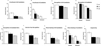
Kruskal Wallis analysis revealed differences across the three groups (KS, AD, NC) for free recall of presidential candidates (H = 20.29, p = .0001; Figure 2a). Follow-up two group comparisons (Mann-Whitney tests) showed that both the KS and AD groups recalled significantly fewer candidates than the NC group (KS vs. NC: z = 2.54, p = .01; AD vs. NC: z = 4.14, p = .0001) but that the KS and AD groups did not differ from each other (z = 1.0, p = .32). Group differences were found for free recall of elected (H = 15.13, p = .0005) and defeated (H = 14.85, p = .0006) candidates (Figure 2b). Both the KS and AD groups recalled fewer elected and defeated candidates than the NC group but again did not differ from each other (elected: z = .59; p = .56; defeated: z = 0; p = 1.0).

(a–i). Bar graphs (M ± SEM) depicting subject groups (Korsakoff syndrome, KS; Alzheimer's disease, AD; and normal control, NC) performance for each subtest of the Presidents Test. Where possible, elected and defeated candidates were also examined separately.
The three subject groups also differed in recognition of presidential candidate names (H = 17.62, p = .0001; Figure 2c). Both the KS and AD groups recognized significantly fewer candidates than the NC group (KS vs. NC: z = 2.14, p = .03; AD vs. NC: z = 3.81, p = .0001). The KS group recognized significantly more candidates than the AD group (z = 2.27, p = .02). In addition, the three groups significantly differed in recognition of defeated candidates (H = 16.25, p = .0003), and a statistical trend was observed for recognition of elected candidates (H = 5.39, p = .07; Figure 2d). In particular, the KS group did not differ from the NC group on recognition of elected candidates (z = .40, p = .69) but did differ on defeated candidates (z = 2.22, p = .026). The AD group performed significantly below the NC group for both recognition of elected (z = 2.31, p = .02) and defeated (z = 3.61, p = .0003) candidates. Relative to the AD group, the KS group recognized significantly more defeated (z = 1.98, p = .05) but not elected candidates (z = 1.32, p = .19).
Recognition of election year and candidate pairs
When examining recognition of election year, only elections for which a subject correctly recognized both presidential candidates were included. We reasoned that unless subjects could accurately identify both candidates, we could not be sure that they were dating the intended election. Although chance level for recognition of election year was 1 out of 3, chance level of candidate pair recognition was 1 out of 15 for each item. The groups differed in ability to recognize candidate pairs (H = 15.06, p = .0005), with the KS and AD groups recognizing significantly fewer pairs than the NC group (KS vs. NC: z = 1.96, p = .05; AD vs. NC: z = 3.59, p = .0003; Figure 2e). Differences between the KS and AD groups were not statistically significant (z = 1.61, p = .11). Similar to free recall and recognition performance, significant group differences were found for recognition of election year (H = 13.82, p = .001; Figure 2f): although the KS group did not differ significantly from either the NC group (z = 1.53, p = .13) or the AD group (z = 1.61, p = .11), the AD group recognized a lower percentage of election years than the NC group (z = 3.57, p = .0004).
Photo naming of presidential candidates
Groups differed on photograph naming in general (H = 13.8, p = .001; Figure 2g) and specifically for elected (H = 11.19, p = .004) and defeated (H = 14.9, p = .0006) candidates (Figure 2h). The KS and AD groups did not differ from one another (z = .95, p = .35) but both groups named fewer presidential candidates than the NC group (KS vs. NC: z = 2.83, p = .005; AD vs. NC: z = 2.83, p = .005). The same pattern of performance was observed when examining elected and defeated candidates separately.
Sequencing of presidential candidates
Groups differed significantly (H = 14.18, p = .0008) on sequencing of presidential candidates, with the KS and AD groups performing worse than the NC group (KS vs. NC: z = 3.06, p = .002; AD vs. NC: z = 2.70, p = .007) but similarly to each other (z = 0, p = 1.0; Figure 2i).
Performance over time periods
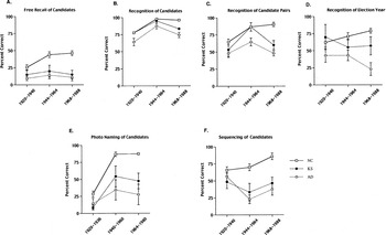
Free recall performance over time (Figure 3a) indicated a significant group difference for the middle (1944–1964: H = 13.99, p = .001) and recent (1968–1988: H = 19.42, p = .0001) time periods. A statistical trend was noted for the most remote (1920–1940: H = 5.52, p = .06) time period assessed. The KS group was impaired compared with the NC group in free recall of candidates on the middle and recent time periods (z = 2.22, p = .03; z = 3.0, p = .003) but not for the remote time period (z = 1.2, p = .23). The AD group was impaired on free recall for all time periods assessed. The KS and AD groups did not differ significantly from one another in any free recall over time comparison.

(a–f). Line graphs depicting performance of all three subject groups (KS, AD, NC) over designated time periods for various subtests. Of particular interest was the rapid decline in performance observed in the KS group from the middle time period (1944–1964) to the recent time period (1968–1988) on candidate pair recognition. Although the KS group performed at the level of the NC group for the middle time period they were as impaired as the AD group on the recent time period.
Similar to free recall performance, recognition of candidate pairs (Figure 3b) indicated a significant group difference for the middle and recent time periods (H = 9.05, p = .01; H = 19.94, p = .001). No group difference was observed, however, for the more remote time period. Follow-up analyses indicated that the AD group performed significantly worse than the KS and NC groups for the middle time period, whereas both the AD and KS groups performed below the NC group for the more recent time period. In contrast to the AD performance, which was significantly below that of the NC group starting with the 1944–1964 time period, the KS group did not differ from the NC group until the more recent time period (1968–1988).
Performance across time periods for election year recognition indicated group differences for the middle (H = 7.73, p = .02) and most recent (H = 16.55, p = .0003) periods (Figure 3c). The KS showed a modest impairment for election year recognition compared with the NC group for the most recent time period (z = 1.85, p = .06), whereas the AD group was impaired on the middle (z = 2.68, p = .007) and most recent (z = 3.85, p = .0001) time periods. Although the NC group was better able to recognize the election year for the more recent elections, this was not the case for the KS or the AD groups. Differences between the KS and AD groups approached significance for the most remote (z = 1.76, p = .08) and most recent (z = 1.68, p = .09) time periods, suggesting that the AD group had even more difficulty than the KS group in correctly identifying the election year for candidate pairs that were correctly recognized.
Group differences were observed for all three time periods for photo naming (Time 1: H = 5.9, p = .05; Time 2: H = 12.6, p = .002; Time 3: H = 15.9, p = .0004; Figure 3d). No differences between the KS and AD groups were observed for any time period (Time1: z = .31, p = .75; Time 2: z = .83, p = .40; Time 3: z = 1.15, p = .25). The KS group named fewer candidates than the NC group on all three time periods (Time1: z = 2.17, p = .03; Time 2: z = 2.45, p = .01; Time 3: z = 2.83, p = .005). The AD group was impaired relative to the NC group on the middle and most recent time period only (Time1: z = 1.47, p = .14; Time 2: z = 2.92, p = .004; Time 3: z = 3.20, p = .001).
Examination of candidate sequencing over time also yielded group differences for the middle (H = 10.72, p = .005) and most recent (H = 14.04, p = .0009) periods (Figure 3e). Both the KS and AD groups differed significantly from the NC group on these two periods (KS vs. NC: z = 2.34, p = .019, z = 2.92, p = .005; AD vs. NC: z = 2.67, p = .008, z = 2.91, p = .004) but did not differ from each other (KS vs. AD: z = .10, p = .92, z = .52, p = .60). The three groups did not differ from each other in sequencing performance on the most remote time period.
Relationship between remote and anterograde memory measures in KS
Anterograde memory performance, as assessed by the general and delayed memory indices of the WMS–R (GMI, DMI), did not predict, at least not in the expected direction, remote memory performance on the Presidents Test [candidate pairs (GMI: re = −.625, p = .11; DMI: re = −.825, p = .05), election year (GMI: re = −.6, p = .12; DMI: re = .125, p = .40), photo naming (GMI: re = −.3, p = .27; DMI: re = −.125, p = .40), sequencing (GMI: re = .125, p = .40; DMI: re = .425, p = .20)]. Due to the restricted range in scores for free recall (4 of 5 subjects scored between 2 and 3 points) and recognition of all presidential candidates (all 5 subjects scored between 20 and 22 points), correlations are not reported for these subtests. Further, better performance on the Warrington Recognition Test (anterograde memory measure) did not predict better performance on recognition of candidate pairs (remote memory measure) even though both were recognition memory tests (Words: re = −.25, p = .33; Faces: re = −.85, p = .07).
MRI correlates of the Presidents Test in KS
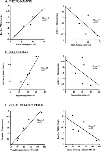
Brain volumes for the KS group are presented in Table 4. Relationships between remote memory scores and regional brain volumes were analyzed with and without the earliest time period included to ensure that the structure-function relationships reported were not due to information reflecting elections that an individual may not have personally experienced. Only those brain–behavior relationships that were significant with and without Time 1 data are reported. Photo naming scores in the KS group were positively correlated (predicted direction; i.e., poorer performance related to smaller brain volume) with posterior parietal–occipital white matter volume (re = 1.0, p = .02) and were negatively correlated with anterior hippocampal volume (re = −.7, p = .08; Figure 4a). Sequencing of presidential candidate names was significantly correlated with prefrontal white matter (re = .83, p = .05), but not with hippocampal volume (re = −.33, p = .26; Figure 4b). In contrast to the remote memory measures, the WMS–R Visual Memory Index, an anterograde memory measure, showed a positive relationship to the anterior hippocampal volume (re = 1.0, p = .02) but was negatively correlated to posterior parietal–occipital white matter volume (re = −.7, p = .08; Figure 4c). Even when the subject who scored highest (VISI = 106) was excluded from the analyses, the Visual Memory Index was still significantly correlated with anterior hippocampal volume (re = 1.0, p = .04) but not with posterior parietal–occipital white matter volume (re = −.4, p = .24).
Brain data for KS group


(a–c). Scatterplots depicting the relationship between remote and anterograde memory measures and selective brain volumes.
DISCUSSION
Both the KS and AD groups showed impairments on remote and anterograde memory measures involving verbal and nonverbal material for past public historical information and for novel information. Relative to controls, both patient groups were impaired on free recall, recognition, photo naming, and sequencing of remote public information. Similarities and differences between KS and AD on nonmnemonic processes were consistent with previous studies and lend evidence to the clinical and neuropathological distinction between these conditions. For example, the KS group made a greater number of perseverative responses on a frontal executive measure (i.e., WCST) compared with the control group or AD group. Furthermore, the AD, but not the KS group, was significantly impaired on a confrontation naming measure (i.e., BNT) compared with the control group.
KS and AD Commonalities in Remote Memory Processing
This study demonstrates that KS and AD subjects, despite their severe memory impairment, were able to recognize remote historical information when material was presented in a forced choice format. This type of remote information (names of presidents) is unique in that it is presented continually through the media for an extended period of time (years in the case of elected presidential candidates). Individual performance patterns for remote memory in the KS subjects were suggestive of a progressive rather than an acute onset (cf. Butters & Cermak, 1980). Memory performance appeared to be inconsistent across elections, with no abrupt demarcation observed. Whether memory loss for the recent past in KS can be partly due to progressive anterograde memory deficits is controversial. In this study, recognition, sequencing, and photo naming performance for the most recent time period were not significantly related to anterograde memory measures. The relationship between free recall of presidential candidates for the most recent time period and anterograde memory measures were not determined due to the limited distribution of free recall scores in the KS group (range of scores = 0–3, with 3 of the 5 subjects recalling only 1 presidential candidate).
The KS and AD groups did not differ significantly from each other on any overall remote memory parameter, a pattern similar to that observed by Kopelman and Stanhope (2002). These groups were equally impaired on overall scores of free recall, recognition, and photo naming of presidential candidates. The KS and AD even performed comparably on the remote memory tasks that are thought to have an additional frontal executive component (i.e., recognition of election years, sequencing of presidential candidates) and that we predicted would be particularly compromised in KS. These findings are consistent with previous reports (cf. Downes et al., 2002) indicating temporal order impairments in cases of medial temporal amnesia (AD) as well as in diencephalic amnesia (KS). Further, the similarities observed between the KS and AD groups are consistent with reports from Aggleton and colleagues and their proposed “extended hippocampal system” theory.
KS and AD Differences in Remote Memory Processing
The two patient groups did differ, however, in time-linked patterns of performance. In particular, the KS showed a more temporally graded impairment for recognition of remote public information than did the AD. The KS group performed at the level of the NC group for the two earliest time periods (1920–1940, 1944–1964) but showed a marked deficit compared to controls for the most recent time period (1968–1988), performing at the level of the AD group and significantly below their own performance level for the previous two time periods. The AD group was impaired on recognition of candidate pairs dating back to the 1940s, but performed similarly to the KS and NC groups on the most remote period. These results are consistent with studies that report moderately extensive and steeply temporally graded remote memory deficits in KS (e.g., Albert et al., 1979, 1981; Butters & Albert, 1982; Kopelman, 1989, 1995; Kopelman et al., 1999; Parkin et al., 1990; Sanders & Warrington, 1971; Squire et al., 1989). Differences observed between the KS and AD groups on select memory parameters may be attributable to nonmnemonic processes that are reflective of cortical damage outside of that supporting declarative memory associated with each condition. For example, the AD group may show a more temporally extensive impairment for remote memory information because of a semantic/lexical knowledge deficit, associated with temporal neocortical dysfunction (Hodges et al., 1992; Kapur et al., 1994; Kensinger et al., 2001), which is likely greater in AD than KS.
Anterograde Memory and Brain Correlates of Remote Memory Impairments in KS
As hypothesized, despite impairment in both anterograde and remote memory in the KS group, these disabilities were not obviously related to one another. Severity of remote memory impairment was not predictive of severity of anterograde memory impairment in the KS subjects. These results provide further support that anterograde and remote memory processes are at least partly subserved by different brain systems (Clark et al., 2002; Fama et al., 2001; Squire et al., 1993). It is generally believed that the hippocampus and related structures are important in anterograde memory and memory consolidation, possibly binding the cortical sites where memories are eventually stored until these memories can be activated and retrieved without the assistance of medial temporal structures (Clark et al., 2002; Squire & Alvarez, 1995).
A series of dissociations successfully distinguished relationships between memory performance and regional brain volumes. In particular, photo naming of presidential candidates was related to posterior parietal–occipital white matter volume but not to hippocampal volume. Sequencing of presidential candidates was related to prefrontal white matter volume but not to hippocampal volume. These relationships endured when considering only the elections from the 1940s–1980s. There was a concern that recall/recognition of information prior to the 1940s may be different because the subjects in this study may have been too young to have personally experienced these events. By contrast, the visual memory index of the WMS–R was related to anterior hippocampal volume but not cortical volumes associated with the remote memory measures. These findings parallel relationships reported previously in AD (Fama et al., 2001) and emphasize the relevance of posterior cortical regions in the mediation of remote memory functioning. In light of the limited sample size, we do not interpret these findings as an indication of a lack of influence of temporal neocortex in the mediation of aspects of remote memory functioning but rather evidence for function of extratemporal cortical areas in the mediation of select component processes of remote memory for historical information.
The selective relationship between recent declarative memory performance and hippocampal volumes provides support for the relevance of the hippocampus (particularly the anterior extents) to the amnesia of KS and calls into question traditional concepts of KS as solely a diencephalic amnesia (Sullivan & Marsh, 2003). These results are consistent with previous reports (cf. Haist et al., 2001) that highlight the role of the anterior hippocampus in anterograde memory processes. In addition, these results comport with in vivo and postmortem findings, suggesting that white matter abnormalities contribute significantly to cognitive deficits associated with KS and other alcohol related impairments.
In conclusion, the degree of similarity between the KS and AD groups on remote memory tasks suggests that common psychological and neural mechanisms underlie particular component processes of remote memory in these two neurological conditions. The similarities between KS and AD on anterograde memory measures provide further support for the concept of an “extended hippocampal system” (Aggleton & Brown, 1999; Aggleton & Saunders, 1997). Volume measures of posterior cortical brain regions were significantly related to memory for remote, historical information, and volume measures of prefrontal cortical regions predicted ability in sequencing such information. Further to this point, the double dissociation observed here in KS and previously in AD (Fama et al., 2001), identifying selective neural support of remote memory from cortical brain systems and of declarative anterograde memory from the hippocampus, supports the dissociation of these cognitive processes and also demonstrates the importance of the hippocampus in the anterograde amnesia of KS (cf. Sullivan & Marsh, 2003).
ACKNOWLEDGMENTS
This research was supported by National Institute on Alcohol Abuse and Alcoholism Grants AA10723 (E.V.S.), AA05965 (A. Pfefferbaum) and National Institute on Aging AG17919 (E.V.S.). A portion of this research was presented at the International Neuropsychological Society conference (2000, Chicago). We thank Adolf Pfefferbaum, M.D., for his continued support and encouragement in this project, Kelvin O. Lim, M.D, for contributions to image acquisition and processing, and Margaret J. Rosenbloom, M.A., for assistance with manuscript preparation.
References
REFERENCES
Agartz, I.,
Momenan, R.,
Rawlings, R.R.,
Kerich, M.J., &
Hommer, D.W.
(1999).
Hippocampal volume in patients with alcohol dependence.
Archives of General Psychiatry,
56,
356–363.Google Scholar
Aggleton, J.P. &
Brown, M.W.
(1999).
Episodic memory, amnesia, and the hippocampal–anterior
thalamic axis.
Behavioral Brain Science,
22,
425–444.Google Scholar
Aggleton, J.P. &
Saunders, R.C.
(1997).
The relationships between temporal lobe and diencephalic structures
implicated in anterograde amnesia.
Memory,
5,
49–71.Google Scholar
Albert, M.S.,
Butters, N., &
Brandt, J.
(1981).
Patterns of remote memory in amnesic and demented patients.
Archives of Neurology,
38,
495–500.Google Scholar
Albert, M.S.,
Butters, N., &
Levin, J.
(1979).
Temporal gradients in the retrograde amnesia of patients with
alcoholic Korsakoff's disease.
Archives of Neurology,
36,
211–216.Google Scholar
American Psychiatric Association.
(1994).
Diagnostic and statistical manual of mental disorders.
Washington, DC.
Beatty, W.W.,
Salmon, D.P.,
Butters, N.,
Heindel, W.C., &
Granholm, E.L.
(1988).
Retrograde amnesia in patients with Alzheimer's disease or
Huntington's disease.
Neurobiology of Aging,
9,
181–186.Google Scholar
Butters, N. &
Albert, M.S.
(1982).
Processes underlying failures to recall remote events. In
L.S. Cermak (Ed.),
Human memory and amnesia (pp.
257–274).
Hillsdale, NJ:
Erlbaum.
Butters, N. &
Cermak, L.S.
(1980).
Alcoholic Korsakoff's syndrome.
New York:
Academic Press, Inc.
Butters, N. &
Stuss, D.T.
(1989).
Diencephalic amnesia. In
F. Boller &
J. Grafman (Eds.),
Handbook of neuropsychology, Vol. 3 (pp.
107–148).
Amsterdam:
Elsevier.
Cermak, L.S.,
Butters, N., &
Goodglass, H.
(1971).
The extent of memory loss in Korsakoff patients.
Neuropsychologia,
9,
307–315.Google Scholar
Clark, R.E.,
Broadbent, N.J.,
Zola, S.M., &
Squire, L.R.
(2002).
Anterograde amnesia and temporally graded retrograde amnesia for a
nonspatial memory task after lesions of hippocampus and subiculum.
Journal of Neuroscience,
22,
4663–4669.Google Scholar
Corkin, S.
(1982).
Some relationships between global amnesias and the memory
impairment of Alzheimer's disease. In
S. Corkin,
K. Davis,
J.H. Growdon,
E. Usdin, &
R.J. Wurtman (Eds.),
Alzheimer's disease: A report of progress in research (pp.
149–164).
New York:
Raven Press.
Corkin, S.,
Cohen, N.J.,
Sullivan, E.V.,
Clegg, R.A.,
Rosen, T.J., &
Ackerman, R.H.
(1985).
Analyses of global memory impairment of different etiologies.
Annals of the New York Academy of Sciences,
44,
10–40.Google Scholar
Damasio, A.R. &
Damasio, H.
(1993).
Cortical systems underlying knowledge retrieval: Evidence from
human lesion studies. In
T.A. Poggio &
D.A. Glaser (Eds.),
Exploring brain functions: Models in neuroscience (pp.
233–248).
New York:
Wiley & Sons.
Davila, M.D.,
Shear, P.K.,
Lane, B.,
Sullivan, E.V., &
Pfefferbaum, A.
(1994).
Mammillary body and cerebellar shrinkage in chronic alcoholics: An
MRI and neuropsychological study.
Neuropsychology,
8,
433–444.Google Scholar
Deweer, B.,
Lehericy, S.,
Pillon, B.,
Baulac, M.,
Chiras, J.,
Marsault, C.,
Agid, Y., &
Dubois, B.
(1995).
Memory disorders in probable Alzheimer's disease: The role of
hippocampal atrophy as shown with MRI.
Journal of Neurology, Neurosurgery and Psychiatry,
58,
590–597.Google Scholar
Downes, J.J.,
Mayes, A.R.,
MacDonald, C., &
Hunkin, N.M.
(2002).
Temporal order memory in patients with Korsakoff's syndrome
and medial temporal amnesia.
Neuropsychologia,
40,
853–861.Google Scholar
Fama, R.,
Shear, P.K.,
Marsh, L.,
Yesavage, J.,
Tinklenberg, J.R.,
Lim, K.O.,
Pfefferbaum, A., &
Sullivan, E.V.
(2001).
Remote memory for public figures in Alzheimer's
disease: Relationships to regional cortical and limbic brain
volumes.
Journal of the International Neuropsychological Society,
7,
384–390.Google Scholar
Fama, R.,
Sullivan, E.V.,
Shear, P.K.,
Marsh, L.,
Yesavage, J.A.,
Tinklenberg, J.R.,
Lim, K.O., &
Pfefferbaum, A.
(1997).
Selective cortical and hippocampal volume correlates of Mattis
Dementia Rating Scale in Alzheimer's disease.
Archives of Neurology,
54,
719–728.Google Scholar
Fama, R.,
Sullivan, E.V.,
Shear, P.K.,
Stein, M.,
Yesavage, J.A.,
Tinklenberg, J.R., &
Pfefferbaum, A.
(2000).
Extent, pattern, and correlates of remote memory impairment in
Alzheimer's disease and Parkinson's disease.
Neuropsychology,
14,
265–276.Google Scholar
Folstein, M.F.,
Folstein, S.E., &
McHugh, P.R.
(1975).
Mini-mental state: A practical method for grading the cognitive
state of patients for the clinician.
Journal of Psychiatric Research,
12,
189–198.Google Scholar
Haist, F.,
Gore, J.B., &
Mao, H.
(2001).
Consolidation of human memory over decades revealed by functional
magnetic resonance imaging.
Nature Neuroscience,
4,
1139–1145.Google Scholar
Hamsher, K. &
Roberts, R.J.
(1985).
Memory for recent U.S. presidents in patients with cerebral disease.
Journal of Clinical and Experimental Neuropsychology,
7,
1–13.Google Scholar
Harding, A.,
Halliday, G.,
Caine, D., &
Kril, J.
(2000).
Degeneration of anterior thalamic nuclei differentiates alcoholics
with amnesia.
Brain,
123,
141–154.Google Scholar
Harper, C.G. &
Kril, J.J.
(1993).
Neuropathological changes in alcoholics. In
W.A. Hunt &
S.J. Nixon (Eds.),
Alcohol induced brain damage: NIAAA Research Monograph No. 22 (pp.
39–69).
Rockville, MD:
National Institute of Health.
Heaton, R.K.
(1981).
Wisconsin Card Sorting Test manual.
Odessa, FL:
Psychological Assessment Resources, Inc.
Hodges, J.R.
(1995).
Retrograde amnesia. In
A.D. Baddeley,
B.A. Wilson, &
F.N. Watts (Eds.),
Handbook of memory disorders (pp.
81–107).
New York:
John Wiley & Sons.
Hodges, J.R.,
Salmon, D.P., &
Butters, N.
(1992).
Semantic memory impairment in Alzheimer's disease: Failure of
access or degraded knowledge?
Neuropsychologia,
30,
301–314.Google Scholar
Hodges, J.R.,
Salmon, D.P., &
Butters, N.
(1993).
Recognition and naming of famous faces in Alzheimer's disease:
A cognitive analysis.
Neuropsychologia,
31,
775–788.Google Scholar
Huff, F.J.,
Collins, C.,
Corkin, S., &
Rosen, T.J.
(1986).
Equivalent forms of the Boston Naming Test.
Journal of Clinical and Experimental Neuropsychology,
8,
556–562.Google Scholar
Hunkin, N.M.,
Parkin, A.J.,
Bradley, V.A.,
Burrows, E.H.,
Aldrich, F.K.,
Jansari, A., &
Burdon-Cooper, C.
(1995).
Focal retrograde amnesia following closed head injury: A case study
and theoretical account.
Neuropsychologia,
33,
509–523.Google Scholar
Jack, C.,
Petersen, R.,
Xu, Y.,
O'Brien, P.,
Smith, G.,
Ivnik, R.,
Tangalos, E., &
Kokmen, E.
(1998).
Rate of medial temporal-lobe atrophy in typical aging and
Alzheimer's disease.
Neurology,
51,
993–999.Google Scholar
Jernigan, T.L.,
Schafer, K.,
Butters, N., &
Cermak, L.S.
(1991).
Magnetic resonance imaging of alcoholic Korsakoff patients.
Neuropsychopharmacology,
4,
175–186.Google Scholar
Kaplan, E.,
Goodglass, H., &
Weintraub, S.
(1976).
Boston Naming Test, Experimental Edition.
Boston University.
Kapur, N.,
Ellison, D.,
Parkin, A.J.,
Hunkin, N.M.,
Burrows, E.,
Sampson, S.A., &
Morrison, E.A.
(1994).
Bilateral temporal lobe pathology with sparing of medial temporal
lobe structures: Lesion profile and pattern of memory disorder.
Neuropsychologia,
32,
23.Google Scholar
Kensinger, E.A.,
Ullman, M.T., &
Corkin, S.
(2001).
Bilateral medial temporal lobe damage does not affect lexical or
grammatical processing: Evidence from amnesic patient H.M.
Hippocampus,
11,
347–360.Google Scholar
Khachaturian, Z.S.
(1985).
Diagnosis of Alzheimer's disease.
Archives of Neurology,
42,
1097–1105.Google Scholar
Killiany, R.J.,
Gomez-Isla, T.,
Moss, M.,
Kikinis, R.,
Sandor, T.,
Jolesz, F.,
Tanzi, R.,
Jones, K.,
Hyman, B.T., &
Albert, M.S.
(2000).
Use of structural magnetic resonance imaging to predict who will
get Alzheimer's disease.
Annals of Neurology,
47,
430–439.Google Scholar
Killiany, R.J.,
Moss, M.B.,
Albert, M.S.,
Sandor, T.,
Tieman, J., &
Jolesz, F.
(1993).
Temporal lobe regions on magnetic resonance imaging identify
patients with early Alzheimer's disease.
Archives of Neurology,
50,
949–954.Google Scholar
Kopelman, M.D.
(1989).
Remote and autobiographical memory, temporal context memory and
frontal atrophy in Korsakoff and Alzheimer patients.
Neuropsychologia,
27,
437–460.Google Scholar
Kopelman, M.D.
(1995).
The Korsakoff syndrome.
British Journal of Psychiatry,
166,
154–173.Google Scholar
Kopelman, M.D. &
Stanhope, N.
(2002).
Anterograde and retrograde amnesia following frontal lobe,
temporal lobe and diencephalic lesions. In
L.R. Squire &
D.L. Schacter (Eds.),
Neuropsychology of memory
(3rd ed., pp.
47–60).
New York:
Guilford Press.
Kopelman, M.D.,
Stanhope, N., &
Kingsley, D.
(1999).
Retrograde amnesia in patients with diencephalic, temporal lobe or
frontal lesions.
Neuropsychologia,
37,
939–958.Google Scholar
Laakso, M.P.,
Soininen, H.,
Partanen, K.,
Helkala, E.-L.,
Hartikainen, P.,
Vainio, P.,
Hallikainen, M.,
Hanninen, T., &
Riekkinen, P.J.
(1995).
Volumes of hippocampus, amygdala and frontal lobes in the MRI-based
diagnosis of early Alzheimer's disease: Correlation with memory
functions.
Journal of Neural Transmission,
9,
73–86.Google Scholar
Maguire, E.A.,
Henson, R.N.,
Mummery, C.J., &
Frith, C.D.
(2001).
Activity in prefrontal cortex, not hippocampus, varies
parametrically with the increasing remoteness of memories.
Neuroreport,
12,
441–444.Google Scholar
McFie, J. &
Thompson, J.A.
(1972).
Picture Arrangement: A measure of frontal lobe function?
British Journal of Psychiatry,
121,
547–552.Google Scholar
McKhann, G.,
Drachman, D.,
Folstein, M., &
Katzman, R.
(1984).
Clinical diagnosis of Alzheimer's disease: Report of the
NINCDS-ADRDA Work Group under the auspices of Department of Health and
Human Services Task Force on Alzheimer's disease.
Neurology,
37,
939–944.Google Scholar
Milner, B.
(1958).
Psychological deficits produced by temporal-lobe excision.
Research Publication of the Association for Research of Nervous and
Mental Diseases,
36,
244–257.Google Scholar
Milner, B.
(1963).
Effects of different brain lesions on card sorting.
Archives of Neurology,
9,
90–100.Google Scholar
Nadel, L.,
Samsonovich, A.,
Ryan, L., &
Moscovitch, M.
(2000).
Multiple trace theory of human memory: Computational, neuroimaging,
and neuropsychological results.
Hippocampus,
10,
352–368.Google Scholar
Nelson, H.E.
(1982).
The National Adult Reading Test (NART).
Windsor, Ontario, Canada:
Nelson Publishing Company.
Parkin, A.J.,
Montaldi, D.,
Leng, N.R., &
Hunkin, N.M.
(1990).
Contextual cueing effects in the remote memory of alcoholic
Korsakoff patients and normal subjects.
Quarterly Journal of Experimental Psychology A,
42,
585–596.Google Scholar
Pfefferbaum, A.,
Mathalon, D.H.,
Sullivan, E.V.,
Rawles, J.M.,
Zipursky, R.B., &
Lim, K.O.
(1994).
A quantitative magnetic resonance imaging study of changes in brain
morphology from infancy to late adulthood.
Archives of Neurology,
51,
874–887.Google Scholar
Pfefferbaum, A. &
Sullivan, E.V.
(2002).
Microstructural but not macrostructural disruption of white matter
in women with chronic alcoholism.
Neuroimage,
15,
708–718.Google Scholar
Pfefferbaum, A.,
Sullivan, E.V.,
Hedehus, M.,
Adalsteinsson, E.,
Lim, K.O., &
Moseley, M.
(2000).
In vivo detection and functional correlates of white matter
microstructural disruption in chronic alcoholism.
Alcoholism: Clinical and Experimental Research,
24,
1214–1221.Google Scholar
Ryan, L.,
Nadel, L.,
Keil, K.,
Putnam, K.,
Schnyer, D.,
Trouard, T., &
Moscovitch, M.
(2001).
Hippocampal complex and retrieval of recent and very remote
autobiographical memories: Evidence from functional magnetic resonance
imaging in neurologically intact people.
Hippocampus,
11,
707–714.Google Scholar
Sagar, H.J.,
Cohen, N.J.,
Sullivan, E.V.,
Corkin, S., &
Growdon, J.H.
(1988).
Remote memory function in Alzheimer's disease and
Parkinson's disease.
Brain,
111,
201–222.Google Scholar
Sanders, H.I. &
Warrington, E.K.
(1971).
Memory for remote events in amnesic patients.
Brain,
94,
661–668.Google Scholar
Shear, P.K.,
Sullivan, E.V.,
Lane, B., &
Pfefferbaum, A.
(1996).
Mammillary body and cerebellar shrinkage in chronic alcoholics with
and without amnesia.
Alcoholism: Clinical and Experimental Research,
20,
1489–1495.Google Scholar
Shimamura, A.P.
(1989).
Disorders of memory: The cognitive science perspective. In
F. Boller &
J. Grafman (Eds.),
Handbook of neuropsychology, Vol. 3 (pp.
35–73).
New York:
Elsevier.
Siegel, S. &
Castellan, N.J.
(1988).
Nonparametric statistics for the behavioral sciences
(2nd ed.).
New York:
McGraw-Hill.
Squire, L.R.
(1982).
Comparisons between forms of amnesia: Some deficits are unique to
Korsakoff's syndrome.
Journal of Experimental Psychology: Learning, Memory and Cognition,
8,
560–571.Google Scholar
Squire, L.R. &
Alvarez, P.
(1995).
Retrograde amnesia and memory consolidation: A neurobiological
perspective.
Current Opinion in Neurobiology,
5,
169–177.Google Scholar
Squire, L.R.,
Amaral, D.G., &
Press, G.A.
(1990).
Magnetic resonance imaging of the hippocampal formation and
mammillary nuclei distinguish medial temporal lobe and diencephalic
amnesia.
Journal of Neuroscience,
10,
3106–3117.Google Scholar
Squire, L.R.,
Haist, F., &
Shimamura, A.P.
(1989).
The neurology of memory: Quantitative assessment of retrograde
amnesia in two groups of amnesic patients.
Journal of Neuroscience,
9,
828–839.Google Scholar
Squire, L.R.,
Knowlton, B., &
Musen, G.
(1993).
The structure and organization of memory.
Annual Review of Psychology,
44,
453–495.Google Scholar
Sullivan, E.V.,
Lane, B.,
Rosenbloom, M.J.,
Deshmukh, A.,
Desmond, J.,
Lim, K.O., &
Pfefferbaum, A.
(1999).
In vivo mammillary body volume deficits in amnesic and nonamnesic
alcoholics.
Alcoholism: Clinical and Experimental Research,
23,
1629–1536.Google Scholar
Sullivan, E.V. &
Marsh, L.
(2003).
Hippocampal volume deficits contribute to the amnesia of alcoholic
Korsakoff's syndrome.
Neurology,
61,
1716–1719.Google Scholar
Sullivan, E.V.,
Marsh, L.,
Lim, K.O., &
Pfefferbaum, A.
(2001).
Equivalent hippocampal volume deficits in Korsakoff's syndrome
and Alzheimer's disease [Abstract].
Journal of the International Neuropsychological Society,
7,
232.Google Scholar
Sullivan, E.V.,
Marsh, L.,
Mathalon, D.H.,
Lim, K.O., &
Pfefferbaum, A.
(1995a).
Age-related decline in MRI volumes of temporal lobe gray matter but
not hippocampus.
Neurobiology of Aging,
16,
591–606.Google Scholar
Sullivan, E.V.,
Marsh, L.,
Mathalon, D.H.,
Lim, K.O., &
Pfefferbaum, A.
(1995b).
Anterior hippocampal volume deficits in nonamnesic, aging chronic
alcoholics.
Alcoholism: Clinical and Experimental Research,
19,
110–122.Google Scholar
Sullivan, E.V.,
Mathalon, D.H.,
Zipursky, R.B.,
Kersteen-Tucker, Z.,
Knight, R.T., &
Pfefferbaum, A.
(1993).
Factors of the Wisconsin Card Sorting Test as a test of
dorsolateral prefrontal cortical function in schizophrenia and
alcoholism.
Psychiatry Research,
46,
175–199.Google Scholar
Sullivan, E.V.,
Rosenbloom, M.J., &
Pfefferbaum, A.
(2000).
Pattern of motor and cognitive deficits in detoxified alcoholic men.
Alcoholism: Clinical and Experimental Research,
24,
611–621.Google Scholar
Sullivan, E.V.,
Sagar, H.J.,
Gabrieli, J.D.E.,
Corkin, S., &
Growdon, J.H.
(1989).
Different cognitive profiles on standard behavioral tests in
Parkinson's disease and Alzheimer's disease.
Journal of Clinical and Experimental Neuropsychology,
11,
799–820.Google Scholar
Ungerleider, L.G.
(1995).
Functional brain imaging studies of cortical mechanisms for memory.
Science,
270,
769–775.Google Scholar
Victor, M.,
Adams, R.D., &
Collins, G.H.
(1989).
The Wernicke-Korsakoff syndrome and related neurologic disorders
due to alcoholism and malnutrition
(2nd ed.).
Philadelphia:
F.A. Davis Co.
Warrington, E.
(1984).
Recognition Memory Test manual.
Windsor, UK:
Nelson Publishing Company.
Wechsler, D.
(1981).
Wechsler Adult Intelligence Scale–Revised.
San Antonio, TX:
The Psychological Corporation.
Wechsler, D.
(1987).
Wechsler Memory Scale–Revised.
San Antonio, TX:
The Psychological Corporation.
Wilson, R.S.,
Kaszniak, A.W., &
Fox, J.H.
(1981).
Remote memory in senile dementia.
Cortex,
17,
41–48.Google Scholar

Subject demographics
Characteristics of the KS patients

Fig. 1a. Axial MR images were 5-mm thick (2.5 mm skip) and acquired in an oblique plane using a dual-echo spin-echo sequence (TE = 20, 80 ms) with a 24 cm field of view and a 256 × 256 matrix. Acquisition was gated to every other cardiac cycle for an effective TR of >2400 ms with one excitation for each of 256 phase encodes. Seven consecutive slices, beginning with the anterior horns of the lateral ventricles, were segmented into gray matter (shown in dark gray), white matter (light gray), and cerebrospinal fluid (black). Each measured MRI slice was divided into an outer 45%, which represented cortical regions, and an inner 55%, which included ventricular regions. The cortical area was then divided into regions that roughly correspond to lobar anatomy: a indicates prefrontal, Slices 1 to 7; b, frontal, Slices 3 to 7; c, anterior superior temporal, Slices 1 to 2; d, posterior superior temporal, Slices 1 to 2; e, anterior parietal, Slices 3 to 7; f, posterior parietal–occipital, Slices 3 to 7.Fig. 1b. Early echo coronal MRI with hippocampi and temporal lobes outlined. With this protocol 22 contiguous 3-mm thick coronal images were acquired with a dual-echo, flow compensated, cardiac gated pulse sequence [TE = 40, 80 ms; effective TR ≈ 2800 ms; field of view = 24 cm, NEX = 1, 256 × 256 matrix; image acquisition oriented perpendicular to the anterior commissure-posterior commissure (AC-PC) line]. The hippocampus was outlined on consecutive slices in each hemisphere and volume was derived by adding the areas of each measured slice.

Group performance on ancillary neuropsychological tests
(a–i). Bar graphs (M ± SEM) depicting subject groups (Korsakoff syndrome, KS; Alzheimer's disease, AD; and normal control, NC) performance for each subtest of the Presidents Test. Where possible, elected and defeated candidates were also examined separately.
(a–f). Line graphs depicting performance of all three subject groups (KS, AD, NC) over designated time periods for various subtests. Of particular interest was the rapid decline in performance observed in the KS group from the middle time period (1944–1964) to the recent time period (1968–1988) on candidate pair recognition. Although the KS group performed at the level of the NC group for the middle time period they were as impaired as the AD group on the recent time period.

Brain data for KS group
(a–c). Scatterplots depicting the relationship between remote and anterograde memory measures and selective brain volumes.
 You have
Access
- 45
- Cited by