Introduction
Lace bugs (Hemiptera: Tingidae) are phytophagous, generally slow-flying insects that live and feed mostly on the underside of the leaves of their host plants. Many kinds of plants, especially terrestrial flowering species, serve as feeding and breeding hosts. As dwellers on the underside of their food leaves, these two lace bugs feed and rest in an upside-down position. The inherent ability of these bugs to “stand” or “sit” still and remain motionless for a long period is vital to their survival (Drake and Davis Reference Drake and Davis1960). Most of the lace bugs are relatively small insects (2–6 mm). The head is without ocelli; the antenna has four antennomeres; the rostrum is four-segmented; and the tarsi have two tarsomeres. Tingidae contains three subfamilies, approximately 250 genera, and 2000 described species (Drake and Ruhoff Reference Drake and Ruhoff1965; Slater and Baranowski Reference Slater and Baranowski1978; Stonedahl et al. Reference Stonedahl, Dolling and du Heaume1992; Neal and Schaefer Reference Neal, Schaefer, Schaefer and Panizzi2000; Schuh et al. Reference Schuh, Cassis and Guilbert2006). The common name “lace bug” comes from the thin outgrowth of pronotum and the forewings that appear lacy (Hurd Reference Hurd1946; Drake and Ruhoff, Reference Drake and Ruhoff1965). Drake and Davis (Reference Drake and Davis1960) provided a useful discussion of classification. Péricart (Reference Péricart1983) published an account on the natural history and distribution of the Mediterranean species of Tingidae. Neal and Schaefer (Reference Neal, Schaefer, Schaefer and Panizzi2000) reviewed the literature and presented an informative summary of species of economic importance. Schuh et al. (Reference Schuh, Cassis and Guilbert2006) gave a description of macropterous Vianaidinae with comments on the phylogenetic relationships of the family within the Cimicomorpha. Cassis and Symonds (Reference Cassis and Symonds2008) studied systematics, biogeography, and host associations of Inoma Hacker. Guidoti et al. (Reference Guidoti, Tallamy and Luiz Marsaro2015) reported on maternal care in Gargaphia decoris Drake, with comments on this behaviour within the genus and family.
The genera Corythucha Stål and Gargaphia Stål are restricted to the Western Hemisphere, and 19 of the 68 species have been reported to occur in Canada (Maw et al. Reference Maw, Foottit, Hamilton and Scudder2000). Many of the Corythucha and Gargaphia species are host-specific and strikingly similar to each other (Bailey Reference Bailey1951; Drake and Ruhoff Reference Drake and Ruhoff1965). The Gargaphia species may be separated from Corythucha species by the transverse carina that interrupts the rostral channel between the mesosternum and metasternum. The genus Gargaphia has a much smaller hood. Multiple generations per year have been determined for species in genera Corythucha and Gargaphia (Neal and Douglass Reference Neal and Douglass1990; Tallamy Reference Tallamy2005). Maternal solicitude is an interesting phenomenon in the genus Gargaphia (Tallamy Reference Tallamy2005; Guidoti et al. Reference Guidoti, Tallamy and Luiz Marsaro2015).
All species of lace bugs are highly specialised and are specific to their host plants. Livingstone (Reference Livingstone and Ananthakrishana1977), Livingstone and Yacoob (Reference Livingstone and Yacoob1987), and Cobben (Reference Cobben1978) concurred with the Drake and Ruhoff (Reference Drake and Ruhoff1965) suggestion that most species are largely monophagous. In the past five decades since the appearance of the Drake and Ruhoff (Reference Drake and Ruhoff1965) world catalogue, there has been an increase in reports concerning the natural history and economic impact of lace bugs as well as an increase in the identification of host plants (Štusák Reference Štusák1958; Jing Reference Jing and Hsiao1981; Tallamy and Denno Reference Tallamy, Denno, Denno and Dingle1981; Tomokuni Reference Tomokuni1983; Vogt and McPherson Reference Vogt and McPherson1986; Qi et al. Reference Qi, Nonnaizab and Schaefer1991; Cassis and Symonds Reference Cassis and Symonds2008). Several are noted tree pests that extract photosynthates from leaves. In heavy infestations, the upper leaf surface whitens or turns brown (Bailey Reference Bailey1951; Baker Reference Baker1972; Neal and Schaefer Reference Neal, Schaefer, Schaefer and Panizzi2000; Javahery Reference Javahery2002; Boucher Reference Boucher2006).
Egg dumping and parental care for eggs and brood of G. tiliae are reported by Tallamy (Reference Tallamy2005) and Guidoti et al. (Reference Guidoti, Tallamy and Luiz Marsaro2015). Corythucha species appear to be of fairly recent origin (Drake and Ruhoff Reference Drake and Ruhoff1965). For these reasons, the morphology of the males, females, eggs, nymphs, and the reproductive systems, particularly the genitalia of male and the ovipositors of female, have been investigated to determine whether their structural features would aid in the separation of species.
The natural history of Tingidae has been neglected in Canada since Provancher (Reference Provancher1886) reported on the Québec species. The objective of this study was to complete a long-term biological, ecological, and morphological investigation of the tingids in Ontario and Québec, Canada.
Materials and methods
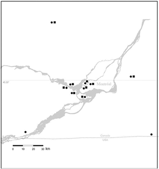
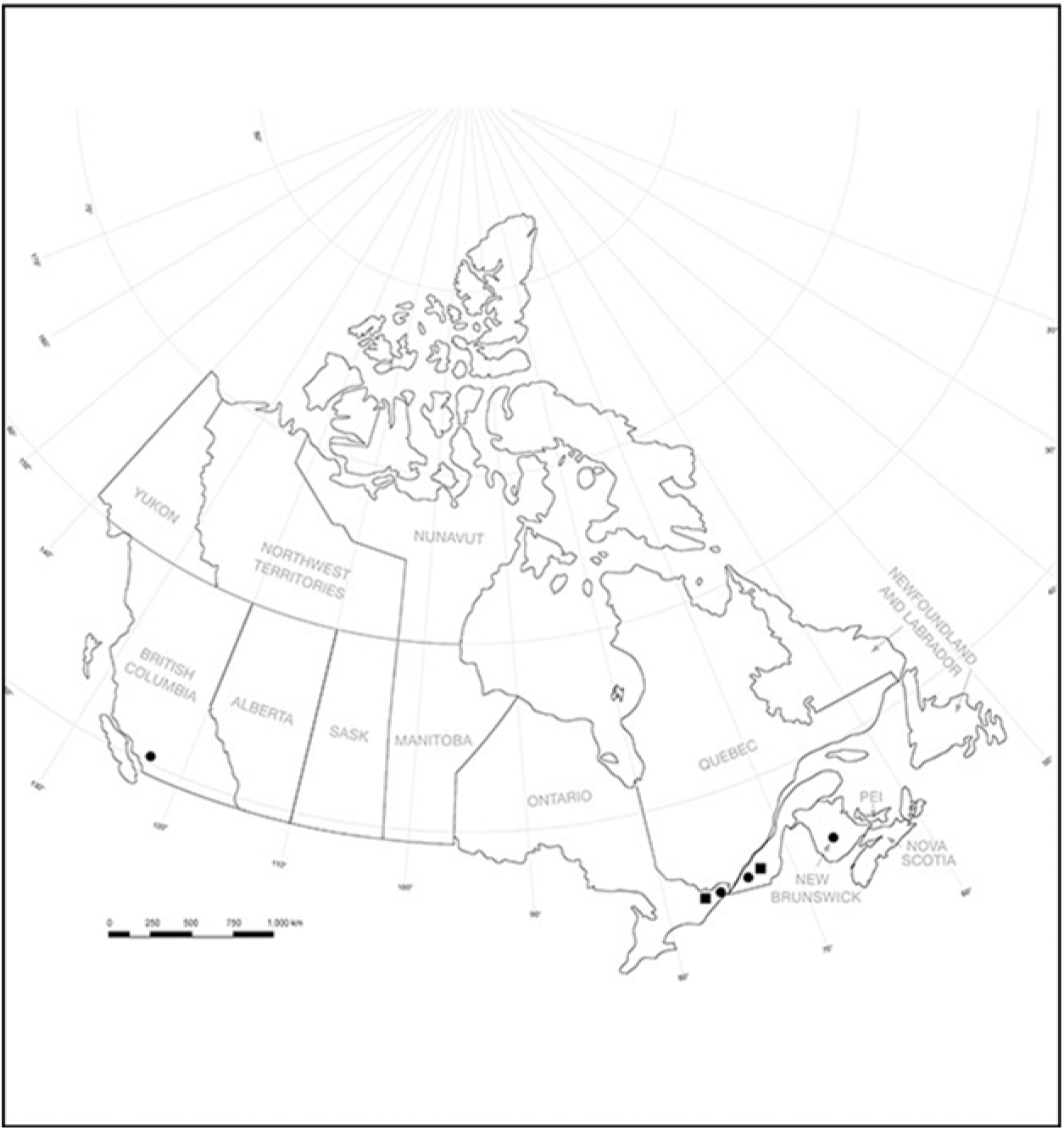
Adults, nymphs, and eggs of C. juglandis and G. tiliae were collected from the underside of the green, heart-shaped leaves of basswood (Tilia americana Linnaeus; Tiliaceae) trees. The leaves of host plants were picked by hand up to 3 m above the ground in the Morgan Arboretum, Macdonald Campus, McGill University, Sainte-Anne-de-Bellevue, Québec. Overwintered and new-generation adults of C. juglandis and G. tiliae were also collected in Ile Perrot, Centennial Park, Ile Bizard, Sainte Lazare, Hudson, Oka, Sainte Agathe, Mont Sainte-Hilaire, and Philipsburg in Québec, and Green Valley, Cornwall, and Richmond in Ontario (Fig. 2). Adults of these species were also recorded from leaf litter samples collected in their overwintering sites in the Morgan Arboretum after the first frost in October and early November. In addition, new-generation adults of C. juglandis were also collected on 26 July 2014 near Sussex, New Brunswick, Canada, and on 12 August 2015 in Manning Provincial Park, British Columbia, Canada (Fig. 3).
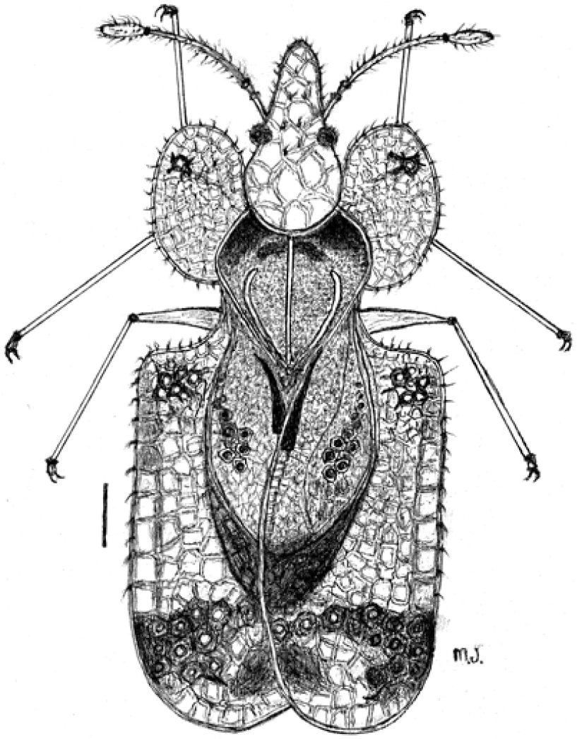
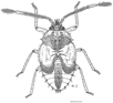
Corythucha juglandis dorsal view of adult showing pronotum and large pronotal hood (scale, 0.5 mm).

Localities of Corythucha juglandis (•) and Gargaphia tiliae (▪) used in this study in Ontario and Québec, Canada.

Localities of Corythucha juglandis (•) and Gargaphia tiliae (▪) used in this study in Canada.

The field study involved observations of feeding and pre-copulation of C. juglandis, mating of G. tiliae, oviposition site selections, fecundity, embryo development, egg hatching, first instar, and the number of generations per year. In addition to damage on leaves, aggregations of C. juglandis and G. tiliae nymphs and adults were observed on the underside of Tilia americana leaves from mid-May to the end of October 2014–2017 in the Morgan Arboretum study sites.
Breeding and rearing in the field and laboratory
To determine the number of adults, eggs, nymphs, and new-generation adults of C. juglandis and G. tiliae, five samples each of small (10 × 8 cm), medium (19 × 15 cm), and large (25 × 19 cm) leaves of Tilia americana were collected at random from near the ground up to 3 m high. Leaves were sampled every two weeks from mid-May to mid-October 2014–2017. The numbers of each stage were then counted using a 10× magnifying lens in the field or by checking them with a microscope in the laboratory. Breeding and rearing of the two lace bug species were carried out in the modified ventilated plastic cages as described in Javahery (Reference Javahery1986, Reference Javahery1990) in the study sites at the Morgan Arboretum. For this study, five pairs of overwintered, non-mated C. juglandis and G. tiliai were collected from small or medium-size green leaves of T. americana on 4 May 2015 and 7 May 2016 and were separated (one pair per cage) in the field. These cages were placed over the free ends of live, young branches with 2–3 green, small-size T. americana leaves about 2 m from the ground in semi-shade at the north side of the Morgan Arboretum. The open end of the cage was tied securely with a string around the branches. Each cage was also tightly fixed to two other neighbouring branches with a strong string. One fresh small leaf was added to each cage every two weeks from mid-June to mid-July. The lace bugs in the cages experienced ambient conditions of temperature, humidity, and light. Feeding, mating, fecundity per female, egg incubation, development of nymphs, and numbers of new adults per female per cage were checked and recorded every week from mid-May to mid-October during 2015–2016 (Tables 1–4). Laboratory breeding and rearing of C. juglandis and G. tiliae were carried out separately on medium-size leaves of host plants in modified plastic boxes as described by Javahery (Reference Javahery1986, Reference Javahery1990).
Fresh eggs of C. juglandis and G. tiliae from laboratory cultures were incubated in petri dishes (2 × 6 cm) as described by Javahery (Reference Javahery1990, Reference Javahery1994). These eggs were kept on 1–2 cm of sterile, moist (75–80% relative humidity) sand in a glass jar (15 × 10 cm) with a screened lid. They were then incubated at 23 °C; 70–75% relative humidity and 14:10 (light–dark hours). Humidity was provided with drops of distilled water added daily to the towel on the sand, and any condensation from the lid of Petri dishes was wiped off.
Dissection and illustrations
Live adults of C. juglandis and G. tiliai were dissected in isotonic solution (0.9% NaCl) as described by Javahery (Reference Javahery1994), washed for 10 minutes in acetic acid, then put in glycerine for five minutes. The reproductive organs, terminalia, aedeagus, paramere, ovipositor, and egg cup were removed with fine watchmaker forceps in a relaxing fluid of 75% ethanol. Illustrations and measurements were made through a Leica MS5 binocular microscope (Wetzlar, Germany), using an ocular micrometre and a drawing tube. For permanent storage, dissected parts from a specimen were transferred to a micro-vial containing glycerine and pinned under the specimen. Colour images of specimens, aggregations on leaves, leaf damage, ectoparasitic mites (Acari), and predators were taken with a Panasonic Lumix GH4 camera (Kadoma, Japan) combined with an Olympus 60 mm f 2.8 macro-lens (Tokyo, Japan) and Kenko extension tubes (Tokyo, Japan).
Voucher specimens of adults (male and female), first to fifth instars, eggs, and egg shells preserved in 75% ethanol, as well as adults and egg-shells of C. juglandis and G. tiliae on pointed tree-angles-cardboard are deposited in the Lyman Entomological Museum, McGill University. Adult specimens (n > 3000) from southern Canada, northeastern United States of America, and western Palearctic Region were examined from the following institutions to help confirm the identification of the specimens used in this study: American Natural History Museum, New York, New York, United States of America; Arthropod Collection of the Natural History Museum, Barcelona, Spain; Canadian National Collection of Insects, Arachnids, and Nematodes, Ottawa, Ontario, Canada; Lyman Entomological Museum, McGill University; National Natuurihistorisch Museum, Leiden, The Netherlands; Plant Protection Research Institute, Tehran, Iran; Royal British Columbia Museum, Victoria, British Columbia, Canada; Royal Ontario Museum, Toronto, Ontario, Canada; Spencer Entomological Collection, Beaty Biodiversity Museum, University of British Columbia, Vancouver, British Columbia, Canada; University of Connecticut, Storrs, Connecticut, United States of America; and Zoological Institute, Insect Collection, Saint Petersburg, Russia.
Results
The natural history and distribution of Corythucha juglandis and Gargaphia tiliae are reported. Results are compared with relevant publications on these two lace bugs in other regions outside of Canada.
Corythucha juglandis Fitch, 1857
Habitat and host plants
The overwintered adults of C. juglandis, eggs, and all stages of nymphs were mainly collected in large numbers from the underside of leaves of their preferred host plant, Tilia americana, in the Morgan Arboretum. They have also been collected on other species and varieties of Tilia Linnaeus in the Morgan Arboretum close to their preferred host T. americana. Adults have rarely been found (1–3 overwintered per 10 medium-size leaves) in June on Juglans cinerea Linnaeus (Juglandaceae), Sorbus americana (Rosaceae), and Fraxinus Linnaeus (Oleaceae).
Male reproductive system
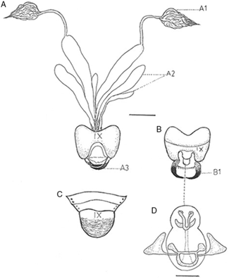
The main parts of the male reproductive system of C. juglandis include a pair of testes, which are enclosed in a peritoneal sheath (Fig. 4A). Within the sheath, the testis consists of a number of testicular follicles that correspond to the ovarioles of the female. The apical region of each follicle is the germarium, which contains the spermatogonia. The other parts of the male reproductive system include a pair of lateral ducts, the vas deferentia, and a median ectodermal exit tube. There are also two pairs of accessory glands. The pygophore is round, broader than long (Fig. 4B dorsal, Fig. 4C ventral). A pair of parameres with developed stem and somewhat sharp pointed blade are closed at rest (Fig. 4A3) and open during copulation (Fig. 4B1). The aedeagus is ovoid shaped, with a pair of developed basal plates (Fig. 4D). The external opening of the exit duct, the male gonopore, is generally situated on or within a median intromittent organ, the penis (Fig. 4B, D).
Corythucha juglandis male reproductive system. A1, testis; A2, accessory glands; A3, parameres: closed; B, terminalia with open parameres; B1, dorsal view; C, terminalia, ventral view (scale, 0.3 mm); D, aedeagus (scale, 0.1 mm).

Female reproductive system
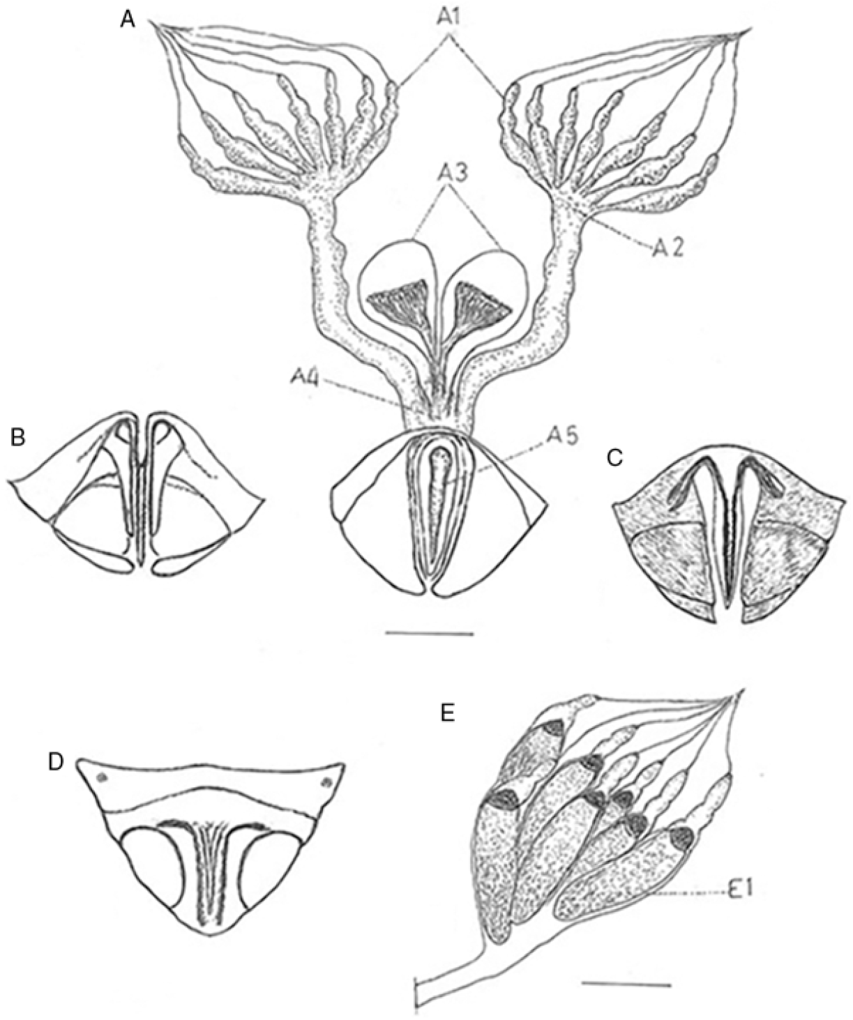
The essential female parts of the reproductive system of C. juglandis consist of a pair of ovaries, two lateral oviducts, and a median oviduct opening to the exterior by the ovipositor (Fig. 5A4–A5). In addition, there are two sac-like pseudospermathecae (Fig. 5A3), which open to the median oviduct, and a tubular exit passage from the median oviduct known as the vagina. Each ovary consists of seven cylindrical ovarioles, each with a delicate terminal filament, an egg tube, and a supporting stalk or pedicel (Fig. 5A1–A2). Eggs develop and mature in the ovarioles (Fig. 5E). The ovipositor (Fig. 5B–D) is connected to the median oviduct and consists of a shaft or egg-tube and basal apparatus. The ovipositor serves for the insertion of eggs on the underside tissue of leaves.
A,Corythucha juglandis female reproductive system. A1, ovariole; A2, calyx; A3, pseudospermathicae; A4, common oviduct; A5, vagina; B–C, female terminalia, dorsal view; D, female terminalia ventral view (scale, 0.3 mm); E, right ovary with seven mature dark-coloured eggs in seven ovarioles (E1) (scale, 0.5 mm).

Description of eggs
Elongate, pointed at end, somewhat curved, cap conical, dark brown or black, chorion smooth towards the apex (Figs. 5E, 6E, 7A, 8A); operculum with smooth collar, the areolae smaller at base; length 0.52 mm; width 0.16 mm; colour dark brown to light black. Eggs are generally laid in odd numbers ranging from 1 to 5, as well as 2, 4, and rarely 7 or 9 eggs.
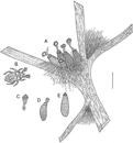
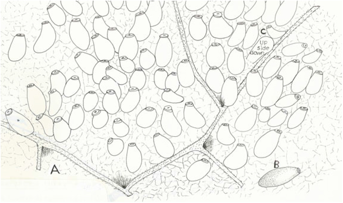
Corythucha juglandis cluster of five hatched egg shells. A, Egg shells with attached egg caps by embryonic membrane on the underside of Tilia americana leaf; B, first instar; C, egg cap attached to embryonic membrane; D, an egg shell with egg cap attached to the embryonic membrane; E, an egg with developed embryo before hatching (scale, 0.3 mm).

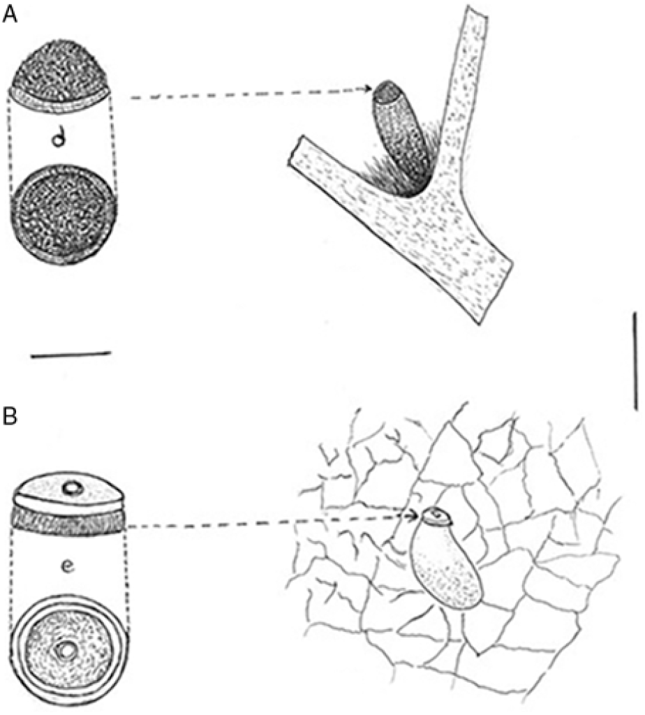
Oviposition pattern, side selection, and chorionic collar cum apparatus. A, Corythucha juglandis; B, Gargaphia tiliae (scales: eggs, 0.3 mm; caps, 0.1 mm); d, operculum conical; e, flat operculum with rim-like collar (scale, 0.1 mm).

Description of first instar
Body elongate, oval; length 0.60 mm, width 0.25 mm (Figs. 6B, 9C); head, thorax, and abdomen elevated dorsally; colour light brown; head and dorsum each with two minute tubercles and one seta; antennae rather stout, with three antennomeres; pale with scattered pubescent setae; antennomere I slender, two times longer than antennomere II; antennomere III wider, equal in length to antennomere I, with long setae and cluster of short setae at apex; beak rather long, four-segmented, light yellow; compound eye with five red ommatidia; abdominal segments 2–8 with a pair of setae posteriorly on lateral margin; abdominal segments 2, 5, 6, and 8 with a pair of setae medially; tarsi with two tarsomeres, with two claws.
Oviposition pattern and side selection on host plants. A, Eggs of Corythucha juglandis are laid on the underside of a Tilia americana leaf and hidden under leaf pubescence between the main rib and a green vein (scale: 1 mm); B, eggs of Gargaphia tiliae are laid in a circular group not attached on the underside of a Tilia americana leaf; d, first instar; e, hatching eggs (scale, 1 mm).

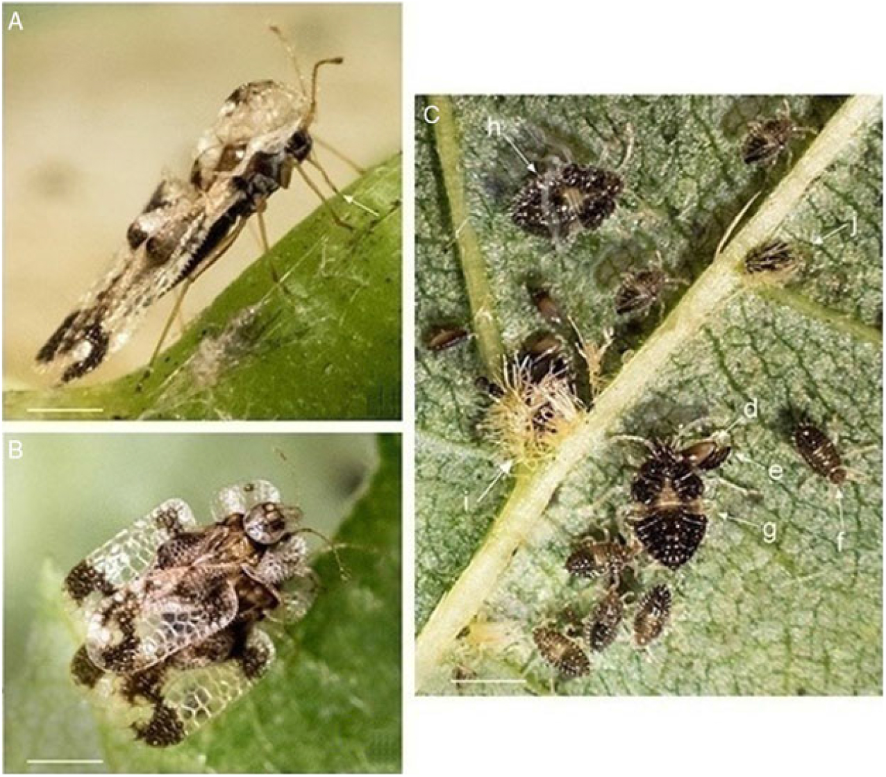
Corythucha juglandis. A, Lateral view feeding on the sap of Tilia americana (arrow) after insertion of stylets into the cells of parenchyma (scale, 1 mm). B, A male mounting a female and touching her antenna during pre-copulation. The female opens her forewings about 35° and extends her genital out, enabling the male to insert its aedeagus (scale, 1 mm). C, Overlapping of immature stages on the underside of a Tilia americana leaf; d, egg; e, first instar; f, second instar; g, third instar; h, fourth instar; i, cluster of eggs laid at the juncture of the midrib and secondary vein under pubescent of plant hairs on the underside of the leaf of host plant; j, single egg laid between two ribs (scale, 1 mm).

Description of second instar
Body less elongate and covered with spinules dorsally; length 0.75 mm, width 0.35 mm (Fig. 9C); colour brown dorsally, light yellow ventrally, antennae and legs light amber; eyes not prominent, consisting of four distinct red ommatidia; two small tubercles with three spines between eyes and one in front of head; dorsal armature similar to that of the first stage; antennae with three antennomeres, one-fourth length of body, pale, with scattered pubescent setae; beak translucent, one-half the length of the body; abdominal segments 2–8 with one seta laterally and a pair of spiracles; tarsi with two tarsomeres, with two claws.
Description of third instar
Body less elongate, more oval, slightly narrow at anterior end; length 1.16 mm, width 0.55 mm (Fig. 9C); colour dark brown dorsally and yellow ventrally, antennae and legs light amber; antennae with four antennomeres, slightly longer than those of the second instar; body armature similar to those of the second instar; median line on dorsal surface of head is widening on thorax and extending across the first three abdominal segments; wing pads slightly developed; tubercle with three spines and two setae between eyes, those in front are more visible; abdomen with one seta laterally on segments 2–8; tarsi with two tarsomeres, with two claws.
Description of fourth instar
Body elevated medially on pronotum and abdomen becoming oval in shape (Fig. 9C); colour dark brown dorsally; length 1.45 mm, width 0.78 mm; bead bears a pair of separated spines on front, a pair on vertex, a pair of separated tubercles each bearing several spines and setae; body armature with three tubercles between the eyes; cluster of setae on abdominal segments 2–8 more developed; wing pads developed, extending onto abdominal segment 2; antennae with four antennomeres, white, one-third length of body; antennomere III slender, about two times length of antennomere IV; eyes red; legs similar to those of the previous stage; rostrum extending to base of third pair of legs; tubercles and spines on abdomen similar to those of previous stage; tarsi with two tarsomeres, with two claws.
Description of fifth instar
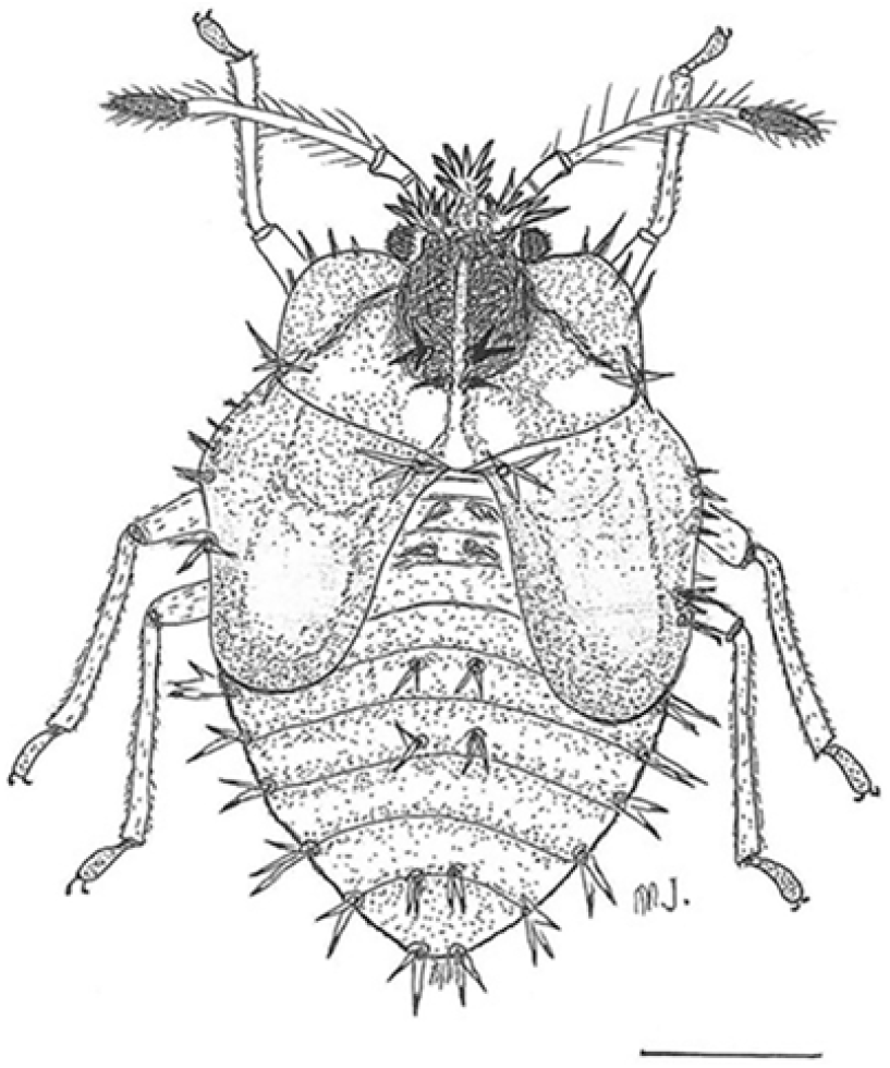
Body broadly oval, somewhat elongate on pronotum and abdomen; length 2.12 mm, width 1.20 mm (Fig. 10); colour dark brown dorsally and ventrally; legs, antennae light amber; wing pads fully developed, extending to about the middle of abdominal segment 5; pronotum with lateral margins yellow; two tubercles and four spines between the compound eyes; abdominal segments 2–9 with three spines dorsolaterally; a pair of spines near the mid-dorsal line on abdominal segments 5, 6, 8, 9; antennae with four antennomeres, one-third length of body, tinged with brown, bearing several long setae; antennomere I equal in length to antennomere II; antennomere III slender, four times the length of antennomere II; antennomere IV half the length of antennomere III; eyes prominent, consisting of a number of ommatidia; rostrum reaching the base of third pair of legs. Tarsi with two tarsomeres, with two claws.
Fifth instar of Corythucha juglandis dorsal view, showing spines and wing pads (scale, 0.35 mm).

Life cycle
Adults of C. juglandis emerged on 29 April 2017 and during May 2014–2016 from leaf litter in the overwintering sites near their host plants when the temperature within litter was about 15 °C. However, 1–3 individuals were also collected on the underside of newly opened small green leaves (12 × 10 cm) of host plants on 25 April 2015. Adults fed on sap of leaves of host plants (Fig. 9A, arrow) for about a week prior to mating at temperatures between 15 °C and 25 °C (Fig. 9B).
Mating behaviour
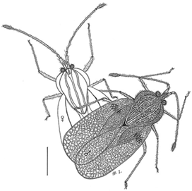
The overwintered adults of C. juglandis are reproductively active in spring and mate after several days feeding on Tilia. Mating in most Heteroptera (Hemiptera) is generally end-to-end (e.g., Eurygaster integriceps Puton (Hemiptera: Scutelleridae); Javahery Reference Javahery1995: 19). However, several exceptions were observed during previous studies as well. In Reduvius personatus Linnaeus (Hemiptera: Reduviidae), mating is side-by-side or in a lateral orientation (Javahery Reference Javahery2013, Fig. 5B). In C. juglandis and G. tiliae, as well as in several other tingids such as Dictyla echii (Schrank) and Stephanitis pyri (Fabricius), mating was observed to be at an angle of about 30–45° (Figs. 11, 12A), which is described as follows.
Gargaphia tiliae pair in copulation, male on top. Male closes its antennae during mating. Note: female is not made lacy in this image (scale, 0.5 mm).

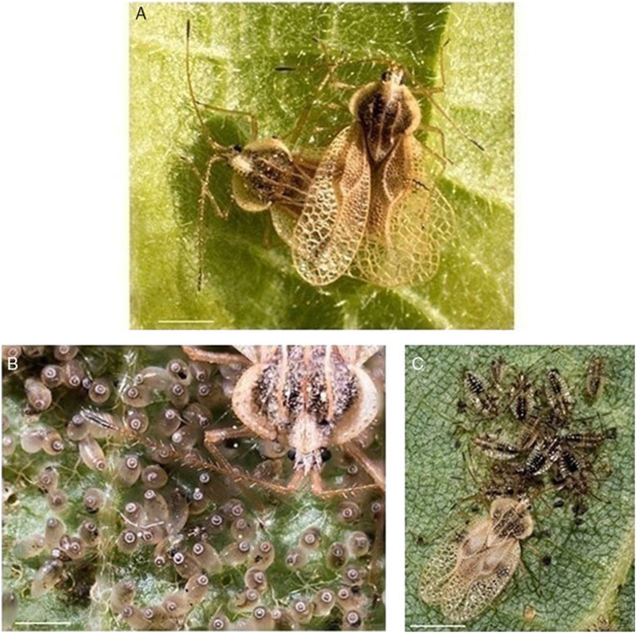
Gargaphia tiliae live photographs. A. In copulation, male on top and usually closes its antennae during mating. The male and female remain end-to-end with an angle about 45° during entire mating. Female may feed or walk around carrying male on her back during copulation (scale, 1.4 mm). B, Female guarding a cluster of her eggs in the field during incubation period, which is about 10 days in May or June. She touches the operculum of the eggs with her antennae three to four times each day and attacks any predators or parasitic insects with flapping wings in order to push them away (scale, 1 mm). C, Female guarding her mixed group of immature third, fourth, and fifth instars on the underside of the host leaf. She also touches immature stages every 4–5 hours and regroups them when they are apart (scale, 1 mm).

Mating in Corythucha juglandis began with a male mounting the female and palpating the antennae, head, or lateral margin of the female with its antennae. A responsive female responds by remaining on the underside of the host leaf, opening her fore wings about half vertically and raising the end of her last abdominal segment at an angle of approximately 35–40° from the substrate with slightly open genital plates. The male then moves a little backward with its two parameres open (Fig. 4B) and, with its pygophore extended, enters its aedeagus into the female open genitalia. The pair remains in contact for 2–5 hours, approximately at an angle of 35–40°.
Pre-copulatory mating behaviour (Fig. 9B) was often observed in both overwintered adults and the newly matured C. juglandis in the morning at about 15–28 °C in the field and the laboratory. Copulated pairs were always observed on the underside of leaves of Tilia. When disturbed, the female would pull the male for a short distance (3–12 cm), or the pair would separate. A female mated 3–5 times.
Oviposition, fecundity, and incubation
The oviposition period of C. juglandis was recorded from third week of May to first week of July in 2014–2017. The number of eggs per cluster per female feeding on T. americana is variable and related to temperature and relative humidity. However, fecundity of five pairs in cages in the field varied from 57 to 94 per female (Table 1). The maximum number of eggs was laid during the second and third weeks of June. Eggs were laid singly at start and then in odd numbers of 1, 3, 5, and 7, as well as in clusters of 2, 4, and rarely 9 towards the base on the underside of leaf at the juncture of the midrib and secondary veins. The leaf pubescence of the usual hosts partly obscures the eggs on the underside of leaf (Figs. 6A, 7A, 9C), and usually top of eggs is visible.
Oviposition period, fecundity, and incubation times of overwintered C. juglandis in the field in 2014–2017.

Note: Food: Tilia americana; n = 5 pairs.
* Females and males collected 6–9 May 2014.
† Females and males collected 18 May 2015.
‡ Females and males collected 12 May 2016.
§ Females and males collected 8–14 May 2017.
Egg incubation was monitored under a binocular by dissecting 2–3 eggs each day. The period of incubation varied with temperature and humidity during 2014–2017, between 11 and 14 days (Table 1). During cold weather and at temperatures below 8 °C (in the field and in the laboratory), the development of embryo slowed down and eggs did not hatch. The best temperatures for egg incubation and embryonic development were between 23 °C and 25 °C, relative humidity of 75%, and a light–dark photoperiod of 10:14.
Development of immature
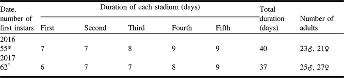
Duration of five stages of nymphs was recorded 2–3 times per week in the field cages and every 1–2 days in the laboratory cultures. The first instars were observed during the first week of June; the second and third instars in the third week of June to the first week of July; the fourth instars from the last week of June to the second week of July; and the fifth instars from second week of July to mid-August in 2016 and 2017 (Fig. 9C). Duration of each stadium, number of males and females, as well as total duration (days) of development of immatures of C. juglandis in 2016–2017 are given in Table 2.
Duration of immature stages and number of adults of Corythucha juglandis reared in the field in 2016–2017.

Note: Food: Tilia americana.
Number of newly hatched first instars of C. juglandis taken from laboratory cultures (23 °C, relative humidity 75%, and a 14:10 light–dark photoperiod) that were reared in field cages, *28 May 2016 and †25 May 2017.
The new-generation adults were recorded from third week of July to last week of October. Corythucha juglandis is apparently univoltine in Ontario and Québec. Both adults and nymphs are gregarious and are usually observed on the underside of leaves of their host plants. Overlapping of various stages is common in this lace bug in late July and during August (Fig. 9C).
Damage on leaves of host plants
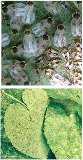
The surface of the leaf above the feeding spots of C. juglandis nymphs, particularly the third to fifth instars and adults of the new generation, becomes discoloured in June, July, and August but gradually turn to brown in September and October as a result of injury to the leaf tissue (Fig. 13B).
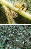
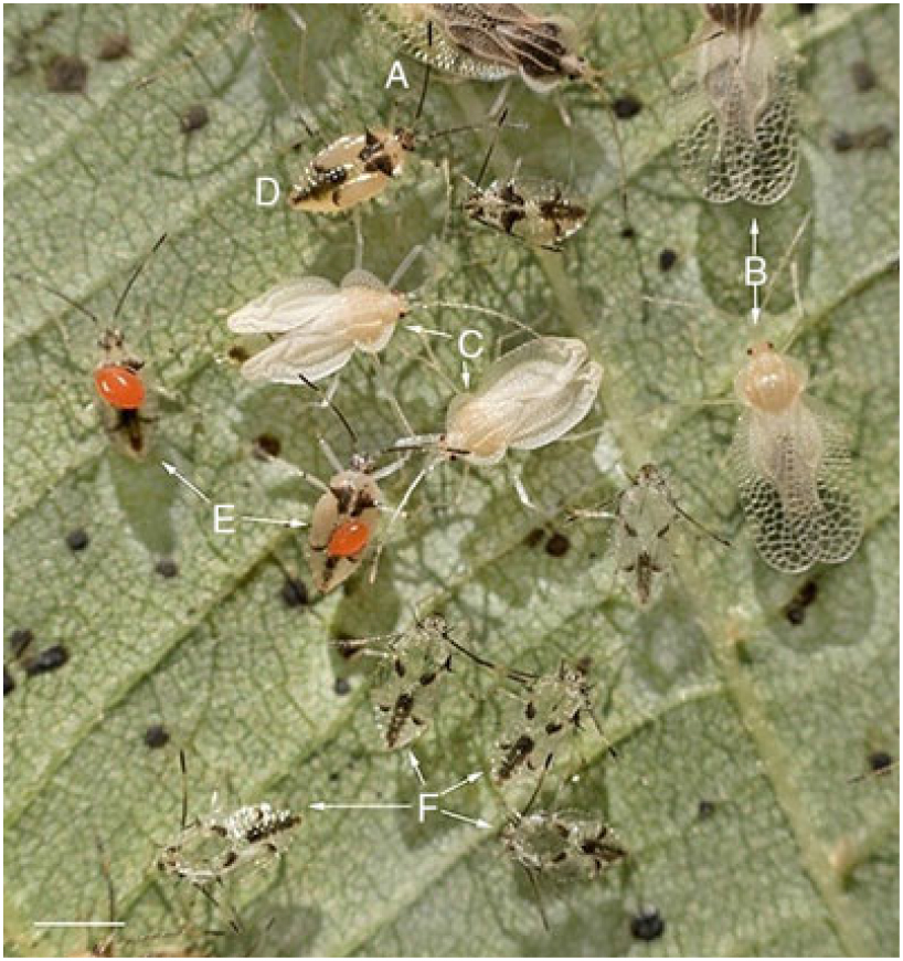
A, Gargaphia tiliae aggregation and overlapping of new-generations adults and fifth instars; B, whitening or discolouration of Tilia americana leaf surface due to feeding by adults and nymphs of Corythucha juglandis and G. tiliae from the underside cells of host plant. Black points on the underside of leaf are excrement of adults and nymphs of both species (scale, 0.5 cm).

Gargaphia tiliae (Walsh, 1984)
Habitat and host plants
The overwintered adults, eggs, and nymphs of G. tiliae were collected from the underside of leaves of Tilia americana. All the food plant records for G. tiliae are species of Tilia. This plant is found near or around the open areas of woodlands and within deciduous plants in the Morgan Arboretum. Ten species and varieties of Tilia were planted close to the main host plants at the study site. All stages of G. tiliae were also collected on the underside of leaves of other Tilia species and varieties in the Morgan Arboretum. This species was also collected on Tilia americana in two woodlands close to Alexandria and Richmond in southeastern Ontario (Fig. 2). Provancher (Reference Provancher1886) reported G. tiliae from Québec without precise details. This species has also been reported in New Brunswick (Maw et al. Reference Maw, Foottit, Hamilton and Scudder2000).
Male reproductive system
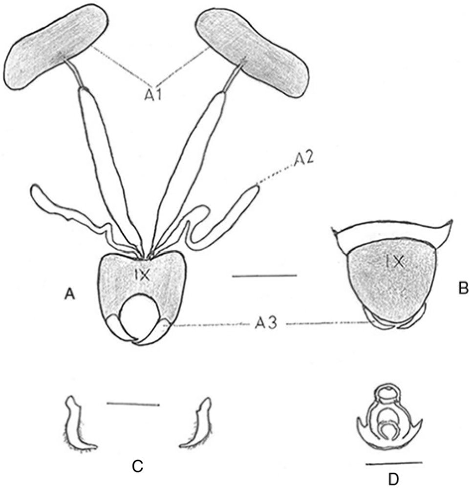
The male reproductive system of G. tiliae consists of a pair of relatively large, bean-shaped testes (Fig. 14A1), which are connected to an elongated wide duct by a very short, thin tube. These ducts, the vas deferentia, as well as a pair of accessory glands open to the median ectodermal tube. Pygophore rounded, almost as wide as long (Fig. 14A–B), and connected at the end to a pair of parameres (Fig. 14A3); parameres slightly curved on stem, the apex pointed (Fig. 14C); aedeagus ovoid, laterally upwardly pointed from the apex (Fig. 14D).
Gargaphia tiliae male reproductive system. A1, Testis; A2, accessory gland; A3, parameres; B, male terminalia, dorsal view (scale, 0.3 mm); C, Right and left parameres (scale, 0.05 mm); D, Aedeagus (scale, 0.2 mm).

Female reproductive system
The female reproductive system of G. tiliae consists of two ovaries each with seven ovarioles, a pair of pseudospermathecae, a pair of accessory glands, a median oviduct, and one ovipositor (Fig. 15A). Each ovary has seven ovarioles that terminate in a fine, tread-like ligament. Eggs develop inside, near the top of each ovariole, and pass down to the common oviduct, then to the median duct towards the exit passage and ovipositor. Up to two developed eggs may develop in one ovariole. The eggs are laid in circular groups in 3–5 days but are not attached to each other (Figs. 8B, 12B, 16).
Gargaphia tiliae female reproductive system. A, Dorsal view with two mature eggs in each ovariole (both the right and left ovaries); ol, ovariole ligament; g, germarium; o, oocyte; fme, first mature egg; sme, second mature egg (scale, 0.3 mm). B, Female terminalia, ventral view; ps, pseudospermatheca; ag, accessory glands. C, An egg before the development of embryo. D, Hatching mechanism with first instar enveloped within embryonic membrane; ca, egg cap. E, First instar emerging from egg shell and embryonic membrane; re, compound red eyes of first instar; em, embryonic membrane (scale, 0.5 mm).

Gargaphia tiliae cluster of eggs. A, 82 eggs laid by one female on the underside of Tilia americana leaf; B, a developed egg, lateral view; C, an upside-down laid egg that did not hatch (scale, 0.3 mm).

Description of eggs
Colour pale, sub-oval in shape, base rounded; length 0.48 mm, width 0.18 mm; operculum rounded, somewhat flat with a slight nipple-like projection in the centre (Figs. 7B, 12B, 16); operculum with a rim-like collar (Fig. 7B); eggs deposited in somewhat circular groups of 49–219 (Figs. 12B, 16) on the underside of the host leaf. The eggs are translucent except for the upper third, which is covered with a varnish-like secretion.
Description of first instar
Oval, elongate (Fig. 8B); length 0.80 mm, width 0.50 mm; thorax and abdomen elevated dorsally, flattened ventrally; labium pale, four-segmented, reaching the middle of the second abdominal segment; antennae with three antennomeres and narrow, long, nearly the length of the body; head and pronotum with four three-branched setae; eyes with five red circular-like dots; two setae between eyes on each side of pronotum; one seta on each dorsoabdominal area of abdominal segments 2–9; tarsi with two tarsomeres, claws light brown.
Description of second instar
Elongate, colour pale; length 1.44 mm, width 0.80 mm; antennae with three antennomeres, long, almost the length of body; labrum reaching the middle of the second abdominal segment; body generally pale except for two triangular brown spots on the lateral margins of the pronotum, and a brown fuscous mid-dorsal line on abdominal segments 2–9; head armed with five spines; pronotum with two light brown spines on the lateral margin, and the end of abdominal segment 2; one clear protuberance laterally on abdominal segments 2–8; tarsi with two tarsomeres, with two claws.
Description of third instar
Oval elongate; length 1.83 mm, width 0.87 mm; similar to the second instar morphology; wing pads slightly developed.
Description of fourth instar
Body oval (Fig. 12C); length 2.32 mm, width 1.12 mm; pronotum somewhat elevated; head with five brown spines, two on the collar and three on frons; eyes red; colour mostly light amber; pronotum with a triangular brown spot with a sharp brown spine on both anterolateral margins as well as one on the lower side; wing pads developed; abdomen with a mid-dorsal fuscous line from abdominal segments 2–8; antennae with four antennomeres; antennomere III the longest; colour of the antennae and legs amber except the terminal antennomere, which is dark brown or fuscous; tarsi with two tarsomeres, tarsomere 2 and claws brown.
Description of fifth instar
Body oval (Figs. 12C, 17); elongate; length 2.56 mm, width 1.28 mm; medial areas of mesometasterum somewhat elevated; two spines at the lower angle of pronotum and at the lateral area of the abdominal segments; head with two brown spines, each near the inner side of eyes and three on the anterior part; antennae with four antennomeres, slender, long, and two-thirds the length of the body; antennomere I twice the length of antennomere II; antennomere III three times the length of antennomeres I plus II combined; antennomere IV one-third the length of antennomere III; legs and antennae with fine setae; rostrum four-segmented, at rest reaching to the midpoint of the second coxal bases; wing pads fully developed; abdominal segments 2–9 with double pale spines laterally and one longer spine between abdominal segments 2–3; colour generally pale but head and thorax brown dorsally; legs and antennae light amber, the last antennomere dark brown; legs, antennae with fine setae; tarsi with two tarsomeres, with two claws.
Fifth instar of Gargaphia tiliae dorsal view, showing spines and wing pads (scale, 0.65 mm).

Life cycle
Gargaphia tiliae is univoltine, with reproductive diapauses (quiescent) in the northern range of its geographical distribution in Ontario and Québec, producing one generation per year. The life history comprises three stages: adult, nymph, and egg (Figs. 11, 12B, 17).
The time spent in each stage varies depending on the weather for the egg stage, and on both the food and weather for the first instars to the fully mature adult. The time for a complete development of a generation requires about two months in breeding sites on Tilia species, particularly T. americana. Overwintered, immature, and new-generation adults are in an active feeding and reproductive phase in May, June, July, and August. Later, the new-generation adults fly slowly to the higher leaves and branches of the same host plant or to nearby deciduous trees, to aestivate before migrating a short distance, or being blown by wind to the leaf litter where they hibernate in a state of almost complete inactivity. During the overwintering period, the adults use their stored reserves (fat) to survive until spring.
Oviposition, fecundity, and incubation
The overwintered adults of G. tiliae appeared on their host plants in spring. Oviposition started about a week after feeding and mating from late May to early July. Eggs were laid within 3–5 days in a circular cluster close to each other and glued at their end to the underside of the leaf, towards the base and exterior lateral margin (Figs. 12B, 16) (Table 3).
Oviposition period, fecundity, incubation times of overwintering Gargaphia tiliae in the field in 2014–2017.

Note: Food: Tilia americana; n = 5 pairs.
* Females and males collected 8 May 2014.
† Females and males collected 16 May 2015.
‡ Females and males collected 12–15 May 2016.
§ Females and males collected 15 May 2017.
Fecundity and the number of eggs per cluster per female varied in the breeding cages in the field between 79 and 124 in four seasons from 2014–2017 (Table 3). However, data from 10 clusters collected at random in other study sites in the field during four seasons (2014–2017) were 67, 70, 76, 81, 118, 119, 124, 147, 209, and 216, with an average 123 eggs per female (Figs. 8B, 12B, 16; Table 3).
The incubation period of eggs of G. tiliae was also variable in the field and related to the temperature and relative humidity in each season. The eggs developed in 12–15 days during 2014–2017 (Table 3).
Maternal behaviour
Gargaphia tiliae demonstrated maternal behaviour during both egg incubation and development of immature stages, guarding them against parasitoids and predators (Fig. 12B–C).
Development of immature stages
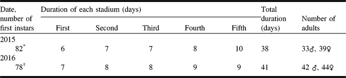
Durations of five stages of nymphs were recorded 2–3 times per week in the field cages and every 1–2 days in the laboratory cultures. First instars of G. tiliae (Fig. 8B) were observed on 29 May 2014 and during the first week of June 2015–2017; the second instars during second week of June; the third instars during third week of June; the fourth instars (Fig. 12C) during fourth week of June; and the fifth instars (Figs. 12C, 18) during first and second weeks of July in 2014–2017. Adults of the new generation (Fig. 18) were observed during second and third weeks of July in 2014–2017 at the study sites, respectively. Duration of immature stages and number of adults reared in the field during 2015–2016 are given in Table 4.
Gargaphia tiliae overlapping generations of adults and fifth instars in the field at Centennial Park, Québec. A, adults of an older generation; B, adults of a new generation with clear lacy forewings; C, adults of a new generation emerging from the fifth instar skins; D, fifth instars; E, two ectoparasitic red mites feeding on fifth instars; F, cast-off skins of fifth instars (scale, 1 mm).

Duration of immature stages and number of adults of Gargaphia tiliae reared in the field in 2015–2016.

Note: Food: Tilia americana.
Number of newly hatched first instars of G. tiliae taken from laboratory cultures (23 °C, relative humidity 75%, and a 14:10 light–dark photoperiod) that were reared in field cages on *6 June 2015 and †2 June 2016.
Adults of the new generation are gregarious and remain in groups of several to > 100 on the underside of host leaves after maturation and before leaving the breeding site (host plants) for aestivation or hibernation sites (Fig. 13A). Gargaphia tiliae is probably univoltine in Ontario and Québec.
Damage on leaves of host plants
The nymphs of G. tiliae are gregarious and are guarded by the female while feeding on the underside of host leaves. The surface of the leaf above the feeding spots of the nymphs, particularly, third to fifth instars, become discoloured in late June and during July. This white discolouration gradually turns to brown in September and October as a result of injury to the leaf tissues (Fig. 13B).
Natural enemies of lace bugs
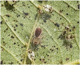
The natural enemies of lace bugs have been rarely mentioned. However, among the biotic factors that reduce the population of Corythucha juglandis and Gargaphia tiliae, the following enemies were observed attacking or feeding on adults, nymphs, or eggs of the above two lace bugs at the study sites: The fourth and fifth instars of Zelus lurichus Stål (Hemiptera: Reduviidae) fed on fourth and fifth instars of G. tiliae on 4 July 2015, 11 July 2016, and 29 June 2017 at the Morgan Arboretum; the adult of Podisus maculiventris (Say) (Hemiptera: Pentatomidae) fed on adults of C. juglandis and fourth and fifth instars of G. tiliae on 14 July 2015 at the study sites; one Coccinella Linnaeus (Coleoptera: Coccinelidae) fed on fifth instars of G. tiliae on 18 July 2014 at the Centennial Park Dollard-Des Ormeaux, west of Montréal, Québec; a Coccinella nymph (Fig. 19) fed on a cluster of eggs of G. tiliae on the underside of leaves of T. americana on 14 June 2016 in Oka, Québec; an ectoparasitic mite (Trombidiformes: Erythraeidae) (Fig. 18) fed on fifth instars of G. tiliae on 14 July 2015 at the Centennial Park; an immature spider (Araneae: Clubionidae or Miturgidae) (Fig. 20) attacked fifth instars of G. tiliae on 8 July 2016 at the Morgan Arboretum.
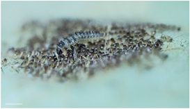
A predatory Coccinella nymph feeding on a cluster of eggs of Gargaphia tiliae (scale, 1 mm).

Gargaphia tiliae on the underside of a Tilia americana leaf. A, an unknown immature spider attacking immature nymphs and adults; B, fifth instar; C–D, cast-off skins of fifth instars; E, black spots on the underside leaf of the host plant (scale, 1.5 mm).

Aestivation and hibernation sites
Lace bugs are not generally considered to be migratory insects. However, both the overwintered adults and the matured adults of C. juglandis and G. tiliae were observed to fly and leave their hibernation or breeding sites. Then they flew slowly a short distance about 50–150 m to land on the early green leaves of their host plants to feed in May and to reproduce during June, July, and August in 2014–2017.
A new generation of adults of C. juglandis and G. tiliae was observed feeding several days on the spongy mesophyll of the underside of leaves of T. americana, on the lower branches of host plants (1–3 m above the ground) before flying slowly to the undersides of the leaves on the higher branches (above 5–6 m) of the same host plants or nearby deciduous trees, such as elm (Ulmus Linnaeus; Ulmaceae), ash (Fraxinus Linnaeus; Oleaceae), beech (Fagus grandifolia Ehrhart; Fagaceae), and maple (Acer Linnaeus; Sapindaceae), to aestivate at cooler temperature during September and a part of October. The aestivation period terminated with cooling weather or the first frost in October. Adults then aggregated, between 10 and > 100, on the undersides of medium leaves (15 × 20 cm) of host plants, and rarely in much higher numbers (n > 1000) under large-size leaves (22 × 26 cm) of host plants at lower branches close to the ground before migrating to their nearest hibernation sites in woodlands.
About two-thirds of the new-generation adults of C. juglandis and G. tiliae left their aestivation sites and flew slowly or were blown by wind during short warmer sunny days (15–25 °C) of October to their hibernation sites and took refuge between fallen autumn leaves, under stones, and on ground in the nearby trees or woodlands. The remaining late-matured populations (about one-third) left their aestivation sites after the first frost in October. Adult feeding was necessary for the two studied lace bugs before migration. The late-maturing individuals did not migrate and dropped to the ground with host leaves.
Discussion
Lace bugs are small insects (2–6 mm). Many species are strikingly similar and are, therefore, difficult to identify (Bailey Reference Bailey1951; Drake and Ruhoff Reference Drake and Ruhoff1965). The external male genitalia, particularly the parameres, have often been used in the identification of species (Pruthi Reference Pruthi1925; Bailey Reference Bailey1951). However, in some groups of similar species in the genus Corythucha, the parameres are not sufficient for species identification. The difficulties in identifying C. juglandis are further complicated by the unusually varied food habits. Other diagnostic techniques such as biomorphology, molecular techniques, and host plant specificity based on co-evolutionary analyses may be helpful in identifying similar species. Biomorphology has previously been used for identifying cryptic Tingidae species (Southwood Reference Southwood1956; Cobben Reference Cobben1968, Reference Cobben1978; Javahery Reference Javahery1968, Reference Javahery1994; Hinton Reference Hinton1981; Livingstone and Yacoob Reference Livingstone and Yacoob1987).
The two species of lace bugs discussed in this study are entirely herbivorous. They feed, breed, and develop on Tilia americana at the study sites. Adults of these lace bugs were also rarely collected on other species and varieties of Tilia, which were planted about 10–15 m away from each other but close to the main and preferred host plant, T. americana, in the Morgan Arboretum. Adults were also collected on native T. americana in other study sites (Fig. 2). Tilia americana was observed to be the first Tilia species to produce buds and small green leaves in spring that attract C. juglandis and G. tiliae in late April and May. Corythucha juglandis and G. tiliae were also collected on the underside of leaves of Ulmus in Oka, Québec, and Richmond, Ontario. Corythucha juglandis has been reported to be common on Ulmus in Ontario and New Brunswick (Twinn Reference Twinn1935). It is also reported that roadside Juglans cinerea were infested in Speerville, New Brunswick (Twinn Reference Twinn1935). Corythucha juglandis was also collected on a Salix Linnaeus (Salicaceae) tree on 16 July 2014 in Manning Provincial Park, British Columbia. This tingid has also been found on Rubus Linnaeus (Rosaceae), Sorbus Linnaeus, and Salix. Twinn (Reference Twinn1935) and Drake and Ruhoff (Reference Drake and Ruhoff1965) reported eggs and nymphs of C. juglandis on Sorbus and Juglans cinerea.
Tilia americana leaves likely emit a semiochemical that attracts C. juglandis and G. tiliae, to feed and to reproduce (Aldrich et al. Reference Aldrich, Neal, Oliver and Lusby1991). In Canada, Tilia americana trees are found in the Saint John River valley in New Brunswick westward to southern Manitoba (Cloutier Reference Cloutier1956).
Corythucha juglandis is also reported from Juglans nigra Linnaeus, J. cinerea, and Carya illinoiensis (Wangenheim) Koch (Juglandaceae) (Drake and Ruhoff Reference Drake and Ruhoff1965; Vogt and McPherson Reference Vogt and McPherson1986). In New England, United States of America, J. cinerea is the usual food plant for C. juglandis (Bailey Reference Bailey1951). Gargaphia tiliae has also been reported from Cercis canadensis Linnaeus (Fabaceae), Prunus serotina Ehrhart (Rosaceae), and Tilia pubescens Aiton (Malvaceae) (Drake and Ruhoff Reference Drake and Ruhoff1965).
Corythucha juglandis and G. tiliae adults hibernate in the winter in leaf litter. They appeared on their food plants in late April or May in Ontario and Québec. The winter hibernation sites and the spring breeding sites of C. juglandis and G. tiliae in Ontario and Québec are similar to the reports for these species in the United States of America by Drake (Reference Drake1917, Reference Drake1928), Weiss (Reference Weiss1919), Weiss and Dickerson (Reference Weiss and Dickerson1918), Blatchley (Reference Blatchley1926) (in New Jersey as C. parshleyi Gibson), and Sheeley and Yonke (Reference Sheeley and Yonke1977) (in Missouri). In New England, C. juglandis and G. tiliae appear in May or early June (Bailey Reference Bailey1951).
Little is generally known about the aestivation period of tingids, including the two species studied. The aestivation period refers to the dormancy time between adult maturation and feeding and flying to the overwintering site in leaf litter. Aestivation starts when the temperature in the breeding sites drops, usually in September. Hibernation starts after the first cold weather or frost in October. Corythucha juglandis and G. tiliae, like many other terrestrial Hemiptera that overwinter as adults, are in a state of low metabolism during aestivation and the first 3–5 months of hibernation (Javahery Reference Javahery1990, Reference Javahery1995). The dormancy period is critical in the life cycle of many terrestrial Hemiptera (Javahery et al. Reference Javahery, Schaefer, Lattin, Schaefer and Panizzi2000) because feeding, mating, and reproducing are influenced by factors such as photoperiod and freezing temperatures. Newly matured C. juglandis and G. tiliae (and other Hemiptera; see Javahery Reference Javahery1995) generally do not mate or reproduce for 3–5 months, which is known as the obligatory diapausing months. Corythucha juglandis and G. tiliae probably produce one generation per year north of 43° latitude, including in Ontario and Québec.
Mating in most terrestrial Hemiptera is usually end-to-end (e.g., Eurygaster integriceps Puton; Hemiptera: Scutelleridae (Javahery Reference Javahery1995)). However, in Reduvius personatus (Linnaeus) (Hemiptera: Reduviidae) mating is side-by-side in a lateral orientation (Javahery Reference Javahery2013). In C. juglandis and G. tiliae, mating was at an angle of about 30–45° (Figs. 11, 12A). This mechanism of mating has been observed in several other tingid species (e.g., Dictyla echii (Schrank)) (personal observation).
The overwintered adults of C. juglandis and G. tiliae appeared on their food plants in May through early June in their reproductive phase. Oviposition occurred from middle of May to end of June for both species. Fecundity and the number of eggs per cluster per female, as well as oviposition site selection, were variable in both species (Figs. 6, 7, 16). Corythucha juglandis and G. tiliae both laid eggs from late May or early June to mid-July. Most eggs were laid during the second and third weeks of June (Tables 1, 3). These observations support those given by Weiss and Dickerson (Reference Weiss and Dickerson1918), Barber and Weiss (Reference Barber and Weiss1922), Bailey (Reference Bailey1951), Sheeley and Yonke (Reference Sheeley and Yonke1977), and Vogt and McPherson (Reference Vogt and McPherson1986). Clusters of six, seven, and even nine eggs of C. juglandis were also collected on the underside of host plant leaves in the Morgan Arboretum. A larger number of eggs per cluster of C. juglandis was probably laid by other females. This phenomenon, called egg dumping, has been studied in Gargaphia species by Tallamy (Reference Tallamy2005) and in Gargaphia decoris by Guidoti et al. (Reference Guidoti, Tallamy and Luiz Marsaro2015).
Oviposition and egg incubation of G. tiliae occurred from mid-May to end of June (Table 3). Eggs were laid in a circular cluster close to each other. The eggs are attached at the end to the tissue to the underside of the host leaf, towards the base and exterior lateral margin. The number of eggs per cluster per female varied from 82 to 124 in the field cages (Table 3). However, 10 clusters of eggs laid by females of G. tiliae near the study sites varied from 67 to 216, with an average of 123 (Figs. 12, 16). Bailey (Reference Bailey1951) reported up to 300 eggs per cluster in New England.
Feeding damage by adults and nymphs of C. juglandis and G. tiliae causes whitening on the upper leaf surface in T. americana. Discoloration on the top of leaf surface, black excrement spots, and cast-off skins of nymphs on the underside of leaf are characteristics of C. juglandis and G. tiliae attacks (Figs. 13B, 18, 20). The time taken for the complete development of a generation was approximately 2–3 months (from May to July) in the breeding sites on T. americana for Corythucha juglandis and Gargaphia tiliae (Tables 2, 4). However, two annual broods have been reported for C. juglandis in New Jersey (Twinn Reference Twinn1935). The first brood adults appear in early July and by late August; and by early September the second brood adults emerge. Therefore, an estimated six weeks is suggested as the time required for the completion of C. juglandis life cycle (Twinn Reference Twinn1935).
Overwintered adults and immature and new-generation adults of C. juglandis and G. tiliae are in an active feeding and reproductive phase from May through August. The new-generation adults feed for about one week on their host plants during July–September before flying to their aestivation sites, and later to hibernation sites in September and October. The life cycle of C. juglandis and G. tiliae is similar to that of Acrosternum hilare (Say) (Hemiptera: Pentatomidae), which appears on Tilia heterophylla Ventenat and Rhamnus cathartica Linnaeus (Rhamnaceae) (Javahery Reference Javahery1990).
Maternal solicitude (guarding eggs and nymphs) is an interesting phenomenon in G. tiliae (Fink Reference Fink1915), which was observed in the field and laboratory during egg incubation and nymph development (Fig. 12B–C). Maternal care is not widespread among tingids, at least not to the extent observed for Gargaphia species (Faeth Reference Faeth1989). Wilson (Reference Wilson1971, Reference Wilson1975) believed that insect parental behaviour is a polyphyletic behavioural solution to exceptional environmental challenges. Maternal care and mode of oviposition in Hemiptera, including Gargaphia, was studied in some detail by Tallamy and Schaefer (Reference Tallamy, Schaefer, Choe and Crespi1997) and Tallamy (Reference Tallamy2005). Guidoti et al. (Reference Guidoti, Tallamy and Luiz Marsaro2015) studied female guarding of eggs and nymphs of Gargaphia decoris Drake in Rio Grande du Sul, Brazil, and referred to four other Gargaphia species and three Corythucha species exhibiting maternal care behaviour.
Acknowledgements
My thanks to C.M. Buddle (McGill University, Sainte-Anne-de-Bellevue, Québec, Canada), C. Cutler (Dalhousie University, Halifax, Nova Scotia, Canada), A. Smith (Canadian Museum of Nature, Ottawa, Ontario, Canada), two anonymous reviewers, R. Manuel and J. Mitchell, for reviewing this paper in manuscript form. I also acknowledge A. Sabouri (Tehran University, Tehran, Iran) and C.M. Buddle for identifying an ectoparasitic mite and a predatory spider, respectively. The late C.W. Schaefer (Connecticut University, Storrs, Connecticut, United States of America), J.E. McPherson (Southern Illinois University, Carbondale, Illinois, United States of America), R.T. Schuh (American Museum of Natural History, New York, New York, United States of America), G.G.E. Scudder (University of British Columbia, Vancouver, British Columbia, Canada), and the late T.A. Wheeler (McGill University) are thanked for comments or providing references. Special thanks to M. Armata and S. Boucher (McGill University), M.M. Goula (University of Barcelona, Barcelona, Spain), J. Ribes, B. Caballero, and G. Masó (Arthropod Collection of the Natural History Museum, Barcelona, Spain), C. Copley (Royal British Columbia Museum, Victoria, British Columbia, Canada), K.M. Needham (University of British Columbia), and A. Grégoire-Taillefer, S. Rochefort, and J.F. Roch for assistance in checking specimens, images, or locality data. I also thank P. Azmayesh Fard (Tehran University), J. Mohaghegh (Plant Protection Institute, Tehran, Iran), R.E. Linnavuori (Zoological Museum, University of Turku, Turku, Finland), and the late I.M. Kerzhner (Zoological Institute, Saint Petersburg, Russia) for helpful comments during the identification of lace bugs or their natural enemies. Finally, I am indebted to my family for their patience and help in collecting, sampling, and field data recording throughout this work.