Introduction
A substernal goitre is defined as a goitre which has most of its mass in the mediastinum.Reference Katlic, Grillo and Wong1 Such goitres represent between 3 and 21 per cent of reported thyroidectomies,Reference Newman and Shaha2 and present a unique technical challenge to the surgeon because of their size and relatively inaccessible location. Most of these goitres can be removed through a cervical incision, but a sternotomy is required in 1 to 2 per cent of cases.Reference Pandya and Sanders3
There is no clear demarcation of which cases will require a thoracic approach. Ahmed et al. Reference Ahmed, Elnazeer and Mahadi4 described the clinical and radiological features of nine cases of substernal goitre requiring median sternotomy. These features included symptoms of dysphagia, audible breathing and nocturnal choking; radiologically, all patients had extension of their goitre to the level of the aortic knuckle, with significant tracheal deviation or compression.
Thyroidectomy is a common operative procedure, but the facilities and expertise required for sternotomy are not available in most hospital settings. Therefore, it is vital to have guidelines which enable relevant cases to be referred for surgery in a setting in which the necessary provisions are available.
We present, from an otolaryngologist's perspective, a case series of substernal goitres requiring sternotomy, and we discuss the clinical and radiological predictors which assisted the pre-operative identification of these patients.
Materials and methods
We retrospectively reviewed the medical records of all patients with substernal goitres who had undergone thyroidectomy, performed by the senior author, over a 10-year period, within the department of otolaryngology and head and neck surgery, St James's Hospital, Dublin. Those requiring sternotomy were identified and their case histories reviewed. The following parameters were then recorded: age, sex, presenting symptoms, length of time from initial onset of symptoms, findings on clinical examination, blood profiles, magnetic resonance imaging (MRI) and computed tomography (CT) scan results, and post-operative complications.
Results
Over a 10-year period, 140 patients with substernal goitre underwent thyroidectomy, performed by the senior author, in our institution. Three cases (2 per cent) required a sternotomy to aid surgical access. Our hospital has a cardio-thoracic department on site. All three patients were identified pre-operatively as requiring sternotomy. Their case histories are presented below.
Case one
A 60-year-old woman presented to the otolaryngology service with a one-year history of increasing shortness of breath on exertion and gradually increasing dysphagia. She denied symptoms of hyper- or hypothyroidism. She had undergone a left partial thyroidectomy 20 years previously, and was an ex-smoker of 12 years.
Clinical examination revealed no palpable goitre, and the rest of the patient's physical examination was unremarkable.
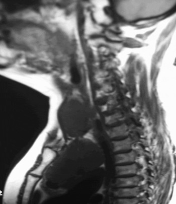
Thyroid function tests showed the patient to be euthyroid. Magnetic resonance imaging (Figure 1) and CT scans of her neck and thorax were performed, and confirmed the presence of a 10 × 10.5 × 8 cm thyroid mass extending into the anterior mediastinum to the level of the carina. There was significant compression of the trachea and the right main bronchus. There was no evidence of pathological lymphadenopathy.
Case 1: T1-weighted, sagittal magnetic resonance imaging scan of the neck and upper thorax, confirming the presence of a large substernal goitre extending to the level of the carina and compressing the trachea.

With the aid of a thoracic surgeon, the patient underwent a total thyroidectomy via a sternotomy. No difficulties were encountered with intubation. Tracheomalacia was noted intra-operatively but presented no complications.
Post-operatively, the patient suffered transient hypocalcaemia. Her vocal folds functioned normally.
Histological examination revealed a benign multinodular goitre.
Two years later, the patient developed metastatic renal cell carcinoma and died from complications associated with immunosuppression secondary to chemotherapy.
Case two
A 53-year-old woman presented with a gradually increasing neck mass which had first been noticed during pregnancy, 27 years previously. She complained of increasing shortness of breath on exertion, dysphagia, dysphonia and symptoms of hyperthyroidism, including sweating, heat intolerance and facial flushing over the previous three months. Her past history included coronary artery bypass grafting (CABG) six years previously. At the time of CABG, a large substernal goitre had been noted, and an attempt was made by the thoracic surgeon to resect this. This was obviously incomplete, as the patient presented with re-growth.
The patient was clinically overweight, with an emphysematous ‘barrel’ chest, making clinical examination extremely difficult.
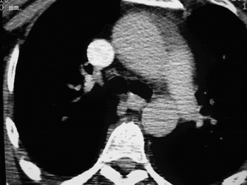
Flexible fibre-optic nasopharyngoscopy revealed normal vocal fold movement. Thyroid function tests revealed the patient to be euthyroid, despite her symptoms. Neck and thorax CT scans (Figure 2) showed a large substernal goitre measuring 10 × 7 × 4 cm. There was extension below the level of the aortic knuckle, with tracheal compression and oesophageal deviation. Also noted on CT were a loss of the facial planes between the substernal goitre and surrounding mediastinal structures.
Case 2: axial computed tomography scan showing a retrosternal goitre extending to the level of the aortic arch, with oesphageal compression.

In conjunction with our cardio-thoracic colleagues, the patient underwent a total thyroidectomy via a sternotomy, with preservation of all parathyroid glands and bilateral recurrent laryngeal nerves. Her intubation was uneventful. Intra-operatively, tracheal softness was noted. It was decided to leave the patient intubated overnight, and she was extubated uneventfully the following day.
Histological examination showed a benign multinodular goitre.
The patient was discharged well from followup, six months post-operatively.
Case three
A 78-year-old woman presented to St James's Hospital accident and emergency department with acute airway compromise. A collateral history revealed a three-day history of increasing shortness of breath with an audible wheeze. The patient was known to the otolaryngology department due to a long-standing substernal goitre, but had refused surgery on multiple occasions. There was no past history of asthma, chronic obstructive airway disease or previous thyroid surgery.
The patient was immediately intubated in the emergency room to secure her airway. There was no goitre palpable in the neck on clinical examination.
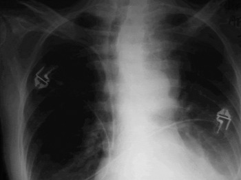
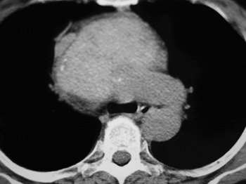
A chest X-ray revealed a large mediastinal mass with significant tracheal deviation (Figure 3). A CT scan of the neck and thorax (Figure 4) confirmed a large substernal goitre with significant tracheal compression. The CT scan also revealed loss of the normal fascial planes between the substernal portion of the goitre and the surrounding mediastinal structures.
Case 3: chest X-ray showing a mediastinal mass, with significant tracheal deviation to the right.

Case 3: axial computed tomography scan of the thorax, confirming a retrosternal goitre with tracheal and oesphageal compression, extending to the arch of the aorta.

The patient became thyrotoxic secondary to the iodine-based contrast used for CT scanning, and underwent emergency thyroidectomy two days later, with the help of a sternotomy performed by a thoracic surgeon. The patient was successfully extubated on the first post-operative day.
Histological examination revealed a multinodular goitre with evidence of thyroiditis.
The patient underwent scar revision surgery six months later for webbing of the T-shaped incision over her sternum.
She was discharged well from follow-up, receiving ongoing thyroid hormone replacement therapy.
Discussion
The otolaryngologist with a keen interest in head and neck endocrine surgery can expect to encounter large substernal goitres that may require sternotomy in order to assist safe removal of the gland. Our incidence of 2 per cent would be in keeping with the literature.Reference Pandya and Sanders3–Reference Gough5 However, larger incidences have been reported, with six of the 19 cases (32 per cent) reported by Hashmi et al. Reference Hashmi, Premachandra, Bennett and Parry6 requiring sternotomy. Referral practices may account for some of the differences in the reported series, if, for example, large intra-thoracic goitres are referred preferentially to thoracic surgeons rather than to thyroid surgeons.
A cervical approach is usually adequate for the majority of large substernal goitres.Reference Wheeler7 Various methods have been described to aid in goitre removal, and thus to avoid the need for sternotomy. The senior author regularly employed the use of a ‘table-spoon’ to assist in the delivery of large goitres into the neck. This technique has previously been described in the literature.Reference Hashmi, Premachandra, Bennett and Parry6 Pandya and SandersReference Pandya and Sanders3 described a novel technique of passing a catheter past the goitre, inflating the balloon, and with steady, gentle traction, delivering the goitre into the neck. Nevertheless, for some patients there will be no alternative but to perform a sternotomy to facilitate safe removal of the gland.
It is the practice of the senior author to pre-operatively inform our thoracic colleagues of all patients with a significant retrosternal goitre. An attempt is always made to resect the goitre through a cervical incision. Only if this was unsuccessful would we employ a sternotomy for safe removal of the gland.
Unfortunately, there are no definitive criteria for selecting patients who are likely to require sternotomy. All three of our patients had extension of their goitre below the level of the aortic knuckle on CT scan. Pre-operative knowledge of this enabled us to predict the subsequent necessity for sternotomy. Therefore, it would appear that gland extension to or beyond the aortic knuckle on CT scan is a strong indication for median sternotomy. This conclusion is confirmed by other authors.Reference Ahmed, Elnazeer and Mahadi4, Reference Wheeler7
• For the otolaryngologist regularly performing thyroid surgery, pre-operative identification of which patients with large substernal goitres will require sternotomy is essential
• Computed tomography evidence of adherence to surrounding mediastinal structures, and extension of a goitre to or below the aortic arch, should raise suspicion of the need for sternotomy
• Such cases need careful selection, as the skill and assistance of a cardio-thoracic surgeon will be required within a specialist centre
Two of our patients (cases two and three) showed loss of the facial planes between the substernal portion of the goitre and the surrounding mediastinal structures on CT scan. One patient had a history of previous thyroid surgery, while the other showed evidence of thyroiditis on histological examination. We would agree with Cho and colleagues' statement that the loss of fascial planes on CT scan is useful in predicting cases which may not be safely accessible through a cervical incision alone, due to the presence of adhesions.Reference Cho, Cohen and Som8
Evidence of tracheal deviation or compression on CT scan is a common finding in patients with substernal goitre. Compression of the trachea of up to 50 per cent can occur with the patient remaining asymptomatic.Reference Michael and Bradpiece9 Again, the literature supports the conclusion that, in these patients and in those with evidence of significant tracheal deviation, suspicion should be raised of a goitre large enough to require a sternotomy for safe delivery of the gland.Reference Torre, Borgonovo, Amato, Azezzo and Ansaldo10
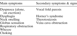
Symptoms associated with substernal goitre are widely described in the literature (Table I). In Hashmi and colleagues' series of six patients,Reference Hashmi, Premachandra, Bennett and Parry6 those who required sternotomy complained of dyspnoea on lying supine, stridor, dysphonia, dysphagia and an inability to sleep comfortably, and two had clinical signs of superior vena caval obstruction. Our patient cohort complained of similar symptoms. However, as seen from Table I, patients with substernal goitre not requiring sternotomy will have the same symptomatology. Therefore, differentiating patients who are likely to require sternotomy on symptoms alone can be difficult. Symptom duration can help predict those patients who are likely to develop complications associated with tracheomalacia. Hedayati and McHenryReference Hedayati and McHenry11 noted that cases presenting with seven to 30 years' duration of symptoms were more prone to airway problems secondary to long-standing compression of the trachea. However, it would appear difficult to predict patients requiring sternotomy from symptom duration alone, especially if there is no enlargement of the goitre on serial CT scanning.
Features of substernal goitres

Other possible indications for sternotomy described in the literature include a bilateral substernal goitre and suspicion of malignancy within a substernal goitre.Reference Gough5, Reference Netterville, Coleman, Smith, Michelle, Day and Burkey12 Although there was no suspicion of malignancy in our cohort, it has been our experience that attempting to deliver a large, bilateral substernal goitre through a cervical incision, with the goitre intact, represents a significant surgical challenge, and that these patients should be considered for sternotomy pre-operatively.
Conclusion
For the otolaryngologist who regularly performs thyroid surgery, pre-operative identification of which cases of large substernal goitre will require sternotomy is essential. In our experience, CT evidence of adherence to surrounding mediastinal structures, and extension of the goitre to or below the aortic arch, should raise suspicion of the need for sternotomy. Other ominous signs include bilateral substernal goitre and suspicion of malignancy within a substernal goitre. Such cases need careful selection, as the skill and assistance of a cardio-thoracic surgeon will be required within a specialist centre.