Introduction
Both visceral (VL) and cutaneous leishmaniasis (CL) are endemic diseases in several countries located in Mediterranean Basin as well as in Turkey (Dujardin et al., Reference Dujardin, Campino, Canavate, Dedet, Gradoni, Soteriadou, Mazeris, Özbel and Boelaert2008). Each year around 2000 CL cases are officially reported in Turkey and more than 95% of them were from nine endemic provinces mainly located in Mediterranean and Southeastern Regions (Gürel et al., Reference Gürel, Yeşilova, Olgen and Ozbel2012). Leishmania infantum MON-1 is the main causative agent for zoonotic human and canine visceral leishmaniasis while anthroponotic L. tropica is the main species causing CL in all regions of Turkey. Besides these agents, L. tropica and L. donovani in VL patients while L. infantum, L. major and L. donovani in CL patients were also reported (Koltas et al., Reference Koltas, Eroglu, Alabaz and Uzun2014; Özbilgin et al., Reference Özbilgin, Töz, Harman, Günaşti Topal, Uzun, Okudan, Güngör, Erat, Ertabaklar, Ertuğ, Gündüz, Çavuş, Karakuş, Östan Ural, Ölgen, Kayabaşi, Kurt and Özbel2019).
Dogs are considered as main reservoirs of L. infantum in Europe as well as in Turkey. So far, canine leishmaniasis (CanL) and reservoir role of the dogs have been studied comprehensively in different VL endemic areas of Turkey. The prevalence of CanL was detected as between 1.45% and 27.5% according to the endemic area (Toz et al., Reference Toz, Nasereddin, Ozbel, Ertabaklar, Culha, Sevil, Alkan and Jaffe2009). The possible role of the cats in the transmission of leishmaniasis is another debating topic in endemic countries. The presence of Leishmania DNA is not the conclusive finding to incriminate cats as a reservoir even though their infectiousness to sand fly was shown (Maroli et al., Reference Maroli, Pennisi, Di Muccio, Khoury, Gradoni and Gramiccia2007). Up to day, several studies performed to screen Leishmania spp. among cats in Turkey and the positivity were ranging from 7% to 33.3% (Paşa et al., Reference Paşa, Tetik Vardarlı, Erol, Karakuş, Töz, Atasoy, Balcıoğlu, Emek Tuna, Ermiş, Ertabaklar and Özbel2015; Can et al., Reference Can, Döşkaya, Özdemir, Şahar, Karakavuk, Pektaş, Karakuş, Töz, Caner, Döşkaya, İz, Özbel and Gürüz2016; Karakuş et al., Reference Karakuş, Arserim, Erişöz Kasap, Pekağırbaş, Aküzüm, Alten, Töz and Özbel2019). The possible role of the rodents was also investigated in Italy by sampling the spleen and various tissues and of the sampled rodents, 45% (n = 20) were found to be infected with L. infantum (Di Bella et al., Reference Di Bella, Vitale, Russo, Greco, Milazzo, Aloise and Cagnin2003). The presence of L. infantum in natural rodent populations was also investigated in Portugal and the infection rate was found to be 33.3% (Helhazar et al., Reference Helhazar, Leitao, Duarte, Tavares and da Fonseca2013).
There are several studies performed in the Old-World countries to investigate the role of rodents in the Leishmania transmission cycle especially for zoonotic cutaneous leishmaniasis (ZCL) caused by L. major. Due to the high endemicity of ZCL in Iran, numerous studies related to rodents were performed. The first study was done in the 1950s by Ansari and Mofidi in Iran and followed by several reports up to today stating their importance in the transmission of ZCL caused by L. major (Ansari and Mofidi, Reference Ansari and Mofidi1950; Akhoundi et al., Reference Akhoundi, Mohebali, Asadi, Mahmodi, Amraei and Mirzaei2013). Leishmania tropica was also demonstrated to infect the Ethiopian wild rodent populations (Kassahun et al., Reference Kassahun, Sadlova, Dvorak, Kostalova, Rohousova, Frynta, Aghova, Yasur-Landau, Lemma, Hailu, Baneth, Warburg, Volf and Votypka2015).
According to those findings, four different Leishmania species present in Turkey was reported to be found in sampled tissues of rodents in different countries. The prevalence rate of Leishmania in various populations of humans, dogs and even cats was investigated in Turkey. However, yet no study was performed to screen leishmaniasis infection in small wild rodents even though at least 65 species are present belonging to seven families in Turkey (Kryštufek and Vohralík, Reference Kryštufek and Vohralík2005, Reference Kryštufek and Vohralík2009; Yiğit et al., Reference Yiğit, Çolak, Sözen and Karataş2006, Reference Yiğit, Çolak and Sözen2016; Wilson et al., Reference Wilson, Lacher and Mittermeier2016, Reference Wilson, Lacher and Mittermeier2017). Therefore, the potential role of rodents in the Leishmania infection cycle was investigated in the present study.
Materials and methods
Sample collection and DNA preparation
The samples collected during epidemiological Hantavirus studies in various provinces of Turkey were used in the present study. For these studies, the wild rodents were collected using Sherman live traps placed with consecutively 10–15 m intervals, and placement points were recorded by GPS. Species identification of rodents was performed according to the phenotypic characteristics of the animals. After taking body measurement of animals, they were sacrificed by cervical luxation method, and tissue samples were preserved in formalin. A total of 906 different tissue samples (spleen, liver, and lung) obtained from 712 rodents caught from 29 provinces were screened. The samples were collected from the individuals belonging to 23 rodent genera and were transferred to Leishmania Laboratory in the Department of Parasitology, Faculty of Medicine of Ege University for molecular screening studies to detect the Leishmania DNA. Samples obtained from Apodemus 431 (47.5%), 68 (7.5%) Mus sp., 68 (7.5%) Myodes sp., 55 (6.0%) Spalax sp., 51 (5.6%) Nannospalax sp., 45 (4.9%) Microtus sp., 42 (4.6%) Meriones sp. and 32 (3.5%) Rattus sp. samples were enrolled in the study. These eight genera made up most of the total samples (87.1%).
Before the DNA extractions, all tissue samples were first cut in small pieces to speed up the evaporation step and dried using Eppendorf Concentrator™ (Thermo Scientific, UK) to remove the formalin. Dried tissue samples were rehydrated using 200-μL of Qiagen® tissue lysis buffer (Qiagen GmbH, Hilden, Germany) and Proteinase K (20-μL) and incubated at 56°C overnight. Tissue samples were later transferred to tubes containing ZR Bashing Bead™ tubes (Zymo Research Corp., USA) and homogenated using Magna Lyser (Roche Molecular Diagnostics, Germany) at 7000 g for 90 s. Homogenates were used in DNA extractions following the manufacturer's instructions of DNeasy Blood & Tissue Kit (Qiagen GmbH).
Realtime kDNA PCR and realtime ITS1 PCR
The detection of Leishmania in rodent tissues and species determination was performed with a combination of Realtime kDNA and ITS1 PCR protocols. The first step was to screen the presence of Leishmania in tissue samples and due to its high copy number, the minicircle region of the kDNA was targeted using genus-specific primers (JW11/JW12) as reported in recent studies (Kassahun et al., Reference Kassahun, Sadlova, Dvorak, Kostalova, Rohousova, Frynta, Aghova, Yasur-Landau, Lemma, Hailu, Baneth, Warburg, Volf and Votypka2015). A Realtime PCR was performed using SYBR Green I Master Kit (Roche Diagnostic, France) and positivity determined according to genus-specific melt peaks.
Leishmania spp. positive samples were used in a second step PCR, which was performed to determine the species of the causative agent. Primers targeting the ITS1 region rRNA gene (LITSR/ITS1R-TR1) was used and species determination were made in Leishmania positive samples using melt analysis. A Real-time PCR protocol was applied by the previously published paper (Toz et al., Reference Toz, Culha, Zeyrek, Ertabaklar, Alkan, Vardarlı, Gunduz and Ozbel2013).
Results
During the years of 2005–2017, 906 tissue samples (spleen, liver, and lung) were obtained from 712 rodent species belonging to 23 genera and 41 species. Sampling was done in 73 different locations and samples were divided according to their geographical locations. The majority of animals (56%) were obtained from urban residential locations in CL endemic areas. Based on the sampled rodents, the majority of them (n = 432; 47.6%) were belonging to Apodemus genera. The mean of whole-body length was 188 mm with a range of 171–220 mm.
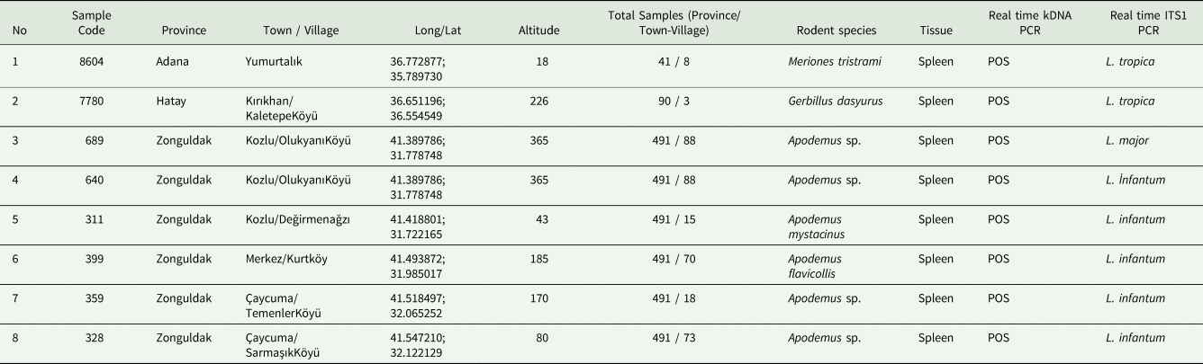
Of the analysed animal samples, eight (8/712; 1.12%) of them were found to be positive for Leishmania spp. DNA by realtime kDNA-PCR (Suppl Fig. 1). The species typing by real-time ITS1 PCR revealed five L. infantum, two L. tropica and one L. major among the positive samples (Suppl Fig. 2).
Leishmania major DNA was detected in the specimen belonging to Apodemus genus from Zonguldak province located in the Western Black Sea Region of Turkey. Leishmania infantum DNA was detected in three different rodent species, Apodemus filavicollis, Apodemus mystacinus and Apodemus sp. (four positive rodent specimens could not be detected in species level) from the same province, Zonguldak. Leishmania tropica DNA was detected in Gerbillus dasyurus (the mean of whole-body length was 84 mm with a range of 73–94 mm) from Hatay province and Meriones tristrami (the mean of whole-body length was 120 mm with a range of 100–155 mm) from Adana province. Both provinces are located in the Eastern Mediterranean Region (Table 1; Figure 1). Leishmania infantum positive rodents were all male while L. major and L. tropica positives were female.
The locations where eight positive small wild rodents were caught and PCR results

The map showing sampling provinces with negative (Blue) and positive results (Red: L. tropica; Purple: L. infantum and L. major DNA positive rodents). Numbers in the bracelets are showing the samples number for each province.

Discussion
Small rodent species in The New World and The Old World are suspicious reservoir hosts for different Leishmania species. Despite the important roles of small rodents for spreading of leishmaniasis, the studies are relatively in low number and mainly carried out in the Middle East and North Africa countries such as Iran, Tunisia, Morocco and Algeria where L. major is the dominant causative agent for CL (Ghawar et al., Reference Ghawar, Toumi, Snoussi, Chlif, Zaatour, Boukthir, Hamida, Chemkhi, Diouani and Ben-Salah2011; Echchakery et al., Reference Echchakery, Chicharro, Boussaa, Nieto, Carrillo, Sheila, Moreno and Bouzmezzough2017; Foroutan et al., Reference Forotuan, Khademvatan, Majidiani, Khalkhali, Hedayati-Rad, Khashaveh and Mohammadzadeh2017; Pourmohammadi et al., Reference Pourmohammadi, Mohammadi-Azni and Kalantari2017; Othman et al., Reference Othman, Ghawar, Chaouch, Ayari, Chemkhi, Cancino-Faure, Tomas-Perez, Alcover, Riera, Ben-Salah, Fisa, Ben-Ismail and Ben-Abderrezzak2018). Also, some reports have indicated the infection to L. donovani, L. infantum and L. tropica in some rodents such as Gerbillus spp., Rattus norvegicus and Mus musculus (Helhazar et al., Reference Helhazar, Leitao, Duarte, Tavares and da Fonseca2013; Kassahun et al., Reference Kassahun, Sadlova, Dvorak, Kostalova, Rohousova, Frynta, Aghova, Yasur-Landau, Lemma, Hailu, Baneth, Warburg, Volf and Votypka2015; Navea-Perez et al., Reference Navea-Perez, Diaz-Saez, Corpas-Lopez, Merino-Espinosa, Morillas-Marquez and Martin-Sanchez2015) suggesting their potential role to act as a reservoir for another Leishmania species.
The comprehensive studies on canine leishmaniasis were done for many years in Turkey, and results are always showing higher prevalence than human infection in VL endemic areas. Leishmania infantum was found as the main causative agent of CanL as well as human VL in these areas (Toz et al., Reference Toz, Culha, Zeyrek, Ertabaklar, Alkan, Vardarlı, Gunduz and Ozbel2013; Sarı et al., Reference Sarı, Limoncu, Balcıoğlu, Aldemir, Tasçı, Kiliç, Toz, Demirci, Demir, Erisoz Kasap, Olgen and Ozbel2015). Because of Leishmania species having zoonotic life cycle have been found in Turkey and no study was carried out on rodents so far (Koltas et al., Reference Koltas, Eroglu, Alabaz and Uzun2014; Özbilgin et al., Reference Özbilgin, Çulha, Uzun, Harman, GünaştıTopal, Okudan, Zeyrek, Gündüz, Östan, Karakuş, Töz, Kurt, Akyar, Erat, Güngör, Kayabaşı, Çavuş, Bastien, Pratlong, Kocagöz and Özbel2016), it was necessary to conduct studies for revealing the reservoir potential of the small wild rodents. In the present study, we have investigated the presence of Leishmania DNA in the tissue samples collected from different rodent species in different provinces of Turkey using molecular techniques. Since we had rodent tissue samples that were previously collected and kept in formalin, the microscopical examination of their smears could not be performed for these samples. Our results revealed the presence of two Leishmania species (L. infantum, L. major) in the collected Apodemus spp. Specimens from one province, Zonguldak located in the Western Black Sea Region of Turkey. DNA of L. tropica was also found in two specimens (Meriones tristrami and Gerbillus dasyurus) from two different provinces, Adana and Hatay, located in the East Mediterranean Region of Turkey.
Although L. tropica is known as anthroponotic species, it is found in different mammalian species as dogs, cats and wild mammals, which play a reservoir role in nature (Töz et al., Reference Toz, Culha, Zeyrek, Ertabaklar, Alkan, Vardarlı, Gunduz and Ozbel2013; Paşa et al., Reference Paşa, Tetik Vardarlı, Erol, Karakuş, Töz, Atasoy, Balcıoğlu, Emek Tuna, Ermiş, Ertabaklar and Özbel2015; Echchakery et al., Reference Echchakery, Chicharro, Boussaa, Nieto, Carrillo, Sheila, Moreno and Bouzmezzough2017; Baneth et al., Reference Baneth, Yasur-Landau, Gilad and Nachum-Biala2017). However, confirmation of these hosts as reservoirs requires xenodiagnostic studies as performed in Italy and Spain for L. infantum (Maroli et al., Reference Maroli, Pennisi, Di Muccio, Khoury, Gradoni and Gramiccia2007; Jimenez et al., Reference Jimenez, Gonzalez, Martin-Martin, Hernandez and Molina2014). Adana and Hatay provinces are highly endemic areas for CL caused by L. tropica and L. infantum (Serin et al., Reference Serin, Daglioglu, Bagirova, Allahverdiyev, Uzun, Vural, Kayar, Tezcan, Yetkin, Aslan, Emekdas and Koksal2005), and recently L. major and L. donovani were also reported as causative agents in both provinces (Koltas et al., Reference Koltas, Eroglu, Alabaz and Uzun2014; Özbilgin et al., Reference Özbilgin, Töz, Harman, Günaşti Topal, Uzun, Okudan, Güngör, Erat, Ertabaklar, Ertuğ, Gündüz, Çavuş, Karakuş, Östan Ural, Ölgen, Kayabaşi, Kurt and Özbel2019). According to the Ministry of Health records, a total of 616 and 207 CL cases were reported from Adana and Hatay provinces between 2013 and 2017, respectively (Özbel Y, personal comm). In a recent study, 21 out of 25 strains (84%) from Adana province were detected as L. tropica, and there were only two isolates from Hatay province and one of them is also found as L. tropica by Realtime ITS1 PCR (Özbilgin et al., Reference Özbilgin, Töz, Harman, Günaşti Topal, Uzun, Okudan, Güngör, Erat, Ertabaklar, Ertuğ, Gündüz, Çavuş, Karakuş, Östan Ural, Ölgen, Kayabaşi, Kurt and Özbel2019). Dog studies carried out in these provinces revealed a high prevalence of CanL as 27.18% by IFAT and 41.74% by conjunctival swap nested PCR in rural areas of Adana (Karakuş et al., Reference Karakuş, Töz, Ertabaklar, Paşa, Atasoy, Arserim, Ölgen, Alkan, Durant and Özbel2015). We analyzed 41 and 90 rodent tissue samples from Adana and Hatay, respectively. Nineteen out of 41 were from Meriones tristrami, caught in five different towns, and one (5.26%; 1/19) of them was found positive for L. tropica. For Hatay province, 12 out of 90 samples from Gerbillus dasyurus, caught in four different towns, and one (8.33%; 1/12) of them was found positive for L. tropica. Although the number of samples included in the present study is not very high, the detection of L. tropica in small rodents in Adana and Hatay provinces will make the control of CL more complicated in these endemic areas. Applying control measures for small wild mammals are more difficult because of the geographical and climatic conditions in the area. More studies are needed to understand better its epidemiological importance. Hatay and Adana provinces are among the six highly endemic provinces in Turkey. Because of four Old World Leishmania species causing CL and suitable vector sand fly species (Alten et al., Reference Alten, Maia, Afonso, Campino, Jimenez, Gonzalez, Molina, Banuls, Prudhomme, Vergnes, Toty, Cassan, Thierry, Sereno, Bongiorno, Bianchi, Khouty, Tsirigotakis, Dokianakis, Antoniou, Christodoulou, Mazeris, Karakus, Ozbel, Arserim, Erisoz Kasap, Gunay, Oguz, Kaynas, Tsetsvadze, Tskhvaradze, Giorgobiani, Gramiccia, Volf and Gradoni2015) are present in both provinces, small wild rodents need to be included to the epidemiological studies related to reservoirs planned in these areas.
Among 65 rodent species recorded in Turkey (Kryštufek and Vohralík, Reference Kryštufek and Vohralík2005, Reference Kryštufek and Vohralík2009; Yiğit et al., Reference Yiğit, Çolak, Sözen and Karataş2006, Reference Yiğit, Çolak and Sözen2016; Wilson et al., Reference Wilson, Lacher and Mittermeier2016, Reference Wilson, Lacher and Mittermeier2017), six of them belonging to genus Meriones (M. persicus, M. tristrami, M. vinogradovi, M. crassus, M. libycus, M. dahli) playing important role as reservoir animal in different countries (Saliba and Oumeish, Reference Saliba and Oumeish1999), are available in most regions of Turkey except Black Sea and Marmara regions (Yiğit et al., Reference Yiğit, Çolak, Sözen and Karataş2006). One endemic species classified in Acomys genus (A. cilicicus) lives only around Silifke and Erdemli towns of Mersin province (Çetintaş et al., Reference Çetintaş, Matur and Sözen2017), where CL caused by L. tropica is endemic. The wide-spreading of these small wild mammals in many areas enhancing the risk for human and canine leishmaniasis, and keeping the circulation of the parasite in nature.
Leishmaniasis tropica was detected in five (0.85%) out of 586 specimens belonging to Gerbillus genus by molecular techniques in Ethiopia (Kassahun et al., Reference Kassahun, Sadlova, Dvorak, Kostalova, Rohousova, Frynta, Aghova, Yasur-Landau, Lemma, Hailu, Baneth, Warburg, Volf and Votypka2015). We also found L. tropica DNA in Gerbillus dasyurus collected from Hatay province, where CL caused by L. tropica and L. infantum is highly endemic (Table 1).
In Central Tunisia, it was reported that the prevalence of L. major in Psammomys obesus and Meriones shawi species ranged from 5% to 33% according to the methods used (Ghawar et al., Reference Ghawar, Toumi, Snoussi, Chlif, Zaatour, Boukthir, Hamida, Chemkhi, Diouani and Ben-Salah2011). Three Leishmania species (L. infantum, L. major, L. tropica) coexist in Morocco and the small rodent Meriones shawi is found to be a rodent reservoir for L. major (Kahime et al., Reference Kahime, Boussaa, Bounoua, Ouanaimi, Messouli and Boumezzough2014). In a recent study carried out in western Morocco, 18 (9.13%) out of 197 animals belonging to ten species were found positive for Leishmania DNA. Sixteen (six Rattus rattus, nine Mus musculus, and one Rattus norvegicus) and two (Mus musculus) positive samples were identified as L. infantum and L. tropica, respectively (Echchakery et al., Reference Echchakery, Chicharro, Boussaa, Nieto, Carrillo, Sheila, Moreno and Bouzmezzough2017). In the present study, we used Realtime kDNA PCR for Leishmania DNA detection and a second Realtime PCR targeting ITS1 region was performed only for positive samples. Eight positives (0.88%) were found among 906 samples. Only one Meriones specimen (M. tristami) was found positive while none of the Mus and Rattus specimens was found positive (Table 1).
Besides dogs are the main reservoirs for L. infantum, CanL itself is an important veterinary health problem in southern European countries. The studies on the reservoir potential of rodents for leishmaniasis are very limited in European countries.
In Greece, 97 small rodents in urban and rural areas were collected and liver, spleen, and blood samples were taken for investigation by parasitological, serological, and molecular techniques. Sixteen (16/66; 24%) Mus musculus and 3 (3/12; 25%) Rattus rattus were found positive by molecular and serological tests while they were negative in smears. The researchers pointed out that the parasite load could be very low in the tissues (Tsakmakidis et al., Reference Tsakmakidis, Angelopoulou, Dovas, Dokianakis, Tamvakis, Symeonidou, Antoniou and Diakou2017). In Portugal, the presence of L. infantum DNA in M. musculus and R. norvegicus with infection rates of 33.3% in both animals was reported. The authors pointed out that Leishmania DNA was found especially in the skin samples taken from the ear lobe and this was a suitable place for sand flies to get infected (Helhazar et al., Reference Helhazar, Leitao, Duarte, Tavares and da Fonseca2013). In a similar study in Italy, the positivity rate was reported as 15.5% among 78 captured R. rattus samples in Montecristo island (Zanet et al., Reference Zanet, Sposimo, Trisciuoglio, Giannini, Strumia and Ferroglio2014). In our study, among 100 specimens belonging to Mus and Rattus genera, all of them were found negative. Because of 56% of these animals were from urban sites of CL endemic areas, the possibility to detect Leishmania DNA was very low. On the other hand, the rodent species of both genera were found positive in different countries, are very wild spread in all regions of Turkey, and this should be considered for the risk estimations and management. In rural areas, considering that the rodent burrows are one of the most suitable living and breeding places for sand flies, it should be known that the parasite is constantly circulated in the environments that can allow limited interventions, and it has a zoonotic cycle among small rodents.
In a study conducted in Granada, Spain, the tissue samples (liver, spleen, blood, skin and bone marrow) from 37 rodent specimens were investigated and ten (27%) animals were found positive by parasitological and/or molecular techniques. Besides three R. rattus and two M. musculus specimens, five Apodemus sylvaticus were also reported as positive (Navea-Perez et al., Reference Navea-Perez, Diaz-Saez, Corpas-Lopez, Merino-Espinosa, Morillas-Marquez and Martin-Sanchez2015). In the present study, 431 Apodemus spp. (62 A. mystacinus) were included in the analysis and only 66 (44 A. mystacinus) of them were from CL endemic areas. However, five L. infantum and one L. major positive Apodemus sp. samples were from CL non-endemic but VL sporadic province, Zonguldak. Only eight CL cases were reported from this province in the last 22 years (species and origin of the cases were unclear, Özbel Y, personal comm) while sporadic human VL cases have been reporting for decades. The prevalence of CanL was also reported as 8% from that region previously (Özbel et al., Reference Özbel, Turgay, Alkan, Babaoğlu, ÖzensoyTöz and Babalıoğlu2002). In Turkey as in other endemic countries, VL circulation among humans continues together with higher percentages of CanL. Therefore, this area is accepted as endemic for CanL. The positivity rate was 1.39% (6/431) in total and 1.70% (6/351) among the samples collected only in Zonguldak province suggesting Apodemus species probably has a role as reservoir animal of Leishmania spp. in the region; even it needs to be supported by further studies such as isolation of the parasites and experimental studies for showing reservoir potentials.
Concluding remarks
This study reports for the first time in Turkey, the natural infection of L. infantum, L. major and L. tropica in small wild rodents, suggesting these animals possibly have a role as reservoirs in the life cycle of all three Leishmania species in Turkey. The molecular detection of three Leishmania species in the present study in highly endemic areas for CL such as Adana and Hatay provinces and also in an endemic area for VL such as Zonguldak shows the possible zoonotic cycle including small wild rodents in addition to the zoonotic cycle of L. infantum in the dogs. Infected rodents with Leishmania may increase the risk not only for humans but also for dogs in rural and urban areas. And this suggests the possible involvement of other wide-spread rodent species in the studies that will be carried out in other endemic areas in Turkey. Further studies are needed not only for planning epidemiological studies but also for developing rodent control measures in potential endemic areas to break the transmission cycle.
Financial support
This study is supported by The Scientific and Technological Research Council of Turkey (TÜBİTAK) Project No: 114S999.
Conflicts of interest
The authors declare there are no conflicts of interest.
Ethical standards
Ethical approval was obtained from Dokuz Eylül University Animal Ethical Committee under the registration number of 2011-24-28/12 at the date of December 28, 2011.
Supplementary material
The supplementary material for this article can be found at https://doi.org/10.1017/S0031182020000803