Introduction
Sodalite-type compounds constitute a versatile family of framework microporous materials of the general formula M 0–8X 0–3[T 12O24]·nH2O (n = 0–8), where T means tetrahedrally coordinated framework-forming components, whereas M and X denote extra-framework cations and anions located inside the so-called β-cages (Peters et al., Reference Peters, Vega-Flores and Depmeier2009).
Sodalite-type materials attract substantial interest because of their luminescent, tenebrescent, cathodochromic, ion conductive, ion exchange, magnetic and catalytic properties, as well as materials for matrix isolation and hydrogen storage (Depmeier, Reference Depmeier2005; Gaft et al., Reference Gaft, Reisfeld and Panczer2005; Finch et al., Reference Finch, Friis and Maghrabi2016; Bolotina et al., Reference Bolotina, Chukanov and Schäfer2022 and references therein).
In recent studies, it has been highlighted that aluminosilicate microporous materials serve as efficient media for the generation of hydroxyl free radicals under UV-B and UV-C irradiation. This plays a crucial role in the development of effective membranes for wastewater treatment and petrochemical catalysis (Chen et al., Reference Chen, Zhang, Shu, Yuan, Yan, Bai, He, Shen, Gong, Zhang, Li, Hu, Li, Wu, Chai, Yu and Wang2021). The presence of halogen ions in the structure of these microporous materials reduces the threshold for free radical formation. Moreover, aluminosilicates possess a suitable structure for applications as getters of radionuclides (Wang et al., Reference Wang, Chen and Ewing2004). Therefore, studying the mechanism of radiation defect formation under irradiation is essential.
Defect generation in halogen-containing microporous materials is primarily attributed to the non-radiative decay of electronic excitations near [Na4X]3+ complexes at room temperature, where X is the Cl, Br or I halogen anion. However, this decay process leads to the emergence of intrinsic luminescence at lower temperatures. The role of alkali halide components [Na4X]3+ clusters (X = Cl–, Br–, I–) in the sodalite-type crystal structure in terms of intrinsic electron excitation, recombination of luminescence and influence of thermostimulation on the above-mentioned physical properties has been described in several works (Denisov et al., Reference Denisov, Denks, Dudel’zak, Osminin and Ruus1977; Lushchik et al., Reference Lushchik, Kirm, Lushchik, Martinson, Nagirnyi and Vasil’chenko2002a, Reference Lushchik, Kirm, Kudryavtseva, Lushchik, Martinson, Nagirnyi and Vasil’Chenko2002b; Zahoransky et al., Reference Zahoransky, Friis and Marks2016 and references therein). Synthetic halide-bearing sodalites are readily synthesized (Trill et al., Reference Trill, Eckert and Srdanov2003 and references therein). In synthetic halide-bearing materials with the sodalite structure, intrinsic luminescence has been observed in samples containing [Na4Br]3+ and [Na4I]3+ in the energy range of 2 to 4 eV. The excitation bands are observed in the energy region of 5 to 11 eV (Denks et al., Reference Denks, Dudel’zak, Lushchik, Ruus, Soshchin and Trofimova1976). Under electron excitation, luminescence at ∼3.6 eV has been observed in materials with a tectosilicate lattice (Garcia-Guinea et al., Reference Garcia-Guinea, Correcher, Sanchez-Muñoz, Finch, Hole and Townsend2007). This luminescence is attributed to the defects in these materials. Further investigations have allowed for the attribution of the observed luminescence to intrinsic recombination processes occurring in sodalite-like systems under vacuum ultraviolet excitation (VUV) (Denks et al., Reference Denks, Murk and Vasil’chenko1996, Reference Denks, Karner and Murk1997). Exciton-like excitations have previously been observed in several minerals with a microporous structure. In cancrinite, Na7Ca[Al6Si6O24](CO3)·2H2O, such excitations were observed in the energy range of 2.8 to 3.8 eV (Kaneva and Shendrik, Reference Kaneva and Shendrik2022). Meionite Ca4[Al6Si6O24]CO3 exhibited excitations at 3.2 eV and fluorcarletonite KNa4Ca4Si8O18(CO3)4F·H2O showed excitations in the range of 2.8 to 3.4 eV (Shendrik et al., Reference Shendrik, Kaneva, Pankratova, Pankrushina, Radomskaya, Gavrilenko, Loginova and Pankratov2024). However, in these materials, the decay of exciton-like excitations occurred near carbonate (CO3)2− complexes, resulting in the formation of a bound pair consisting of (CO3)∙− hole and F-centre. The photochromic properties were also described for Se- and S-bearing sodalites (Kirk et al., Reference Kirk1954; Deb et al., Reference Deb and Gallivan1972; Schalaich et al., Reference Schalaich, Lindner, Feldmann, Göbel and Reinen2000; Armstrong et al., Reference Armstrong, Dann, Neumann and Marco2003; Sidike et al., Reference Sidike, Sawuti, Wang, Zhu, Kobayashi, Kusachi and Yamashita2007; Curutchet et al., Reference Curutchet and Le Bahers2017; Colinet et al., Reference Colinet, Gheeraert, Curutchet and Le Bahers2020). Diversity of sulfur-bearing anions, radical anions and neutral sulfur molecules (SO42–, SO32–, HS–, Sn 2– with n from 1 to 4, S2∙–, S3∙–, cis- and trans-S4∙–, SO4∙–, COS, cis- and trans-S4, S6, etc.) in minerals and synthetic compounds with the sodalite-type structures is quite wide (Chukanov et al., Reference Chukanov, Shchipalkina, Shendrik, Vigasina, Tauson, Lipko, Varlamov, Shcherbakov, Sapozhnikov, Kasatkin, Zubkova and Pekov2022a and references therein). However, the luminescence of these compounds is attributed to the presence of S2∙–. Generally, the most interesting physical properties are peculiar to nonstoichiometric sodalites (SOD) and chemically and structurally related cancrinites (CAN), because of the facilitated formation of defects, for example (Sn)xCl–V xCl defect complexes, where (Sn)x is a polysulfide radical anion and V xCl is chlorine vacancy which has captured an electron (F-centre) (Stoliaroff et al., Reference Stoliaroff, Schira, Blumentritt, Fritsch, Jobic and Latouche2021).
In this paper, we present data on the vibrational spectra, luminescence properties and thermal stability of a series of sodalite-type feldspathoids with mixed halogen-bearing (Сl–, Br–, I–) and S-bearing anions and K+, Na+ and Ca2+ as extra-framework cations in comparison with previous works (Hassan et al., Reference Hassan, Antao and Parise2004a, Reference Hassan, Antao and Parise2004b, Reference Hassan, Antao and Parise2006). In this study, we investigate the intrinsic luminescence from the recombination of electronic excitations near halogen ions positioned within various cages.
Materials and methods
Synthesis
Sodalite-type aluminosilicates were synthesized using highly dispersed metakaolin (VMK-45, ArmMiks, Russia) powder as a source of Al and Si, and KI, KBr, CaCl2, NaCl, Na2S·8H2O, and Na2S2O3 (analytical grade, Calibre Chemicals Pvt. Ltd, India) as the sources of extra-framework components. The charges composed of metakaolin powder (4 g), halogen-bearing and/or S-bearing salts, and 8M aqueous NaOH (8 ml) were loaded into 25 ml PTFE-lined autoclaves, stirred for 10 min and placed into the muffle furnace. After the completion of the experiments, the samples were washed with distilled water and dried in a cabinet dryer oven. The experimental data is given in Table 1.
The conditions and the products of hydrothermal synthesis

Table 1 Long description
The table lists nine hydrothermal experiments, giving the salt mixtures and their masses, treatment temperature and duration, powder colour, and crystallographic results including phase type, space group, lattice parameters, and unit-cell volume. Experiments 1 through 5 use halide salts only and all produce light pink powders identified as a SOD-type phase with lattice parameter a around 8.90 to 8.96 angstroms and volumes about 704 to 719 cubic angstroms. Within these SOD-only runs, the smallest volume is in experiment 5 at about 704.2 cubic angstroms, and the largest is in experiment 1 at about 719.3 cubic angstroms. Experiments 6 through 9 include thiosulfate and, in three cases, hydrated sodium sulfide, and they yield white or greyish powders with two phases reported: a CAN-type phase and a SOD-type phase. For these mixed-phase runs, the CAN-type phase has a near 12.67 angstroms and c near 5.18 angstroms, with volumes around 720 to 722 cubic angstroms, while the accompanying SOD-type phase spans roughly 8.90 to 9.00 angstroms with volumes about 704 to 729 cubic angstroms. The highest SOD-type volume appears in experiment 7 at about 728.6 cubic angstroms, which also notes residual KI alongside the SOD-type phase.
Scanning electron microscopy and electron microprobe analysis
The morphology and chemical composition of feldspathoids were studied using electron microprobe. The dense aggregates of microcrystalline aggregates were pressed in epoxy resin and then polished with diamond suspensions. The analyses from polished aggregates were carried out with a JEOL JXA-8230 instrument (WDS and EDS modes) at the Laboratory of Analytical Techniques of High Spatial Resolution, Dept. of Petrology, Moscow State University. The operating conditions included an accelerating voltage of 20 kV and a beam current of 10 nA; the beam was rastered on an area 4 μm × 4 μm. The data reduction was carried out by means of the INCA Energy 300 software package. The following standards were used for quantitative analysis: albite (Na), orthoclase (K), NaCl (Cl), KI (I), KBr (Br), anorthite (Al and Si), diopside (Ca and Mg), FeS2 (S) and Fe (Fe). Contents of other elements with atomic numbers higher than that of carbon are below detection limits. The analytical data are given in Supplementary data (Tables S1 and S2). The types of S-bearing anions in studied samples are determined, based on the results of Raman and infrared spectroscopies.
Raman, infrared and luminescence spectroscopy
The Raman spectra of synthetic SOD- and CAN-type feldspathoids synthesized in systems with I–, Cl–, Br–, [S2O3]2– and S2– (Experiments 1–9) have been obtained for powder samples using an EnSpectr R532 spectrometer based on an OLYMPUS CX 41 microscope (Enhanced Spectrometry, San Jose, USA) coupled with a diode laser (λ = 532 nm) at room temperature. The spectra were recorded in the range from 100 to 4000 cm–1 with a diffraction grating (1800 gr mm–1) and spectral resolution of ∼6 cm–1. The output power of the laser beam was in the range from 5 to 13 mW. The diameter of the focal spot on the sample was 5–10 μm. The back-scattered Raman signal was collected with a 40× objective; signal acquisition time for a single scan of the spectral range was 1 s, and the signal was averaged over 50 scans. Crystalline silicon was used as a standard. The products of Experiments 2 and 3 are strongly luminescence under a green laser. Thus, Raman spectra for these experiments were obtained using a red laser.
The Raman spectra of products of Experiments 2 and 3 were also acquired with a confocal Raman microscope JY Horiba XPloRA at the Department of Petrology and Volcanology (Geological Faculty, Lomonosov Moscow State University). Laser beam with 785 nm wavelength and 12 mW power was used as an excitation source. Spectra were collected in the range 100–2000 cm–1 with a 1800 gr/mm diffraction grating, 10× objective and 40 seconds accumulation time per each window.
The infrared spectra of suspensions of powdered samples in Nujol (Figures S1 and S2) were acquired using a Fourier infrared spectrometer FS-02 in absorption mode in the range of 400–4000 cm−1 and processed with the software Fspec. 20 scans were collected for each spectrum at a resolution of 4 cm–1. The IR spectrum of an analogous pellet of pure KBr was used as a reference.
The luminescence spectra were recorded using an MDR2 (LOMO, Russia) monochromator. The emission was detected by a H6780-04 Hamamatsu photomodule, which operated in the photon-counting regime. For measurements in the VUV spectral region, a VM-2 monochromator (LOMO) and a Hamamatsu deuterium lamp L7292 were employed. The powder samples were deposited onto high purity single crystal LiF plates. These plates were then placed on a cryofinger and evacuated. The luminescence under VUV was recorded for samples of Experiments 1–6, whereas samples of Experiments 7–8 did not exhibit detectable luminescence. At room temperature, no luminescence was observed when excited with energies below 6 eV and only very weak luminescence was observed under 8 eV excitation. However, during the cooling process, the luminescence intensity significantly increased. The spectra provided below were measured at 80 K.
Powder X-ray diffraction (PXRD)
The PXRD data of samples obtained in Experiments 1–9 were collected using a Rigaku R-AXIS Rapid II diffractometer equipped with image plate detector and rotating anode with the microfocus optics, CoKα radiation, 40 kV, 15 mA, Debye-Scherrer geometry, d = 127.4 mm and exposure of 15 min. The unit cell parameters for each phase were refined with the TOPAS v4.2 program package and given in Table 1. The conversion of raw data to profile was performed with the osc2xrd program (Britvin et al., Reference Britvin, Dolivo-Dobrovolsky and Krzhizhanovskaya2017).
High-temperature powder X-ray diffraction (HTPXRD) patterns for the products of Experiments 2, 3 and 5 were obtained using a Rigaku Ultima IV (CuKα radiation, 40 kV/30 mA, DtexUltra PSD, θ/θ Bragg-Brentano geometry) powder diffractometer equipped with a Rigaku R-300 high-temperature attachment in the temperature range of 25–1000°C in air. These experiments were chosen as the pure probes as they did not contain the small amounts of unreacted KI and KBr that were detected in Experiments 1 and 4. A powder sample was placed on a Pt holder from a heptane suspension. The average time of an experiment was ∼20 hours. The unit cell parameters were calculated at every T step via the TOPAS v4.2 program. The values of the thermal expansion coefficients (TECs) were estimated using the second-order polynomial approximations of the unit cell parameters by ThetaToTensor program (Bubnova et al., Reference Bubnova, Firsova and Filatov2013).
Results
In the sodalite-type feldspathoids synthesized using equimolar pairs of salts KI + KBr and KI + CaCl2, taken in excess with respect to metakaolin, the contents of iodine do not exceed 0.46 atoms per formula unit (apfu) whereas the Br:Cl ratios in the charge and in the reaction products are similar. In all products, the contents of Ca and K do not exceed 1.14 and 1.09 apfu, respectively. The presence of Fe2O3 in all samples studied is due to the initial composition of metakaoline, that contains ∼1 wt.% of Fe2O3 and, generally, determined its pink colour and the same colour of products of Experiments 1–5.
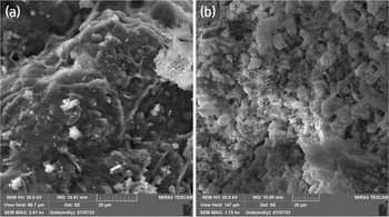
As shown in Fig. 1, the obtained compounds form agglomerates of small particles. Variations of the synthesis conditions did not result in the growth of well-shaped crystals, however, reactions of metakaolin fine powder with mixtures of salts in alkali medium were completed and sodalite- or cancrinite-type aluminosilicates crystallized (Supplementary data, Tables S1 and S2).
Morphology of aggregates in samples 7 and 9. SEM photo, SE mode.

The assignment of Raman bands is given in Supplementary data (Table S3) according to the earlier published data (Chukanov et al., Reference Chukanov, Shchipalkina, Shendrik, Vigasina, Tauson, Lipko, Varlamov, Shcherbakov, Sapozhnikov, Kasatkin, Zubkova and Pekov2022a, Reference Chukanov, Shendrik, Vigasina, Pekov, Sapozhnikov, Shcherbakov and Varlamov2022b; Ariai and Smith, Reference Ariai and Smith1981; Creighton et al., Reference Creighton, Deckman and Newsam1994; Hettmann et al., Reference Hettmann, Wenzel, Marks and Markl2012; and references therein). Thus, in the samples studied the bands at 155, 213 cm–1 (Experiment 2) and 152, 210 cm–1 (Experiment 3) appear as cationic sublattice vibrations and low frequency lattice modes, respectively. In other Experiments these modes are not observed which may be due to the influence of Ca and its interactions with the lattice. Bands at 252–261 cm–1 and 281–298 cm–1 (Experiments 1–5) correspond to the bending vibrations of the [HalNa4]3+ clusters and mixed modes involving Na+ cations, respectively. The stretching vibrations of the [HalNa4] clusters appear in all discussed Experiments at 460–463 cm–1. The most intensive band in Raman spectra of Experiments 1–5 belong to the symmetric Al/Si-tetrahedra stretching (A1 stretching frequency of ν1) of sodalite-type framework at 986–998 cm–1. The identification of S-bearing species in the products of Experiments 6–9 was the main purpose. The assignment of bands in obtained Raman spectra of these samples made in accordance with previous data (Gabelica Reference Gabelica1979, Reference Gabelica1980; Lindner et al., Reference Lindner, Massa and Reinen1995; Bindi et al., Reference Bindi, Nestola, Kolitsch, Guastoni and Zorzi2011; Mofrad et al., Reference Mofrad, Peixoto, Blumeyer, Liu, Hunt and Hammond2018; Chukanov et al., Reference Chukanov, Vigasina, Zubkova, Pekov, Schäfer, Kasatkin, Yapaskurt and Pushcharovsky2020a, Reference Chukanov, Sapozhnikov, Shendrik, Vigasina and Steudel2020b; Pekov et al., Reference Pekov, Chukanov, Shcherbakov, Vigasina, Shendrik, Sandalov, Vyatkin and Turchkova2024 and references therein) is given in Supplementary data (Table S3). The bands in the ranges 449–454 and 666–672 cm–1 correspond to the S–S stretching and O–S–O bending modes, respectively. The bands at 1076–1083 cm–1 and 1137–1141 cm–1 can be assumed as mixed S–O mode and S–O antisymmetric mode of the S2O32– anion. In addition, stretching and bending modes of cis-S4 were detected at 342–347 and 686–699 cm–1, respectively.
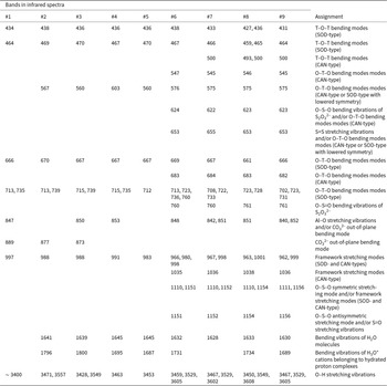
The assignment of IR bands is given in Table 2 in accordance with the recently published data for numerous sodalite and cancrinite (CAN)-type compounds (Chukanov et al., Reference Chukanov, Shchipalkina, Shendrik, Vigasina, Tauson, Lipko, Varlamov, Shcherbakov, Sapozhnikov, Kasatkin, Zubkova and Pekov2022a, Reference Chukanov, Shendrik, Vigasina, Pekov, Sapozhnikov, Shcherbakov and Varlamov2022b, Reference Chukanov, Aksenov and Pekov2023 and references therein) The IR spectra of Experiments 1–5 are typical for halogen-bearing sodalites and, thus, given as supplementary data (Figure S1). In the IR spectra of the products of Experiments 6–9, bands of both SOD- and CAN-type structure types exist (Figure S2), that was also confirmed by powder X-ray diffraction (see below). In all spectra studied (Experiments 1–9), bands in the ranges 1500–1700 cm−1 and 3300–3700 cm−1 are observed, due to the presence of H2O molecules.
The assignment of bands in the IR spectra of Samples 1–9

Table 2 Long description
The table lists infrared band positions for nine experiments and assigns each band to a vibration type. All samples share low-wavenumber T–O–T bending bands around 427 to 438 and 459 to 470, plus an O–T–O bending band near 661 to 670. Mid-range O–T–O bending bands occur broadly from about 702 to 760, with several samples showing multiple peaks in this region. CAN-type features appear mainly in samples 6 through 9, including bands near 493 to 500 and around 545 to 547, and additional bands near 622 to 624 and 653 to 655 that are attributed to sulfate-related bending or sulfur stretching and may overlap with framework modes. Carbonate-related out-of-plane bending is reported near 840 to 853 in several samples, and a separate carbonate band near 873 to 889 appears only in samples 1 to 3. Framework stretching bands are present in every sample near 962 to 1001, while higher framework or sulfate-related stretching bands near 1035 to 1038 and around 1110 to 1156 are reported only for samples 6 to 9. Water bending bands occur near 1628 to 1645 in samples 2 to 9, and hydronium-related bending bands appear in samples 2 to 6, 8, and 9 but are absent in sample 7. Broad O–H stretching bands occur for all samples, ranging from about 3400 to multiple peaks between roughly 3450 and 3608. Assignments note that some bands may reflect mixed origins or lowered symmetry.
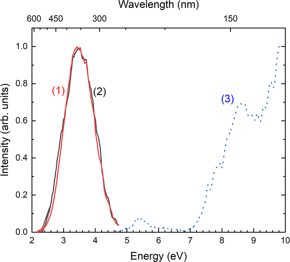
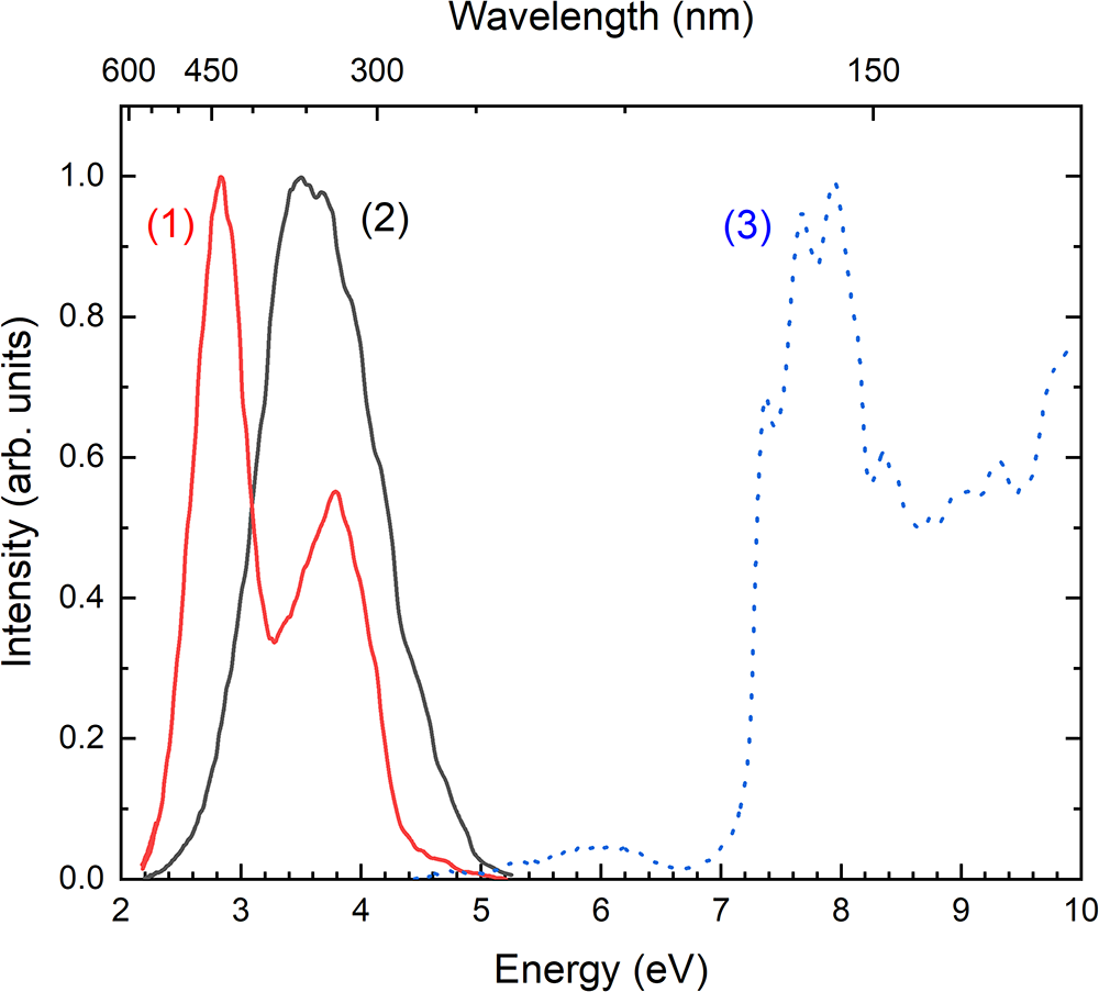
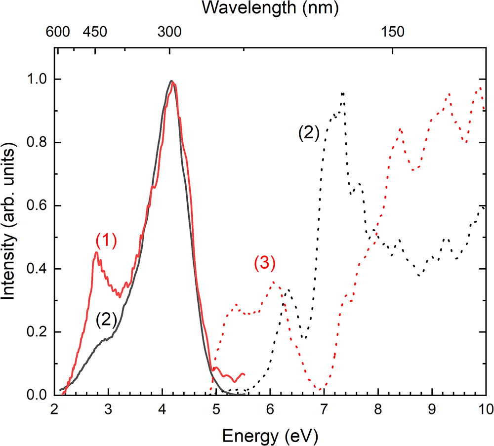
The luminescence and excitation spectra of samples containing iodine (Experiments 2 and 5) are shown in Fig. 2. A luminescence band with a peak at 3.5 eV (355 nm) was observed. The excitation spectrum of this band displays a low-intensity peak with a maximum at 5.4 eV (230 nm), as well as a strong peak at 8.6 eV (144 nm), and a rise in the energy above 10 eV (124 nm). Figure 3 illustrates the luminescence spectra of samples containing bromine (experiments 3 and 4). Sample 3 displayed two bands with peaks at 2.82 and 3.8 eV (440 and 325 nm), whereas the sample obtained in Experiment 4 exhibited a broad band with a peak at 3.6 eV (345 nm). The luminescence excitation spectra of the bands at 2.8 and in the region of 3.6–3.8 eV are similar. They reveal a weak band with a maximum at 6 eV (205 nm), as well as a strong band at ∼7.8 eV (158 nm), and a slight increase in the energy region above 10 eV. Figure 4 presents the luminescence and excitation spectra of samples with mixed bromine and iodine stoichiometry (Experiments 1 and 6). The luminescence spectrum of Experiment 1 consists of a wide band peaking at 4.18 eV, with excitation bands at 6.3 and 7.3 eV. The emission spectrum of the sample from Experiment 6 displays two bands, at 2.48 and 4.2 eV. The excitation spectrum of this luminescence shows bands at 5.5 and 6.0 eV, along with a rise at 7.3 eV to a higher energy region.
Luminescence spectra of Experiments 2 (red curve 1) and 5 (black curve 2) under 8.5 eV excitation. The excitation spectra monitored at 3.5 eV for both samples are the same. It is shown by curve 3.

Luminescence spectra of Experiments 3 (red curve 1) and 4 (black curve 2) under 8 eV excitation. The excitation spectra monitored at 3.2 eV of the both samples are the same and it is given by curve 3.

The luminescence spectra of Experiments 1 (solid curve 1) and 6 (solid curve 2) under 8 eV excitation. The dotted lines demonstrate excitation spectra of sample 1 monitored at 2.78 eV (curve 3) and sample 6 monitored at 4.2 eV (curve 4).

In Experiments 1–5 with only halogen anions, single-phase products with the sodalite-type crystal structure (sp.gr P  $\bar{4}$3n) were obtained whereas the products of Experiments 6–9 are mixtures of sodalite- and cancrinite-type phases (Figure S3). In the Experiments 2, 3 and 5 the partial decomposition was observed and the reflections of nepheline appeared at temperatures ≥ 810°C (Fig. 5). Noteworthy is that the sodalite-type compound obtained in Experiment 5 totally decomposes at 900°C, whereas those from Experiments 2 and 3 are stable up to 1050°C. The thermal evolution of the unit cell parameters of studied sodalite phases is shown in Fig. 6a. All three phases expand almost identically; the expansion intensity increases in the whole temperature range and especially above 750°C, when the sodalite phase starts to decompose and the nepheline reflections appear. Calculated TECs differ slightly for three sodalities in the range from 3–7 × 10⁻⁶ °C–1 (30°C) up to 28–32 × 10–6 °C–1 (730°C) (Fig. 6b) with an average value of TEC for all three compositions of ∼18 × 10–6 °C–1. The sodalite phase from Experiment 5 shows the most intensive increase of TEC from 3 up to 32 × 10–6 °C–1.
The XRD patterns of the sodalite sample (Experiment 5) at different temperatures. The reflections of the sodalite-type phase (sp.gr. P  $\bar{4}$3n) are marked as S, nepheline (sp.gr. P63) as N, unidentified impurity as * and Pt sample holder as Pt.

Figure 5 Long description
The x-axis label is “2Th, CuKa”. The x-axis has labelled ticks at 20, 30 and 40. The y-axis label is “cps”. Multiple overlaid line traces are shown, each representing a different temperature condition indicated by the label “T degree C” with values 30, 300, 600 and 900. Across the x-axis span, each trace contains many narrow peaks. Several peaks are annotated with the letter “N” above the peak positions at multiple x values. The letter “S” appears near the baseline at multiple x values. The text “Pt” labels a tall peak near the highest x values and also labels another peak around the low 40s on the x-axis. One asterisk symbol appears near the baseline at an x value below 20. The peak positions are largely aligned across the different temperature traces, while peak heights vary between traces. The tallest labelled peak is the one marked “Pt” near the highest x values.
Unit-cell parameter of sodalites from Experiments 2, 3 and 5 at different temperatures (a) and their TECs in the range of 30–730°C (b). Errors are smaller than symbols.

Discussion
The influence of the type of cation and anion on the formation and stability of SOD- and CAN-type structures
As our experiments for powder samples show, Ca-bearing SOD-type compounds with halogen-bearing anions are stable up to the temperature of 1050°C, whereas compounds with only alkaline metals (Na, K) decompose in the range 850–900°C. TECs of the latter’s increase more intensively which also indicate their lower stability. It is noteworthy that the relatively low temperature of decomposition among studied sodalite-type compounds was detected for so-called hydroxysodalite Na8[Al6Si6O24](OH)2·4H2O and tetrahydroxyborate sodalite Na8[Al6Si6O24][B(OH)4]2. Their total decomposition occurs at the temperatures of 738°C and 700°C, respectively. The cause of the easier decomposition of these compounds is due to the influence of the dehydration process on the stability of the aluminosilicate framework (Buhl and Luger, Reference Buhl and Luger1990; Khavi et al., Reference Khavi, Sartipi, Gascon, Jansen and Kapteijn2010). Despite the presence of water molecules in synthetic aluminosilicates obtained in our work (Figures S1 and S2), their loss did not decrease drastically the temperature of transformation to nepheline (or its polymorph carnegieite, as was mentioned for hydroxysodalite by Khavi et al. (Reference Khavi, Sartipi, Gascon, Jansen and Kapteijn2010)). In our compounds the removal of extra-framework halogen anions from aluminosilicates should affect the transformation to nepheline (or carnegieite).
In the case of SOD-type compounds synthesized in this work, we suggest that Ca in SOD-type halogen-bearing compounds can play a role in the component that has an influence on the temperature range of their stability. Natural nepheline is usually Ca-depleted (the Ca rarely exceeds 0.25 apfu), that indicates that the nepheline structure has a low affinity towards Ca2+ cations. This may be a cause for the inhibitory role of calcium in the processes of the transformation of SOD-type compounds to nepheline. The research of the influence of different extra-framework cations (K+, Na+ and Ca2+) on the stability and flexibility of the Zeolite A framework (its crystal structure includes so-called SOD-type β-cages), shows that Na and Ca possess no sterical influence on the compression or extension mechanisms of the aluminosilicate framework, whereas K affects the decrease of framework flexibility, and as a result the decrease of its stability in the wide temperature range (Cornelius et al., Reference Cornelius, Price, Wells, Petrik and Sarbaeva2019). In our studied samples, no explicit correlation between the Ca and the Al contents as a suggested charge-balancing mechanism was found; moreover, the calcium content varies in a wide range in the products studied of Experiments 2 and 3. The suggested charge balancing mechanism in these compounds involves incorporation of CO32– and OH– anions in the β-cages as affirmed by infrared and Raman spectroscopy. Thus, we propose the formation of stronger M–O bonding (Ca–O bonds at six-ring sites in the SOD-type crystal structure) in experiments with Ca, that affects the stability of the SOD-type crystal structure. Generally, it should be noted that fine-dispersed powders are more reactive than larger crystals, as was determined, for example, in thermal experiments for the synthetic analogue of sapozhnikovite and its comparison with data on thermal behaviour of sapozhnikovite itself (Chukanov et al., Reference Chukanov, Zubkova, Pekov, Shendrik, Varlamov, Vigasina, Belakovskiy, Britvin, Yapaskurt and Pushcharovsky2022c; Shchipalkina et al., Reference Shchipalkina, Vereshchagin, Chukanov, Gorelova, Bocharov and Pekov2023). Thus, direct comparison of well-shaped crystalline SOD-type compounds with their fine powders in terms of thermal stability would be incorrect.
Our experiments reveal that the type of anion plays an important role in the formation of SOD- or CAN-type feldspathoids synthesized under low-temperature hydrothermal conditions in a highly alkaline medium. In particular, the addition of Na2S2O3 to the initial mixture of halogen-bearing salts results in the appearance of a cancrinite-type compound as the main phase. Despite the low temperature synthesis of the SOD-type phases without S-bearing components, no CAN-type products were formed. The order of Na, K and Ca, that results in formation of the topology of a framework and lowering of symmetry, did not happen. This confirms the suggestion that anionic groups with a special crystallographic direction as S2O32–, C2O42–, CO32– and molecular sulfur of complicated forms as S4, S6 stabilize the CAN-type Al/Si-framework with a wide channel. The small monoatomic anions as Cl–, I– and Br– and isometric tetrahedral groups like SO42– stabilize SOD-type cages.
Luminescence properties
Bromine (Br–) or iodine (I–) ions occupy sodalite cages where they are tetrahedrally coordinated by four sodium (Na+) ions. Violations of stoichiometry can create cavities containing only three Na+ ions and a pair of vacancies, cation and anion ones.
Upon excitation within the interband transition region of sodalite-like materials, exciton-like states form. These states decay, generating free electrons and holes. Subsequent exciton autolocalization leads to defect pair formation: the electron localizes on an anion vacancy, forming an F centre, while the hole localizes on a Br– or I– centre coordinated by either three or four Na+ cations (Denks et al., Reference Denks, Karner and Murk1997). This process typically occurs at low temperatures.
As temperature increases, hole centres migrate through six-membered oxygen rings. At the boundary between cages containing (Na3□Br)3+ and (Na4Cl)3+ units, the bromine atom and the Cl– anion come closer together and a specific hole centre, (BrCl)–, forms. Similar phenomena were previously documented in non-stoichiometric sodalites (Denks et al., Reference Denks, Murk and Vasil’chenko1996) and in crystals with mixed-anion alkaline earth halides (Shalaev, Reference Shalaev, Shendrik, Myasnikova, Bogdanov, Rusakov and Vasilkovskyi2018).
Consequently, a ‘genetic’ pair of electron (F-centre) and hole defects forms, located either in adjacent cavities or within a single cavity. These complexes can manifest as excimer-like defects, such as (Na3□I)3+, or as a paired F-centre and (BrCl)– hole centre, denoted (BrCl)–e. These metastable excimer-like defects can either radiatively recombine or diffuse at elevated temperatures, potentially forming stable colour centres.
Such defect pairs exhibit electron paramagnetic resonance (EPR) activity. F-centres coordinated by four Na+ ions produce EPR spectra with 13 lines, whereas those coordinated by three Na+ ions and a cation vacancy yield spectra with 10 lines (Denks et al., Reference Denks, Vasil’chenko, Korsakov, Kyarner and Seman1988). Hole centres are also detectable by EPR; at liquid helium temperatures, their geometric parameters can be determined from hyperfine splitting measurements (De Pinillos, Reference De Pinillos and Weltner1976).
The observed earlier luminescence at 4.2 eV in iodine-containing and 4.5 eV in bromine-containing sodalites can be attributed to triplet recombination in I− and Br− ions located within [Na4X]3+ clusters, where X is a halogen ion (Kirm et al., Reference Kirm, Demidenko, Denks, Feldbach, Lushchik, Lushchik and Martinson1999). The lower energy luminescence at 3.6 eV in I-sodalites and 3.55 eV in Br-sodalites is attributed to recombinations in the Na3X 2+ complexes. In these cases, the absence of a Na ion between two halogen ions allows for the formation of (I2)−e excimer molecules. Photons with energies of 5.8 and 6.75 eV lead to the formation of triplet and singlet excitations of I− ions. Higher energy photons in the range of 7.0 to 8.5 eV cause direct ionization of I− ions (Lushchik et al., Reference Lushchik, Kirm, Lushchik, Martinson, Nagirnyi and Vasil’chenko2002a). Photons in the range of 8.5 to 19 eV provide excitation and ionization of the aluminosilicate host (Lushchik et al., Reference Lushchik, Kirm, Kudryavtseva, Lushchik, Martinson, Nagirnyi and Vasil’Chenko2002b; Shendrik et al., Reference Shendrik, Kaneva, Pankratova, Pankrushina, Radomskaya, Gavrilenko, Loginova and Pankratov2024). The mixing halogen stoichiometry (Br/I, Br/Cl etc) systems are not well studied. In some mixed Br/I halides the recombination luminescence attributed to (BrI)− complexes has been observed in the region 2.55 eV (Shalaev et al., Reference Shalaev, Shendrik, Myasnikova, Bogdanov, Rusakov and Vasilkovskyi2018).
The luminescence observed in our samples is most probably associated with exciton-like excitations near the halogen anions. This is supported by the fact that the luminescence is excited at relatively low energies, ≈ 5.5−6 eV. These energies are too low to excite the framework, but sufficient to ionize I− or Br− anions. It is worth noting that the high-energy bands of the luminescence spectra of samples 1 – 6 (Figs 2–4) can be attributed to the luminescence of halogen (X −) centres, which are located within a regular sodalite or cancrinite cage. In the samples studied, there is a defect-free cavity Na4X where direct excitation of X − anions occurs.
The low-energy luminescence bands at 2.8−3.2 eV are associated with radiation-induced recombination of the Na3□I complex that arises during synthesis. In the vacancy position, Br or I anions can be present, leading to the subsequent formation and recombination of (BrI)−e excimer-like species in the samples obtained in Experiments 1 and 6 (Fig. 4). In the sample with a sodalite cage (Experiment 1), the formation of (BrI)−e is more efficient compared to samples with a cancrinite cage (Experiment 6), as evidenced by the lower intensity of this band in the sample obtained in Experiment 6. Noteworthy is that the admixture of the salt Na2S·8H2O in experiments on synthesis of Na8(Al6Si6O24)(HS)2 (Chukanov et al., Reference Chukanov, Shchipalkina, Shendrik, Vigasina, Tauson, Lipko, Varlamov, Shcherbakov, Sapozhnikov, Kasatkin, Zubkova and Pekov2022a) results not only in formation of (HS)–, but also S2– and S2∙– radical anions; the latter stimulates the strong orange luminescence. However, in Experiments 6–9, despite the addition of this salt, no evidence of the formation of S2∙– radical anions, the luminescence centres, was detected. In the sample from Experiment 3, the luminescence band at 2.8 eV may be associated with the formation of (BrCl)−e excimer-like molecules (Fig. 3). The peak excitation positions at 8.6 eV for I-containing samples (Experiments 2 and 5), and 7.8 eV for Br-containing samples (Experiments 3 and 4), are related to the excitation of the framework. Thus, it is possible to estimate the width of the bandgap for the microporous samples investigated.
Therefore, altering the anion, cations and synthesis conditions can result in a shift in the position of the intrinsic luminescence band and the threshold for excitation of the luminescence. This suggests the potential for tuning the threshold for defect formation and the positions of the luminescence bands. The samples obtained from Experiments 2 and 5 exhibited the highest excitation threshold for the intrinsic luminescence, while the samples from Experiments 3 and 4, which had the lowest value of a certain parameter, showed the lowest energy excitation threshold. Previous studies have indicated that the excitation threshold for the intrinsic luminescence falls within the range of defect formation in microporous materials, attributed to an excitonic-like mechanism of defect creation (Lushchik et al., Reference Lushchik, Kirm, Lushchik, Martinson, Nagirnyi and Vasil’chenko2002a, Reference Lushchik, Kirm, Kudryavtseva, Lushchik, Martinson, Nagirnyi and Vasil’Chenko2002b; Kaneva et al., Reference Kaneva, Bogdanov, Radomskaya, Belozerova and Shendrik2023). Therefore, the sodalities with a lower value of the specified parameter hold promise as potential generators of free anion radicals, which may arise from the non-radiative decay of electronic excitations (Lushchik et al., Reference Lushchik, Kirm, Kudryavtseva, Lushchik, Martinson, Nagirnyi and Vasil’Chenko2002b; Shendrik et al., Reference Shendrik, Kaneva, Pankratova, Pankrushina, Radomskaya, Gavrilenko, Loginova and Pankratov2024).
The intrinsic luminescence observed in studied aluminosilicates lies in the region of excitation of the 5d-4f emission of Eu2+ centres in sodalite-like systems (Gaft et al., Reference Gaft, Panczer, Nagli and Yeates2008; Lezhnina et al., Reference Lezhnina, Jordan, Klimin, Löns, Koller, Mavrin and Kynast2009). It can be expected that an efficient resonant energy transfer from electronic excitations to Eu2+ ions would occur, especially considering that it is possible to obtain compounds where Eu2+ ions are located in the same cage during synthesis. The intrinsic luminescence effectively excited in a higher energy region and multiplication of electronic excitation occurs (Lushchik et al., Reference Lushchik, Kirm, Kudryavtseva, Lushchik, Martinson, Nagirnyi and Vasil’Chenko2002b). Therefore, the systems investigated in this study are likely to be promising for Eu2+ doping to create effective X-ray phosphors.
Conclusions
Our work clearly reveals that the type of anion plays an important role in the formation of SOD- or CAN-type feldspathoids synthesized under low-temperature hydrothermal conditions in a highly alkaline medium. Anionic groups such as S2O32–, C2O42–, CO32–, as well as molecular sulfur species S4 and S6 stabilize the CAN-type framework with wide channels. The monoatomic anions – Cl–, I–, Br– and equant tetrahedral SO42– stabilize SOD-type cages. Ca-bearing SOD-type feldspathoids with halogen-bearing anions (Cl, I, Br) are stable up to the temperature of 1050°C, whereas such compounds with only alkaline metals (Na, K) decompose in the range 850–900°C. The observed luminescence at 80 K is most likely associated with exciton-like excitations near the halogen anions. In studied samples the luminescence is excited at 5.5−6 eV. The low-energy luminescence bands at 2.8−3.2 eV are associated with radiation-induced recombination of a Na3□I complex that arises during synthesis. In the vacancy position, Br or I anions can be present, leading to the subsequent formation and recombination of (BrI)−e excimer-like species. The luminescence band at 2.8 eV may be associated with the formation of (BrCl)−e excimer-like molecules. We propose the systems investigated in this study are likely to be promising for Eu2+ doping to create effective X-ray phosphors.
Supplementary material
The supplementary material for this article can be found at https://doi.org/10.1180/mgm.2025.10158.
Acknowledgements
XRD studies were done at the Center for X-Ray Diffraction Studies of the Research Park of St. Petersburg State University within the project AAAA-A19-119091190094-6. Luminescence spectra were studied as part of the state assignment of A.P. Vinogradov Institute of Geochemistry SB RAS (Irkutsk, Russia) project No. 0284-2021-0004.
Competing interests
The authors declare none.