I. INTRODUCTION
Titanium (Ti) and its alloys are extensively used in orthopedic, dental, and other hard tissue replacement applications, although the low bone bonding ability of Ti is an important concern for the stability of these implants.Reference Gomez-Vega, Saiz and Tomsia1 Many clinical studies showed that due to limited bioactivity of these metallic implants, dense fibrous tissue forms at the implant–bone interface,Reference Gomez-Vega, Saiz and Tomsia1,Reference Baino and Verné2 which hinder direct bone–implant bonding. To improve the bioactivity and osseointegration, Ti implants are generally coated with bioactive materials such as calcium phosphates and bioactive glasses.Reference Gomez-Vega, Saiz and Tomsia1–Reference Nag, Paital, Nandawana, Mahdak, Ho, Vora, Banerjee and Dahotre5 These bioactive surfaces promote formation of carbonate-substituted hydroxyapatite (HA)-like layer and ions dissolute from surface encourage osteoblast-like cells proliferation and promote osseointegration.Reference Baino and Verné2,Reference Cacciotti3 The clinical application of bioactive glass coatings is limited compared to HA coating, which is due to its degradation over time in physiological conditions.Reference Baino and Verné2,Reference Cacciotti3 Nowadays laser-based surface modification of Ti with calcium phosphate and bioactive glass coatings have been studied by several researchers.Reference Cacciotti3–Reference Yang, Serpersu, He, Paital and Dahotre7 Addition of such bioactive reinforcements can potentially improve the osteoconductivity of bioinert metallic implants. Similar observations have been reported with laser-processed calcium phosphate coatings on Ti and 316L stainless steel.Reference Roy, Balla, Bandyopadhyay and Bose8,Reference Balla, Das, Bose, Janaki Ram and Manna9 Laser deposition is also being applied to fabricate porous coatings of niobium and tantalum on Ti with high adhesion and bioactivity.Reference Zhang, Cheng, Yao, Wei, Han, Shi, Wei and Zhang10,Reference Balla, Banerjee, Bose and Bandyopadhyay11 Laser-processed coatings found to have unique advantages like strong metallurgical bonding, absence of sharp interfaces, controllable concentration of reinforcements, homogeneous, fine and nonequilibrium microstructures with minimum dilution.Reference Balla, Das, Bose, Janaki Ram and Manna9,Reference Balla, Banerjee, Bose and Bandyopadhyay11
Since 2004, strontium (Sr) has gained attention due to its positive role in bone remodeling. Sr is an alkaline earth metal, present nearly 0.035% of total calcium content of the mineral phase of bone.Reference Bose, Fielding, Tarafder and Bandyopadhyay12 Sr is also a trace element in the human body and follows almost similar metabolic pathway of calcium. Several studies showed that Sr ions stimulate the calcium sensing receptor (CaSR) in osteoblasts which activate the production of osteoprotegerin (OPG).Reference Bose, Fielding, Tarafder and Bandyopadhyay12 Thus the Sr ion stimulates the bone forming osteoblasts and suppresses the bone resorption and osteoclastogenesis.Reference Bose, Fielding, Tarafder and Bandyopadhyay12–Reference Braux, Velard, Guillaume, Bouthors, Jallot, Nedelec, Laurent-Maquin and Laquerrière14 Due to its dual mode of action in bone remodeling, Sr ranelate, a Sr-based drug, has been used as an effective treatment for osteoporosis.Reference Bose, Fielding, Tarafder and Bandyopadhyay12 Several investigations have been carried out to utilize the stimulatory effects of Sr on bone healing and increased implant osseointegration by incorporating Sr into various biomaterials.Reference Capuccini, Torricelli, Sima, Boanini, Ristoscu, Bracci, Socol, Fini, Mihailescu and Bigi15–Reference Xia, Lindahl, Lausmaa, Borchardt, Ballo, Thomsen and Engqvist17 Sr incorporation found to promote the attachment and proliferation of osteoblast cells on calcium phosphate bone substitutes and HA coatings in vitro.Reference Capuccini, Torricelli, Sima, Boanini, Ristoscu, Bracci, Socol, Fini, Mihailescu and Bigi15,Reference Ni, Chiu, Lu, Wang, Zhang, Hao, Li, Lam, Lu and Luk16 In another approach, Sr was incorporated on the surface of metallic implants to enhance the osteoconductivity of bone interfacing implants.Reference Xia, Lindahl, Lausmaa, Borchardt, Ballo, Thomsen and Engqvist17–Reference Zhao, Wang, Huo, Zhang, Wang, Zhang, Wu and Chu20 Forsgren et al. showed different release rates of Sr ions from a film of strontianite (SrCO3) formed on sodium titanate.Reference Forsgren and Engqvist18 Recent studies showed that strontium titanate (SrTiO3) nanotube arrays can be fabricated on Ti using a simple hydrothermal treatment of anodized titania nanotubes.Reference Xin, Jiang, Huo, Hu and Chu19,Reference Zhao, Wang, Huo, Zhang, Wang, Zhang, Wu and Chu20 This SrTiO3 nanotube array released Sr at a slower rate for long time and demonstrated good biocompatibility.Reference Zhao, Wang, Huo, Zhang, Wang, Zhang, Wu and Chu20 Andersen et al. prepared Ti–Sr–O coatings by a magnetron co-sputtering process and demonstrated that the Sr released from the coating can significantly enhance the peri-implant bone formation after four weeks of implantation in the Wistar rat model.Reference Andersen, Offermanns, Sillassen, Almtoft, Andersen, Sørensen, Jeppesen, Kraft, Bøttiger, Rasse, Kloss and Foss21 Some studies showed that perovskite structure-like calcium titanate (CaTiO3) act as the apatite growth promoter.Reference Coreno and Coreno22 The CaTiO3 phases were commonly observed in laser-deposited HA coating on Ti.Reference Roy, Balla, Bandyopadhyay and Bose8 As per literature, no report is available till date on the use of lasers to incorporate Sr into Ti for biomedical applications. Due to its high reactivity, pure Sr cannot be incorporated in Ti. We hypothesize that the high temperature stability of Sr containing compound namely SrTiO3 can be effectively incorporated into the Ti surface using direct laser deposition. In this work, Ti and SrTiO3 premixed powders were melted using a high power laser to form a SrTiO3-reinforced Ti matrix coatings on the Ti substrate. Such composite coatings can have reacted and unreacted Sr–Ti–O phases. In the presence of physiological solution, SrTiO3-incorporated Ti coating can release Sr ions, which can potentially improve the bioactivity of Ti implants in vivo. Therefore, in the present article, we have investigated the feasibility of laser deposition of SrTiO3-reinforced Ti coatings and evaluated their microstructures, electrochemical behavior, and cell–materials interactions in vitro.
II. EXPERIMENTAL
A. Composite coatings fabrication
Commercially pure Ti (CP-Ti) powder (ATI Powder Metals, Pittsburgh, Pennsylvania) of 50–150 μm size and SrTiO3 powder of 2–10 μm particle sizes were used as feedstock powders. In the present work, the SrTiO3 powder was prepared using strontium carbonate (SrCO3, Sigma-Aldrich, Steinheim, Germany) and Ti dioxide (TiO2, Sigma-Aldrich, Steinheim, Germany) powder. The SrCO3 and TiO2 powders in stoichiometric ratio were mixed thoroughly using ball-mill in ethyl alcohol medium and zirconium balls were used as grinding media. The milled powders were dried and fired at 1200 °C for 2 h in air. After calcinations, powders were ground and sieved to achieve SrTiO3 powder with a size between 2 and 20 µm. The synthesized SrTiO3 powders at varying concentrations, from 5 to 20 wt%, were thoroughly mixed with Ti powder using dilute polyvinyl alcohol solution to create premixed powders. The premixed powders were dried in a vacuum oven and sieved through 100 and 270 mesh. The premixed powders were then poured into the powder feeder of laser engineered net shaping (LENS™). A 0.5 kW continuous wave ytterbium-doped fiber laser equipped with LENS™-MR7 (Optomec Inc., Albuquerque, NM) was used to deposit 2 layers on the Ti substrate using a constant powder feed rate of 4.3 g/min. In the laser melt injection process, argon (Ar) carrier gas was used to inject the premixed powders into the molten pool created by the laser. The molten Ti and premixed powder solidify rapidly as the laser head moves on. A detailed description of the operation and capabilities of LENS™ can be found elsewhere.Reference Das, Balla, Kumar and Manna23 The coatings containing 10 wt% SrTiO3 found to be defect-free at optimized laser powers and scan speeds as shown in Table I. All depositions were carried out in a glove box containing an Ar atmosphere with O2 content less than 10 ppm.
Premixed powder compositions, laser parameters, and deposited coating thickness.

B. Microstructural, mechanical, and electrochemical behavior
Microstructural characterization of the coatings was performed using a scanning electron microscope (Phenom proX, Eindhoven, Netherlands) equipped with an energy-dispersive spectrometer. An optical microscope (BX51, Olympus, Tokyo, Japan) was used to study defects in the coatings. An X’Pert Pro MPD diffractometer with X’Celerator (PANalytical, Almelo, the Netherlands) operating at 45 kV and 40 mA with Ni-filtered Cu Kα radiation was used to determine the constituent phases in the coatings. Silicon was used as a standard for instrumental broadening correction. Vickers microhardness was measured (ESEWAY, W4303, Camberley, United Kingdom) on the top surface and cross-section of the coatings using a load of 500 g for 15 s and an average value of 15 measurements were reported. The profile of hardness across the coating thickness was determined by applying 500 g load for 10 s, and an average of ten measurements for each sample were reported. The wettability of the composite coating surfaces was determined by measuring contact angles using the sessile drop method. For contact angle measurement, the samples were mirror polished on velvet cloth using 0.05 µm Al2O3 suspension. An average of 10 measurements are reported for each sample.
Potentiodynamic polarization tests, conforming to ASTM G59, were performed in Hank’s balanced salt solution, at room temperature, using a potentiostat (SP300, Bio-Logic SAS, Seyssinet-Pariset, France). A set of composite coating and CP-Ti surfaces were polished with 1 μm suspension of Al2O3 powder on velvet cloth and the polarisation behavior was compared with the as-deposited coating surface. All potentials were measured with respect to a saturated calomel electrode (SCE) reference electrode and platinum was used as the counter electrode for current measurement. An initial delay of 30 min prior to polarization was applied to the specimen to reach a steady-state condition at open-circuit potential (OCP). After that, the potential was increased at a rate of 10 mV/min, starting from −0.25 V to +1.6 V versus OCP. From the polarization curve, the corrosion potential (E corr versus SCE) and corrosion current density (I corr) were extracted by Tafel extrapolation.
C. In vitro Sr2+ release and cell–material interactions
The Sr2+ and Ti2+ ion release from the composite coatings were measured using inductively coupled plasma (ICP; Spectra Analytical Instrument GmbH, Kleve, Germany). The coated samples, of size 10 × 10 mm square, were ground using fine SiC grinding papers followed by ultrasonic cleaning in alcohol. After cleaning, the samples were immersed in phosphate-buffered solution (PBS) for 1, 2, 4, and 6 days at 37 °C and the extracted solutions were used for ion release study.
In vitro cytotoxicity and cell proliferation was evaluated on SrTiO3-reinforced composite coatings and CP-Ti samples (control) by direct cell viability assay. Before the cell culture study, all the samples (10 × 10 mm square) were ground using 1200 series of SiC grinding papers to eliminate surface roughness effect. The polished samples were washed with water followed by alcohol and sterilized in autoclave at 121 °C for 30 min. In direct cell viability assay, human osteoblast-like cells (MG63) (NCCS, Pune, India) were seeded on polished surfaces. Cells were incubated for 2, 4, and 6 days of culture periods in a minimum essential medium, supplemented with 10% of fetal calf serum, 1% antibiotic–antimycotic solution in a humidified atmosphere at 37 °C and with 5% CO2. The MTT [3-(4,5-dimethylthiazol-2-yl)-2,5-diphenyl tetrazolium bromide] assay was used to evaluate the cell viability. The details of MTT assay protocol can be seen elsewhere.Reference Das, Bhattacharya, Dittrick, Mandal, Balla, Kumar, Bandyopadhyay and Manna24
The morphology of the cells was observed using SEM after 2 days of incubation on the test samples. The cells adhering to the samples were washed with 0.1 M PBS and fixed with 2% paraformaldehyde/2% glutaraldehyde in 0.1 M phosphate-buffered saline overnight at 4 °C. Fixed samples were then dehydrated in an ethanol series 30, 50, 70, 95, and 100% three times. Dried samples were then gold-coated and observed under an SEM. Statistical analysis was performed on the data using Student’s t-test and P < 0.05 was considered statistically significant.
III. RESULTS AND DISCUSSION
A. Microstructure and phase analysis
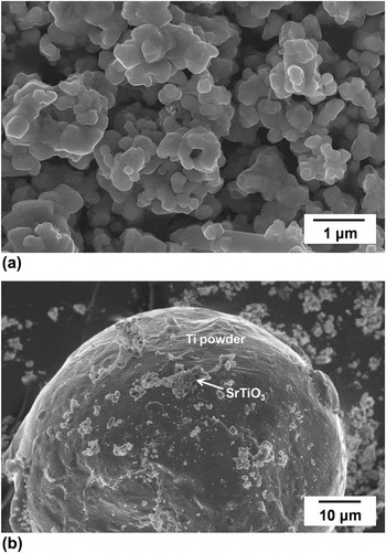
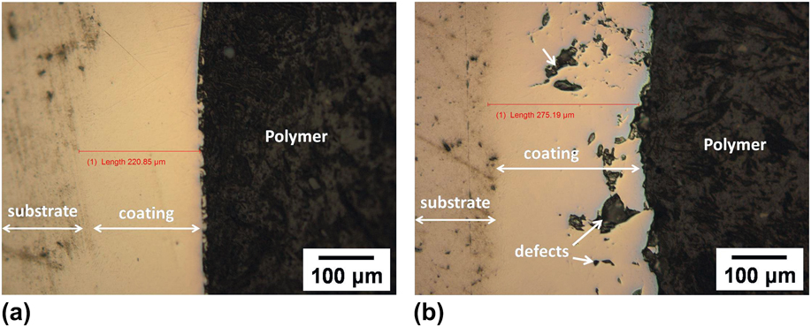
Typical SEM micrographs of SrTiO3 powder and Ti–10 wt% SrTiO3 premixed powders showing their morphologies are presented in Fig. 1. The premixed powder consists of coarse spherical CP-Ti powder coated with fine SrTiO3 powder. Earlier studies showed that laser deposition of premixed powders can improve homogeneous dispersion of the ceramic phase in the coating.Reference Das, Balla, Kumar and Manna23,Reference Das, Bhattacharya, Dittrick, Mandal, Balla, Kumar, Bandyopadhyay and Manna24 Figure 2 shows the cross-sectional optical microstructure of laser-processed Ti–SrTiO3 composite coatings. The microstructure of the composite coatings containing 15 wt% SrTiO3, shown in Fig. 2(b), exhibited cracks and gross defects. It is obvious that with increasing ceramic concentration in the metal matrix, toughness and thermal shock resistance decreases. The laser-processed coatings experience thermal shock due to fast heating and cooling rate, which generates high residual stresses. Due to deteriorating toughness of the coating containing 15 wt% SrTiO3 reinforcement, unable to withstand the high residual stresses leads to cracks and gross defects. However, the composite coatings containing 10 wt% SrTiO3 were relatively defect-free and showed good metallurgical bonding between the deposits and the substrate. It can be inferred from these microstructures that increasing the SrTiO3 concentration from 10 to 15 wt% in the premixed powder, the coatings become brittle therefore less endurance to absorb thermal shock-related stresses, resulted in formation of cracks. Therefore, in the present investigation, Ti–10 wt% SrTiO3 powder was used to fabricate composite coatings using different laser powers and scan speeds as shown in Table I. The average composite coating thickness was measured using cross sectional microstructures and the thickness was 210 ± 40 and 620 ± 50 μm for Ti-10ST-200/20 and Ti-10ST-250/15 coatings, respectively. In general, the laser-deposited layer thickness varies with laser energy input which is influenced by the laser power and scan speed.Reference Das, Bhattacharya, Dittrick, Mandal, Balla, Kumar, Bandyopadhyay and Manna24 In the present investigation, the coating thickness increased with laser energy input and the composite coatings with 10 wt% SrTiO3 prepared using 200 W and 20 mm/s (Ti-10ST-200/20) with a laser energy input of 20 J/mm2 (Table I) showed a coating thickness of 210 ± 40 μm, which increased to 620 ± 50 μm when the laser energy input was increased to 33.3 J/mm2 in Ti-10ST-250/15 coatings. Since all the coatings were prepared with a constant powder feed rate of 4.3 g/min, increasing the laser energy input results in bigger melt pools with higher melt temperatures on the substrate, which can accommodate and melt more amount of premixed powder that is fed to the melt pool leading to thicker coatings than observed with the coatings prepared at lower energy inputs.
SEM image of powder morphology (a) SrTiO3 powders and (b) Ti–10 wt% SrTiO3 premixed powders.

Cross-sectional optical microstructures of Ti-based composite coatings with varying SrTiO3, deposited at 200 W laser power and 20 mm/s scan speed (a) nearly defect-free coating, containing 10 wt% SrTiO3, (b) pores and cracks in coating containing 15 wt% SrTiO3.
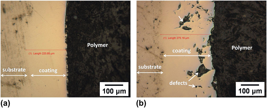
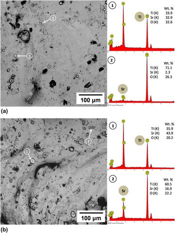
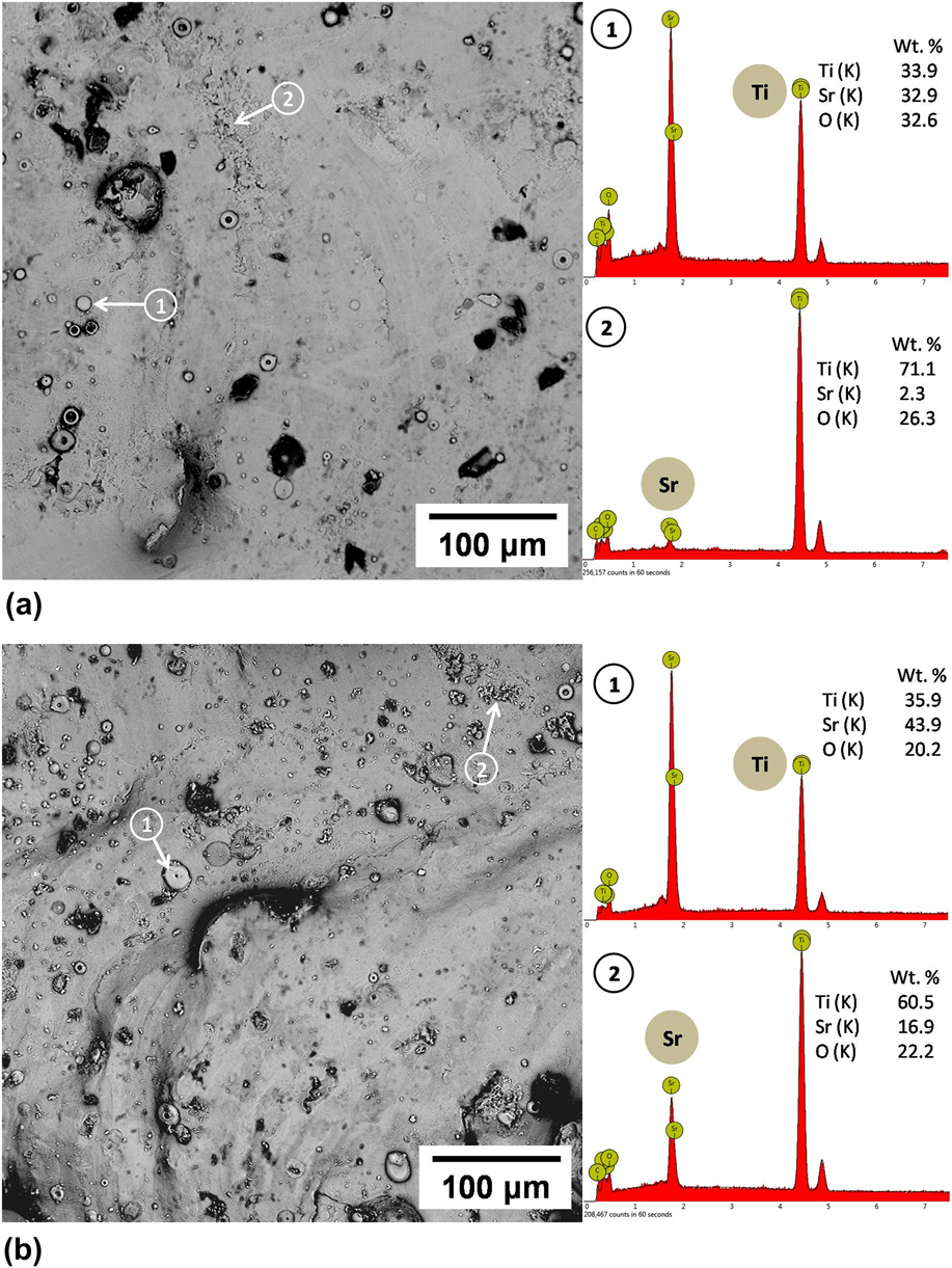
Figure 3 shows the top surface morphology of the 10 wt% SrTiO3 reinforced composite coatings deposited using different laser powers and scan speeds. The surface roughness of these composite coatings varied between 11.6 ± 1.6 μm and 18.3 ± 1.6 μm for Ti-10ST-200/20 and Ti-10ST-250/15 coatings, respectively. The EDX analysis clearly showed the presence of Sr containing ceramic reinforcement phases on the coating’s surface. The reinforcing phases, marked as spot “1” in both micrographs, were found to be depleted SrTiO3 phase surrounded by the Ti matrix phase. The top surface micrograph of the Ti-10ST-250/15 coating showed high concentration of such reinforcing phases compared to Ti-10ST-200/20 coatings due to the decrease in the scan speed. The amount of powder injected into the molten pool increases with the decrease in the laser scan speed leading to high concentration of reinforcements in the composite coatings deposited at slow scan speeds.
Top surface microstructures of laser-processed 10 wt% SrTiO3-reinforced composite coating (a) Ti-10ST-200/20 and (b) Ti-10ST-250/15.
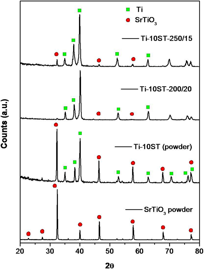
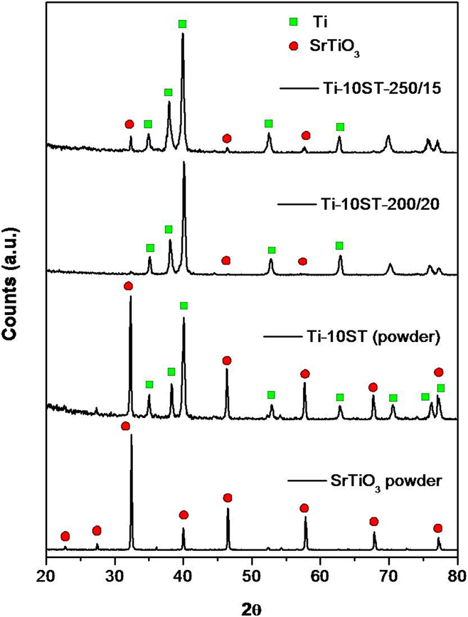
The X-ray diffraction (XRD) patterns of synthesized SrTiO3 powder, premixed powder, and laser-processed composites coatings containing 10 wt% SrTiO3 are shown in Fig. 4. The XRD analysis confirmed that the synthesized powder contains pure SrTiO3 phase, with no residue of reactant chemicals. The premixed powder showed both SrTiO3 and Ti phases. The XRD results of polished surfaces of laser-deposited composite coatings showed predominant peaks corresponding to α-Ti and small peaks of SrTiO3. The SrTiO3 peaks in the Ti-10ST-250/15 coatings were more intense than those observed in the Ti-10ST-200/20 coatings and were primarily due to the increase in the concentration of the reinforcing phase in these coatings. The crystallite size of initial SrTiO3 powder and Ti crystallite size in the as-deposited coating was measured using the Scherrer equation. The crystallite size of SrTiO3 powder was 340 nm and Ti crystallite size was 366 and 414 nm for Ti-10ST-200/20 and Ti-10ST-250/15 coatings, respectively.
XRD patterns of synthesized SrTiO3 powder, Ti–SrTiO3 premixed powder and laser-processed SrTiO3-reinforced Ti composite coatings.
B. Hardness, contact angle, and electrochemical behavior
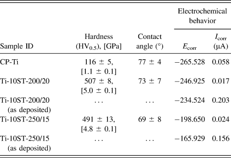
Table II summaries the average top surface hardness of laser-processed SrTiO3-reinforced Ti composite coatings. The experimental data indicate that the hardness of the coated surface was significantly higher than bare Ti. The high hardness in the coating is due to the presence of the hard ceramic phase in the metal matrix. Irrespective of laser parameters, the average hardness of 10 wt% SrTiO3-reinforced coatings was between 491 and 507 HV. The laser parameters had no significant influence on the hardness of these composite coatings.
Average top surface hardness (HV0.5), contact angle, and electrochemical properties of CP-Ti and SrTiO3-reinforced Ti matrix composite coatings.
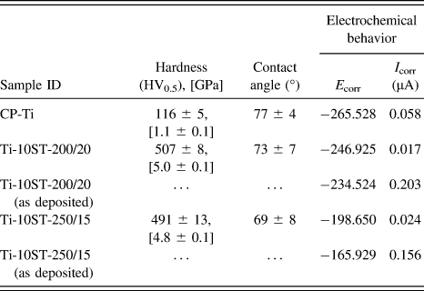
The wettability behavior of the biomaterial surface influences the fluid film layer and cell–material interactions. The hydrophilic nature of the surface is beneficial for cell attachment and proliferation.Reference Balla, Banerjee, Bose and Bandyopadhyay11 The contact angles of water on CP-Ti and composite coatings are shown in Table II. The contact angle values measured on the coated surfaces were marginally lower than the Ti surface. The lower contact angles of composite coatings indicate their hydrophilic nature which is beneficial for cell attachment and proliferation.
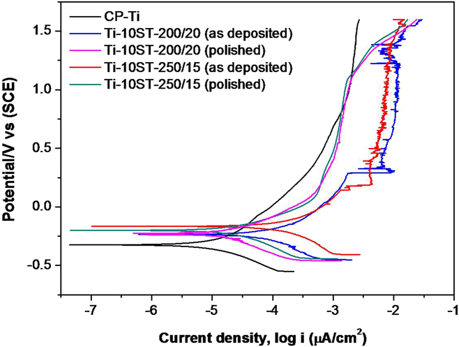
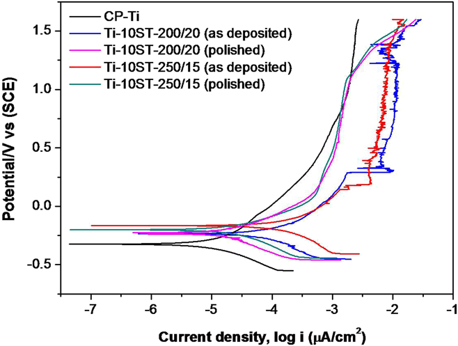
The electrochemical behavior of laser-processed SrTiO3-reinforced Ti matrix composite coatings was assessed using Hank’s solution. Figure 5 shows the potentiodynamic polarisation curves of Ti and composite coatings in polished and as-deposited conditions. The results showed higher corrosion potential (E corr) for composite coatings compared to the bare CP-Ti. Higher E corr of composite coatings indicates their better corrosion resistance than the CP-Ti substrate. Such an improvement in the corrosion resistance of Ti was also observed by several others when a laser-based ceramic-reinforced composite coating was applied.Reference Weng, Chen and Yu25,Reference Das, Balla, Kumar and Manna26 The as-deposited coatings also showed higher corrosion current densities (I corr) suggesting their high corrosion rates compared to respective polished surfaces. The high corrosion rate and pitting of the as-deposited coatings could be due to their rough surface leading to high surface area and leaching of Sr ions from the reinforcing ceramic phase. However, the laser parameters had no significant influence on the electrochemical behavior of these composite coatings.
Potentiodynamic polarization curves of bare Ti and 10 wt% SrTiO3-reinforced composite coatings, obtained in Hank’s solution.
C. In vitro Sr2+ release and cell–material interactions
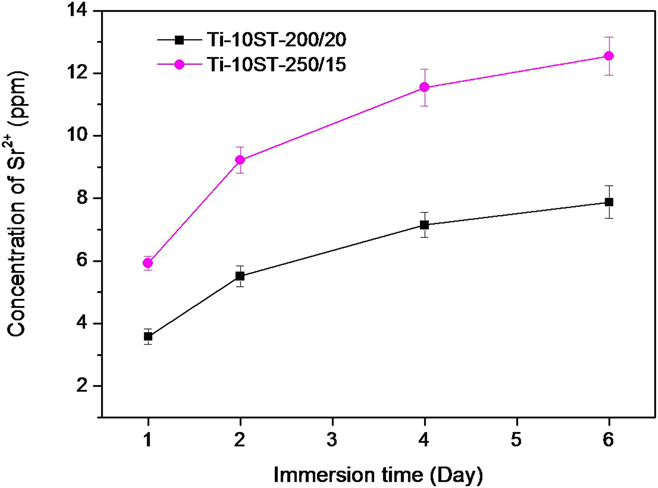
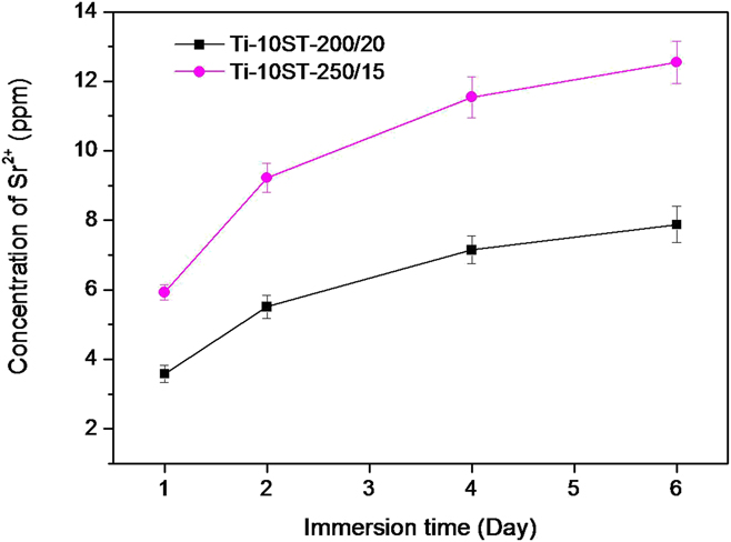
Figure 6 shows the in vitro ion release from the composite coatings. The ICP analysis showed the presence of Sr2+ but no Ti2+ present in the PBS solution. The Sr2+ ion release was sharp up to 2 days and the release rate found less after 4 days of immersion. The Sr2+ ion release from Ti-10ST-250/15 composite coating was more than Ti-10ST-200/20. In case of Ti-10ST-250/15, the coating was deposited at slower scan speed leads to more amount of powder injection into the molten pool. Due to more powder, the unreacted SrTiO3 phases was more in Ti-10ST-250/15 coating which was observed in SEM micrographs also. The higher amount of Sr2+ release in PBS solution is due to the presence of more SrTiO3 in Ti-10ST-250/15 coating. The release of Sr2+ in vivo is desirable which can significantly influence the osteoconductivity of these coatings.Reference Andersen, Offermanns, Sillassen, Almtoft, Andersen, Sørensen, Jeppesen, Kraft, Bøttiger, Rasse, Kloss and Foss21
Concentration of Sr2+ ion release from composite coatings in PBS solution at different immersion times.
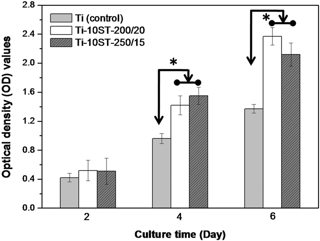
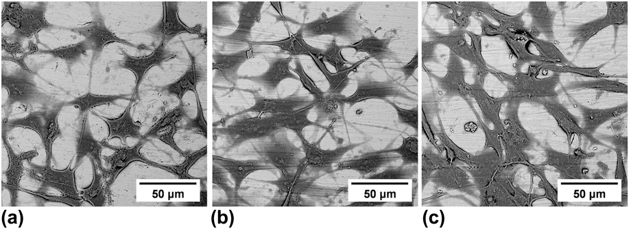
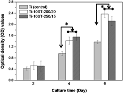
Cell viability and cell proliferation on SrTiO3-reinforced composite coatings were carried out using human osteoblast-like cells (MG63). MTT assay was carried out to assess the cell viability on different surfaces. CP-Ti, one of the most widely used metallic biomaterial, was used as a control sample to compare the cell–material interactions. In MTT assay, optical density of the solution was measured to quantify the cell viability/living cell count. A higher optical density represents a higher concentration of living cells on the surface. Figure 7 compares the MG63 cell viability on Ti and the composite coatings surface after 2, 4, and 6 days of culture. The results indicate that the cells are viable on the composite coatings surface, irrespective of laser processing parameters. The amount of living cells on the coating surfaces was significantly higher (P < 0.05) than that observed on the CP-Ti only after 4 days of culture. The cell proliferation was significantly higher on the SrTiO3-reinforced composite coatings compared to the control surface. The number of cells increased significantly on composite coating surfaces with culture duration. The results indicate that the presence of Sr significantly enhanced the differentiation and proliferation of osteoblast cells. Furthermore, the MG63 cell morphologies on the Ti and coating surfaces were studied after 2 days of culture, shown in Fig. 8. The cells found to grow on the Ti surface as well as on the composite surfaces and showed good adhesion and spreading. The filopodia and lamellipodia were well-established and the cells covered the entire surface. The composite coating surfaces also revealed the presence of ceramic particles which might influence the cellular activity on the coated surface. The earlier studies showed that in aqueous medium, Sr can leach from SrTiO3 as partially soluble SrO and insoluble TiO2.Reference Xin, Jiang, Huo, Hu and Chu19,Reference Anderson, Kahn, LaBone, Brown and Harris27
 $${\rm{SrTi}}{{\rm{O}}_3} + {{\rm{H}}_2}{\rm{O}} \to {\rm{S}}{{\rm{r}}^{2 + }} + 2{\rm{O}}{{\rm{H}}^ - } + {\rm{Ti}}{{\rm{O}}_2}\quad .$$
MTT assay of MG63 cells observed on Ti and composite coating surfaces as a function of culture time.

SEM micrographs of MG63 cells after 2 days of culture (a) Ti control, (b) Ti-10ST200/20 coating, and (c) Ti-10ST250/15 coating.

It is clear from in vitro results that the SrTiO3-reinforced composite coatings have no cytotoxicity and showed significantly high bioactivity compared to the commercial Ti. Numerous in vitro and in vivo studies have reported the stimulatory effects of Sr on osteoblastic cell responses.Reference Andersen, Offermanns, Sillassen, Almtoft, Andersen, Sørensen, Jeppesen, Kraft, Bøttiger, Rasse, Kloss and Foss21,Reference Park, Kim, Kim, Jang, Song and Hanawa28 Sila-Asna et al. observed that osteoblastic differentiation of human bone marrow stromal cells enhanced in the presence of Sr in the cell culture medium.Reference Sila-Asna, Bunyaratvej, Maeda, Kitaguchi and Bunyaratavej29 Several researchers have suggested the probable stimulatory mechanism where Sr ion stimulates the CaSR in osteoblasts which increase the production of OPG.Reference Bose, Fielding, Tarafder and Bandyopadhyay12,Reference Park, Kim, Kim, Jang, Song and Hanawa28 Through a CaSR-mediated mechanism, Sr stimulates bone formation by increasing preosteoblastic cell proliferation and differentiation and by suppressing osteoclast differentiation. The sustain release of Sr2+ ions from the SrTiO3-reinforced composite coating is believed to enhance the biological response of Ti-based implant for orthopedic and dental applications.
IV. CONCLUSIONS
In the current study, SrTiO3-incorporated Ti matrix composite coatings were successfully deposited on the CP-Ti substrate using LENS™. The composite coating containing 10 wt% SrTiO3 was defect-free and exhibited good metallurgical bonding with the substrate. EDX analysis on the top surface of the as-deposited coatings showed the presence of SrTiO3 phases strongly bonded with the Ti matrix. XRD of the polished coating sample showed Ti as the main phase and small peaks of SrTiO3. The average hardness of composite coatings was significantly higher than the Ti substrate. Electrochemical behavior of the composite coatings was slightly better than the Ti surface. All the coated samples showed relatively more hydrophilic compared to the Ti surface. The SrTiO3-reinforced composite coatings also found to be nontoxic and showed significantly high bioactivity compared to the commercial Ti.
ACKNOWLEDGMENTS
The authors would also like to acknowledge the financial support from the Council of Scientific and Industrial Research (CSIR), India, through project ESC 0103. Mr. Souvik Sahoo is grateful to the Director, CSIR-CGCRI, Kolkata, for granting permission to carry out the entire experimental work at CSIR-CGCRI.