Implications
Orexin A (OXA) and B (OXB) are believed to be a link between energy metabolism and the reproductive system. Our recent studies indicated expression of the orexin system in the porcine uterus. It was also demonstrated that the uterus is a steroidogenic organ producing steroids which affect uterine functions. This paper provides pioneering information about the influence of OXB on testosterone (T), oestrone (E1) and oestradiol (E2) production in the porcine uterus during early gestation, a crucial period for implantation. The data obtained in this study provide a better understanding of the mechanisms that control energy homoeostasis and reproduction, which may be helpful in animal breeding practice.
Introduction
Orexin A and B – also known as hypocretin 1 and 2 – are hypothalamic neuropeptides which were discovered simultaneously in 1998 by two independent laboratories. Both orexins are derived by proteolytic cleavage from a common 130-amino acid precursor molecule, prepro-orexin (PPO), which is mainly synthesized in neurons within and around the lateral and posterior hypothalamus. Orexin A is a 33-amino acid peptide, whereas OXB consists of 28 amino acids of which 46% are identical with OXA (de Lecea et al., Reference Dehan, Canon, Trooskens, Rehli, Munaut, van Criekinge and Delvenne1998; Sakurai et al., Reference Sakurai, Amemiya, Ishii, Matsuzaki, Chemelli, Tanaka, Williams, Richardson, Kozlowski, Wilson, Arch, Buckingham, Haynes, Carr, Annan, McNulty, Liu, Terrett, Elshourbagy, Bergsma and Yanagisawa1998). The amino acid sequence of OXA is highly conserved among mammalian species. The amino acid sequence of OXA is identical in pigs, humans, rats and cows, and the sequence of OXB in pigs and humans differs by only one amino acid residue (Sakurai et al., Reference Sakurai, Amemiya, Ishii, Matsuzaki, Chemelli, Tanaka, Williams, Richardson, Kozlowski, Wilson, Arch, Buckingham, Haynes, Carr, Annan, McNulty, Liu, Terrett, Elshourbagy, Bergsma and Yanagisawa1998; Dyer et al., Reference Dyer, Touchette, Carroll, Allee and Matteri1999). Orexin B has much lower lipophilicity and crosses the blood–brain barrier slower than OXA (Kastin and Akerstrom, Reference Kastin and Akerstrom1999). Their biological action is mediated by binding two G-protein-coupled receptors: orexin receptor type 1 (OX1R) and orexin receptor type 2 (OX2R). Orexin A is a more selective ligand for OX1R, whereas OX2R binds both orexins with similar affinity (de Lecea et al., Reference Dehan, Canon, Trooskens, Rehli, Munaut, van Criekinge and Delvenne1998; Sakurai et al., Reference Sakurai, Amemiya, Ishii, Matsuzaki, Chemelli, Tanaka, Williams, Richardson, Kozlowski, Wilson, Arch, Buckingham, Haynes, Carr, Annan, McNulty, Liu, Terrett, Elshourbagy, Bergsma and Yanagisawa1998).
Functionally, both orexins have been related to the control of food intake and energy balance as well as arousal and sleep regulation (de Lecea et al., Reference Dehan, Canon, Trooskens, Rehli, Munaut, van Criekinge and Delvenne1998; Sakurai et al., Reference Sakurai, Amemiya, Ishii, Matsuzaki, Chemelli, Tanaka, Williams, Richardson, Kozlowski, Wilson, Arch, Buckingham, Haynes, Carr, Annan, McNulty, Liu, Terrett, Elshourbagy, Bergsma and Yanagisawa1998). Moreover, it seems that orexins create an integrative link between energy homoeostasis and reproduction. This hypothesis is supported by numerous studies indicating their involvement in the control of the hypothalamic–pituitary–gonadal (HPG) axis (Porka-Heiskanen et al., Reference Porka-Heiskanen, Kalinchuk, Alanko, Huhtaniemi and Stenberg2004; Silveyra et al., Reference Silveyra, Lux-Lantos and Libertun2007; Nitkiewicz et al., Reference Nitkiewicz, Smolinska, Przala and Kaminski2010, Reference Nitkiewicz, Smolinska, Maleszka, Chojnowska and Kaminski2014; Kaminski et al., Reference Kaminski, Smolinska, Nitkiewicz and Przala2010a and Reference Kaminski, Smolinska, Nitkiewicz and Przala2010b) suggesting both indirect orexin effect on reproductive functions through regulation of higher branches of the HPG axis – the hypothalamus and pituitary, and direct influence on gonadal activity. Both in vivo and in vitro studies strongly suggest the effect of orexins on gonadoliberin and LH secretion in rats, pigs and humans (Russell et al., Reference Russell, Small, Kennedy, Stanley, Seth, Murphy, Taheri, Ghatei and Bloom2001; Kok et al., Reference Kok, Roelfsema, Overeem, Lammers, Frolich, Meinders and Pijl2004; Barb and Matteri, Reference Barb and Matteri2005; Cataldi et al., Reference Cataldi, Lux Lantos and Libertun2014). In addition, the engagement of orexins in the control of testicular and ovarian steroidogenesis in rats and pigs (Barreiro et al., Reference Barreiro, Pineda, Navarro, Lopez, Suominen, Pinilla, Senaris, Toppari, Aguilar, Diéguez and Tena-Sempere2004; Cataldi et al., Reference Cataldi, Lux-Lantos and Libertun2012; Nitkiewicz et al., Reference Nitkiewicz, Smolinska, Maleszka, Chojnowska and Kaminski2014) has also been described. However to date, less attention has been focused on the role of the peptides in the uterus. The presence of OXA and OXB and their receptors has been documented in the porcine cyclic and pregnant uteri as well as conceptuses and trophoblasts (Nitkiewicz et al., Reference Nitkiewicz, Smolinska, Maleszka, Kiezun and Kaminski2012; Smolinska et al., Reference Smolinska, Dobrzyn, Kiezun, Szeszko, Maleszka and Kaminski2015). The expression of the peptides and receptors at the gene and protein levels was dependent on the hormonal milieu of animals. In other species, OXA was noted in human, canine and feline placenta, and OX1R was observed in feline placenta (in syncytiotrophoblast and decidual cells derived from the endometrium), whereas OX2R was identified in human endometrial glands and the placenta of the dog (Nakabayashi et al., Reference Nakabayashi, Suzuki, Takahashi, Totsune, Muramatsu, Kaneko, Date, Takeyama, Darnel, Moriya and Sasano2003; Dall’Aglio et al., Reference Dall’Aglio, Polisca, Troisi, Zelli and Ceccarelli2012 and Reference Dall’Aglio, Pascucci, Mercati, Polisca, Ceccarelli and Boiti2014). The presence of the complete orexin system (the hormones and receptors) in the porcine uterus suggests some physiological role of orexins in this organ. In view of these findings, it has been hypothesized that orexins, similar to the ovaries and testes, can affect uterine steroidogenesis. In particular, the aim of this study was to investigate the effect of one of the orexins (orexin B) on E2, E1 and T secretion by porcine endometrial and myometrial slices as well as the gene expression of key steroidogenic enzymes responsible for steroid production (CYP17A1, CYP19A3) during the luteal phase of the oestrous cycle and critical periods of early pregnancy related to maternal recognition of pregnancy and implantation.
Material and methods
Experimental animals and tissue collection
Mature gilts (Large White×Polish Landrace) at the age of 7 to 8 months and weight of 120 to 130 kg were descended from private breeding farm. In total, 25 animals were assigned to one of five experimental groups (n=5 per group) as follows: days (after insemination) 10 to 11 (migration of the embryos within the uterus), 12 to 13 (the maternal recognition of pregnancy), 15 to 16 (implantation), 27 to 28 (the end of implantation) and days 10 to 11 of the oestrous cycle (the mid-luteal phase connected with the period of fully active corpora lutea, which corresponds to the activity of corpora lutea during pregnancy). Cyclic gilts were monitored daily for an oestrus behaviour in the presence of an intact boar. The day of onset of the second oestrus was designated as day 0 of the oestrous cycle. The phase of the oestrous cycle was also confirmed on the basis of ovaries morphology (Akins and Morrissette, Reference Akins and Morrissette1968). Insemination was performed on days 1 to 2 of the oestrous cycle. Uteri from cyclic and early-pregnant animals were collected immediately after slaughter and placed in ice-cold sterile phosphate-buffered saline (PBS) supplemented with 100 IU/ml penicillin and 100 µg/ml streptomycin and transported to the laboratory on ice within 1 h for in vitro explants tissue culture. Pregnancy was confirmed by the presence and morphology of conceptuses. On days 10 to 11 and 12 to 13 of pregnancy, the uterine horns were flushed with 20 ml of sterile PBS to recover conceptuses. On days 15 to 16 and 27 to 28 of pregnancy, conceptuses/throphoblasts were dissected from the endometrium. All slices of the uteri from days 15 to 16 and 27 to 28 of pregnancy used in this study were collected at the implantation sites. All studies were carried out in accordance with the ethical standards of the Animal Ethics Committee at the University of Warmia and Mazury in Olsztyn, Poland (ethical number approval: 91/2011/DTN).
Endometrial and myometrial tissue explants culture
Endometrial and myometrial tissue explants cultures were performed based on technique described by Franczak and Kotwica (Reference Franczak and Kotwica2008) with the modification of Smolinska et al. (Reference Smolinska, Kiezun, Dobrzyn, Szeszko, Maleszka and Kaminski2016). In brief, endometrial and myometrial tissues from the uterine horns were dissected, cut into small slices (100 mg) and washed three times in medium M199 (Sigma-Aldrich Co., MO, USA). The obtained individual slices were placed into culture glass vials with 2 ml of medium M199 containing 0.1% bovine serum albumin (MP Biomedicals Co., CA, USA), 5% dextran/charcoal-stripped newborn calf serum (Sigma-Aldrich Co.), penicillin (100 IU/ml) and streptomycin (100 µg/ml). Cultures were preincubated for 2 h (37°C, 95% O2, 5% CO2). In order to determine the effect of OXB on the expression of CYP17A1 and CYP19A3, as well as on the secretion of E2, E1 and T, endometrial and myometrial tissue slices were treated with OXB at the concentration of 1, 10 and 100 nM alone or in the combination with 1 µM of JNJ10397049 (N-(2,4-Dibromophenyl)-N′-[(4S,5S)-2,2-dimethyl-4-phenyl-1,3-dioxan-5-yl]-urea; OX2R antagonist; Johnson and Johnson Pharmaceiticals, NJ, USA). Tissues incubated without any treatments were used as controls. The tissue explants were incubated with the treatments for another 24 h at the same conditions. The doses of experimental treatments were chosen according to Barreiro et al. (Reference Barreiro, Pineda, Navarro, Lopez, Suominen, Pinilla, Senaris, Toppari, Aguilar, Diéguez and Tena-Sempere2004) and Barb and Matteri (Reference Barb and Matteri2005). All cultures were performed in duplicates in five independent experiments. At the end of experiment, culture media were collected and stored at −20°C until E1, E2 and T concentrations were measured by radioimmunoassay (RIA). Endometrial and myometrial slices were snap-frozen in liquid nitrogen and stored at −80°C for further RNA extraction and gene expression analysis. The viability of tissue explants was monitored by measuring lactate dehydrogenase (LDH) activity in medium at the end of 2 hour-preincubation as well as at the end of the treatment period. The release of LDH was measured using Liquick Cor-LDH kit (Cormay, Poland), following the manufacturer’s instructions. Lactate dehydrogenase activity during the tissue explants culture was compared with its activity in the medium obtained after the destruction of endometrial and myometrial cells by homogenization (positive control for the cell death and the maximal LDH release). Mean activity of LDH in the cultured slices after treatment period was 55.1±4.5 U/l for endometrium (1.8% of maximal release of LDH after total endometrial cells destruction) and 34.1±4.9 U/l for myometrium (1.7% of maximal release of LDH after myometrial cell destruction).
Quantitative real-time polymerase chain reaction
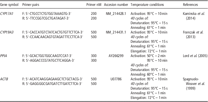
Total RNA was isolated from endometrial and myometrial tissue samples using peqGold TriFast isolation system (Peqlab, Germany). Ribonucleic acid quantity and quality were determined spectrophotometrically (Infinite M200 Pro, Tecan, Switzerland). The entire total RNA was intact with high quality, that is optical density 260/280 and 260/230 ratios were between 1.8 and 2.0 and 1.8 or greater, respectively. One microgram of RNA was reverse transcribed into cDNA in a total volume of 20 µl with 0.5 µg oligo (dT)15 primer (Roche, Basel, Switzerland) using the Omniscript RT Kit (Qiagen, MD, USA) at 37°C for 1 h and was terminated by the incubation at 93°C for 5 min. Quantitative real-time PCR analysis was performed using a PCR System 7300 (Applied Biosystems, CA, USA) with SYBR Green as described previously (Smolinska et al., Reference Smolinska, Kiezun, Dobrzyn, Szeszko, Maleszka and Kaminski2016). Specific primer pairs used to amplify parts of CYP17A1, CYP19A3, cyclophilin A (PPIA) and β-actin (ACTB) genes as well as temperature conditions of the real-time PCR reactions are detailed in Table 1. The constitutively expressed genes, PPIA and ACTB, were used as the internal control to verify the quantitative real-time PCR. Preliminary study indicated that the expression of PPIA and ACTB was similar in the endometrium and myometrium and stable during the oestrous cycle and pregnancy, as well as with or without treatments. The PCR reaction mixtures were containing: 10 ng of cDNA, primers, 12.5 µl Power SYBR Green PCR Master Mix (Applied Biosystems, CA, USA), and RNase free water at the final volume of 25 µl. Negative controls were performed using RNase free water instead of cDNA. All samples were prepared in duplicates. The specificity of amplification was tested at the end of the reaction by the analysis of melting-curve. Products purity was confirmed by agarose gel electrophoresis. Calculation of the relative expression levels of CYP17A1 and CYP19A3 were calculated using comparative cycle threshold method (ΔΔC T ) and normalized using the geometrical means of the reference gene expression levels: PPIA and ACTB.
Primer sequences and qPCR temperature conditions

Radioimmunoassay of steroid hormones
Concentrations of E1, E2 and T were analyzed by RIA according to the method described by Grzesiak et al. (Reference Grzesiak, Knapczyk-Stwora, Ciereszko, Wieciech and Slomczynska2014). Cross-reactivity of the antisera against E1, E2 and T have been published previously (Szafranska et al., Reference Szafranska, Ziecik and Okrasa2002). The extraction yields for E1, E2 and T were 86.93±0.26%, 90.67±0.73% and 87.12±0.29%, respectively. The sensitivities of the assays for E1, E2 and T were as follows: 0.5, 1 and 0.5 pg/ml. The standard curve range for E1 and E2 was from 0.5 to 200 pg/ml and for T from 0.5 to 250 pg/ml. The intra- and inter-assay CV were 1% and 6.6% for E1, 5% and 5% for E2, and 2% and 10,8% for T, respectively.
Statistical analysis
All data were analyzed by one-way ANOVA followed by the Fisher’s least significant difference post hoc test and are reported as means±standard error of the mean from five independent observations. Statistical analysis was performed using Statistica program (StatSoft Inc., USA). Values for P<0.05 were considered statistically significant.
Results
The effect of orexin B on CYP17A1 gene expression in the endometrial and myometrial tissue explants
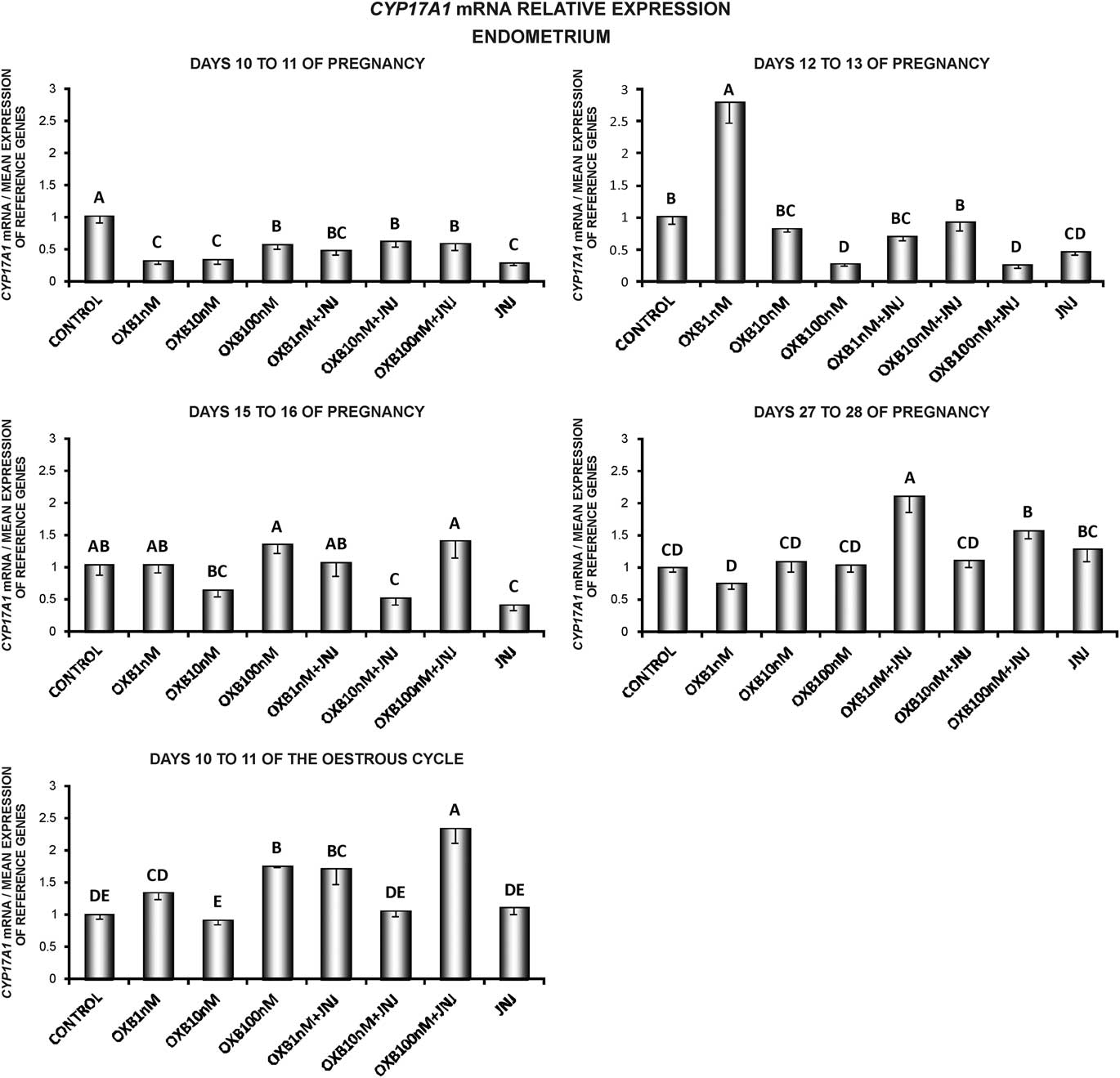
In the endometrium, OXB at the doses of 1 and 100 nM caused an increase in the expression of CYP17A1 on days 12 to 13 of pregnancy and 10 to 11 of the oestrous cycle, respectively. In this tissue, OXB decreased CYP17A1 mRNA content on days 10 to 11 (1, 10, 100 nM) and 12 to 13 (100 nM) of gestation. In the endometrial samples, cultured in the presence of OX2R antagonist (N-(2,4-Dibromophenyl)-N′-[(4S,5S)-2,2-dimethyl-4-phenyl-1,3-dioxan-5-yl]-urea (JNJ)), OXB at the doses of 1 and 100 nM stimulated CYP17A1 expression on days 27 to 28 of pregnancy and 10 to 11 of the oestrous cycle, whereas an inhibitory effect of OXB was observed on days 10 to 11 (1, 10, 100 nM), 12 to 13 (100 nM) and 15 to 16 (10 nM) of gestation. The supplementation of media with JNJ caused a decrease in CYP17A1 expression in the endometrium on days 10 to 11, 12 to 13 and 15 to 16 of gestation (P<0.05; Figure 1).
The effect of orexin B (OXB) on CYP17A1 gene expression. The influence of OXB (1, 10, 100 nM) in the presence or absence of the selective antagonist of orexin receptor type 2 (JNJ10397049 (JNJ)) on the relative expression of P450c17 cytochrome (CYP17A1) gene in endometrial tissue explants harvested from pigs on days 10 to 11, 12 to 13, 15 to 16 and 27 to 28 of pregnancy, and on days 10 to 11 of the oestrous cycle preincubated for 2 h and subsequently incubated for 24 h. The expression levels were normalized with the geometric mean expression of reference genes: cyclophilin A and β-actin. Data are expressed as the mean±standard error of the mean (n=5). A,B,C,D,EBars with different letters are significantly different at P<0.05.

In the myometrial tissue explants, an increase in the CYP17A1 mRNA content caused by OXB was observed on days 12 to 13 (1 nM) and 27 to 28 (1, 10 nM) of pregnancy, whereas a decrease in the CYP17A1 expression was noted on days 10 to 11 of gestation (100 nM) and on days 10 to 11 of the cycle (1, 10 nM). The addition of JNJ to the media caused an increase in the concentration of CYP17A1 mRNA under the influence of OXB on days 10 to 11 (10 nM), 12 to 13 (10 nM), 15 to 16 (10 nM) and 27 to 28 (1, 100) of pregnancy, and a decrease on days 10 to 11 (1, 100 nM) and 12 to 13 (100 nM) of gestation, and 10 to 11 of the cycle (1, 100 nM). JNJ caused an increase in the expression of CYP17A1 on days 15 to 16 and 27 to 28 of pregnancy (P<0.05; Supplementary Figure S1).
The effect of orexin B on testosterone secretion by the endometrial and myometrial tissue explants
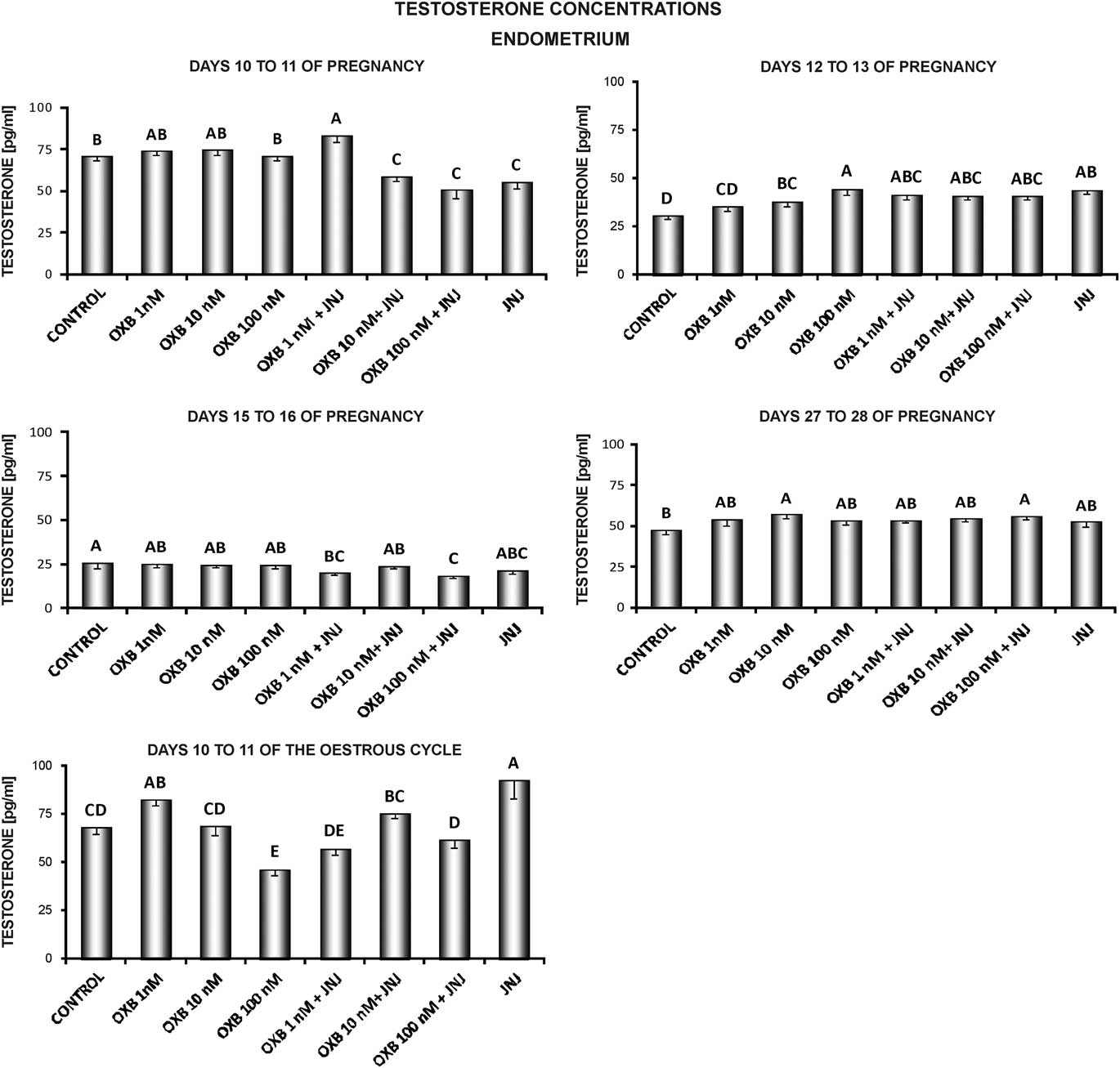
In the endometrial slices, an increase in the secretion of T under the influence of OXB was observed on days 12 to 13 (10, 100 nM) and 27 to 28 (10 nM) of pregnancy and on days 10 to 11 of the cycle (1 nM), whereas a decrease on days 10 to 11 of the cycle (100 nM). In samples cultured in media supplemented with OX2R antagonist, OXB caused an increase in the production of T on days 10 to 11 (1 nM), 12 to 13 (1, 10 ,100 nM) and 27 to 28 (100 nM) of pregnancy, whereas a decrease was observed on days 10 to 11 (10, 100 nM) and 15 to 16 (1, 100 nM) of pregnancy. JNJ stimulated T release on days 12 to 13 of pregnancy and 10 to 11 of the cycle, and inhibited on days 10 to 11 of gestation (P<0.05; Figure 2).
The effect of orexin B (OXB) on testosterone (T) secretion. The influence of OXB (1, 10, 100 nM) in the presence or absence of the selective antagonist of orexin receptor type 2 (JNJ10397049 (JNJ)) on in vitro T secretion by endometrial tissue explants harvested from pigs on days 10 to 11, 12 to 13, 15 to 16 and 27 to 28 of pregnancy, and on days 10 to 11 of the oestrous cycle preincubated for 2 h and subsequently incubated for 24 h. Data are expressed as the mean±standard error of the mean (n=5). A,B,C,D,EBars with different letters are significantly different at P<0.05.

In the myometrium, T secretion was enhanced by OXB (1 nM) on days 27 to 28 of pregnancy, whereas on days 10 to 11 of gestation and 10 to 11 of the cycle this orexin (1, 10, 100 nM) inhibited T release. In the myometrial slices cultured in the presence of JNJ, OXB stimulated T production on days 12 to 13 (1, 10 nM) of pregnancy, whereas a decrease of T secretion under the influence of OXB (1, 10, 100 nM) was observed on days 10 to 11 of pregnancy and 10 to 11 of the cycle. An inhibitory effect of JNJ on T release was observed on days 10 to 11 and 27 to 28 of pregnancy and on days 10 to 11 of the oestrous cycle (P<0.05; Supplementary Figure S2).
The effect of orexin B on CYP19A3 gene expression in the endometrial and myometrial tissue explants
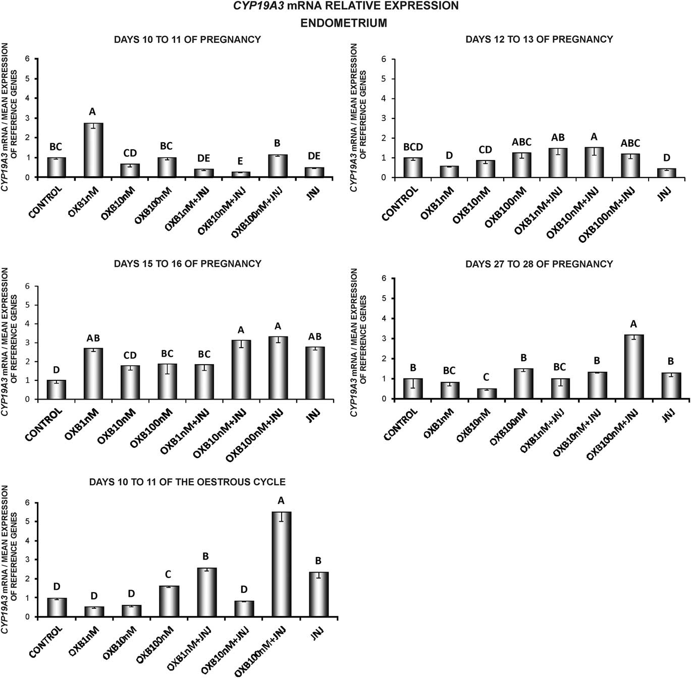
In the endometrial tissue explants, OXB stimulated CYP19A3 expression on days 10 to 11 (1 nM) and 15 to 16 (1, 100 nM) of pregnancy and on days 10 to 11 of the oestrous cycle (100 nM). An inhibitory effect of OXB on CYP19A3 expression was observed on days 27 to 28 of gestation (10 nM). In samples incubated in the presence of JNJ, OXB increased CYP19A3 mRNA level on days 12 to 13 (10 nM), 15 to 16 (1, 10, 100 nM) and 27 to 28 (100 nM) of pregnancy and 10 to 11 of the cycle (1, 100 nM), and decreased on days 10 to 11 of gestation (1, 10 nM). Orexin receptor type 2 blocker stimulated CYP19A3 expression on days 15 to 16 of pregnancy and 10 to 11 of the cycle, whereas inhibited on days 10 to 11 of gestation (P<0.05; Figure 3).
The effect of orexin B (OXB) on CYP19A3 gene expression. The influence of OXB (1, 10, 100 nM) in the presence or absence of the selective antagonist of orexin receptor type 2 (JNJ10397049 (JNJ)) on the relative expression of P450arom cytochrome (CYP19A3) gene in endometrial tissue explants harvested from pigs on days 10 to 11, 12 to 13, 15 to 16 and 27 to 28 of pregnancy, and on days 10 to 11 of the oestrous cycle preincubated for 2 h and subsequently incubated for 24 h. The expression levels were normalized with the geometric mean expression of reference genes: cyclophilin A and β-actin. Data are expressed as the mean±standard error of the mean (n=5). A,B,C,D,EBars with different letters are significantly different at P<0.05.

Orexin B increased CYP19A3 mRNA content in the myometrium on days 12 to 13 (1, 10 nM) and 15 to 16 (10 nM) of pregnancy and on days 10 to 11 of the oestrous cycle (1, 10 nM). An inhibitory effect of OXB (1, 100 nM) on CYP19A3 expression in this tissue was observed on days 10 to 11 of gestation. In samples incubated in the presence of JNJ, OXB stimulated CYP19A3 expression on days 12 to 13 (1, 10 nM) and 27 to 28 (1 nM) of pregnancy and on days 10 to 11 of the cycle (10 nM), whereas inhibited on days 10 to 11 (1, 100 nM) and 15 to 16 (1 nM) of gestation. The supplementation of media with JNJ caused an increase in the level of CYP19A3 mRNA on days 12 to 13 of pregnancy and 10 to 11 of the cycle, and a decrease on days 10 to 11 of gestation (P<0.05; Supplementary Figure S3).
The effect of orexin B on oestrone secretion by the endometrial and myometrial tissue explants
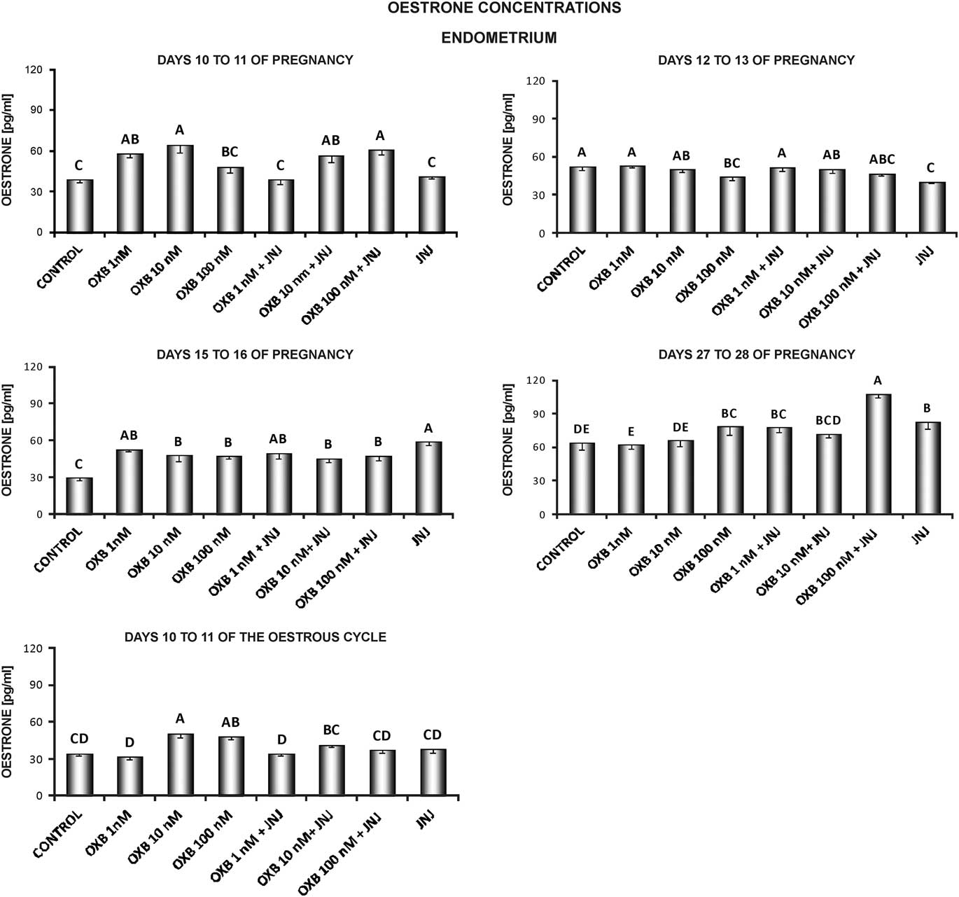
Secretion of E1 by the endometrial tissue slices was enhanced under the influence of OXB on days 10 to 11 (1, 10 nM), 15 to 16 (1, 10, 100 nM) and 27 to 28 (100 nM) of pregnancy and on days 10 to 11 of the cycle (10, 100 nM). A decrease in E1 secretion by the endometrium samples was caused by OXB (100 nM) on days 12 to 13 of gestation. In samples incubated in the presence of JNJ, OXB stimulated E1 release on days 10 to 11 (10, 100 nM), 15 to 16 (1, 10, 100 nM) and 27 to 28 (1, 100 nM). Orexin receptor type 2 antagonist caused an increase in E1 secretion on days 15 to 16 and 27 to 28, and a decrease on days 12 to 13 of pregnancy (P<0.05; Figure 4).
The effect of orexin B (OXB) on oestrone (E1) secretion. The influence of OXB (1, 10, 100 nM) in the presence or absence of the selective antagonist of orexin receptor type 2 (JNJ10397049 (JNJ)) on in vitro E1 secretion by endometrial tissue explants harvested from pigs on days 10 to 11, 12 to 13, 15 to 16 and 27 to 28 of pregnancy, and on days 10 to 11 of the oestrous cycle preincubated for 2 h and subsequently incubated for 24 h. Data are expressed as the mean±standard error of the mean (n=5). A,B,C,D,EBars with different letters are significantly different at P<0.05.

In the myometrial tissue slices, E1 secretion was stimulated by OXB at all doses used on days 10 to 11 and 15 to 16 of pregnancy, and 10 to 11 of the cycle, whereas on days 12 to 13 at the concentration of 1 and 100 nM. In the myometrial slices incubated in the presence of OX2R blocker, OXB at the doses of 1, 10 and 100 nM stimulated E1 secretion on days 15 to 16, 27 to 28 of pregnancy and 10 to 11 of the cycle, whereas at the dose of 1 nM on days 10 to 11 of gestation. JNJ increased E1 release on days 15 to 16 and 27 to 28 of pregnancy, and 10 to 11 of the cycle (P<0.05; Supplementary Figure S4).
The effect of orexin B on oestradiol secretion by the endometrial and myometrial tissue explants
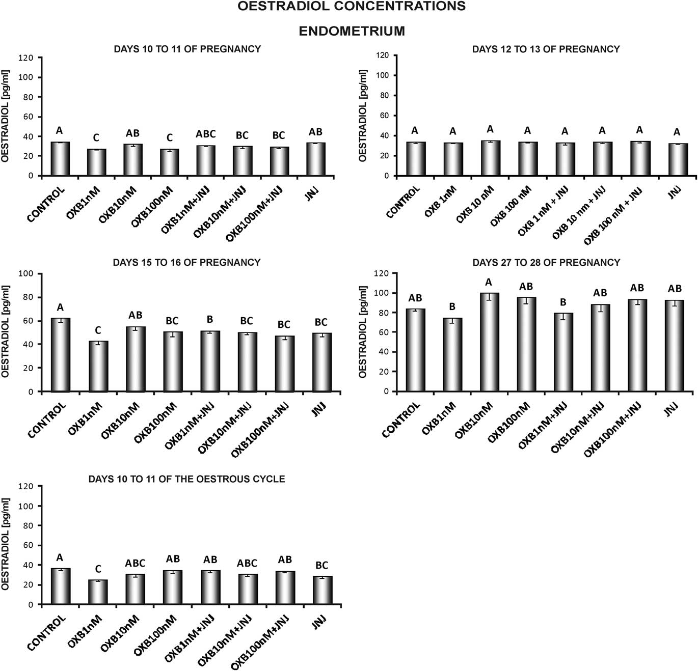
Orexin B at the doses of 1 and 100 nM inhibited E2 secretion by the endometrial tissue explants on days 10 to 11 and 15 to 16 of pregnancy, whereas on days 10 to 11 of the oestrous cycle at the dose of 1 nM. In samples incubated in the presence of OX2R antagonist, OXB caused a decrease in the secretion of E2 on days 10 to 11 (10, 100 nM) and 15 to 16 (1, 10, 100 nM) of pregnancy. JNJ inhibited the secretion of E2 on days 15 to 16 of pregnancy and 10 to 11 of the cycle (P<0.05; Figure 5).
The effect of orexin B (OXB) on oestradiol (E2) secretion. The influence of OXB (1, 10, 100 nM) in the presence or absence of the selective antagonist of orexin receptor type 2 (JNJ10397049 (JNJ)) on in vitro E2 secretion by endometrial tissue explants harvested from pigs on days 10 to 11, 12 to 13, 15 to 16 and 27 to 28 of pregnancy, and on days 10 to 11 of the oestrous cycle preincubated for 2 h and subsequently incubated for 24 h. Data are expressed as the mean±standard error of the mean (n=5). A,B,CBars with different letters are significantly different at P<0.05.

In the myometrial samples, the stimulatory effect of OXB on E2 release was observed on days 12 to 13 of pregnancy (1, 10 nM), whereas a decrease of E2 production was noted on days 10 to 11 of gestation (100 nM). The addition of JNJ and OXB (1 nM) to the media stimulated the production of E2 on days 10 to 11 of the cycle, whereas on days 10 to 11 of pregnancy the inhibitory effect of OXB (10, 100 nM) was observed. JNJ decreased E2 secretion on days 10 to 11 of pregnancy (P<0.05; Supplementary Figure S5).
Discussion
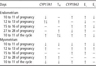
This is the first report on OXB involvement in the regulation of the secretory activity of the uterus. In this study we have shown that OXB modulates E1, E2 and T secretion from porcine myometrial and endometrial slices as well as mRNA concentration of steroidogenic enzymes directly responsible for oestrogen and androgen production during the luteal phase of the cycle and early pregnancy (Table 2). Slightly earlier we described similar effect of OXA on porcine uterine steroidogenesis (Kiezun et al., Reference Kiezun, Smolinska, Dobrzyn, Szeszko, Rytelewska and Kaminski2017).
The simplified scheme of orexin B (OXB) influence on CYP17A1 and CYP19A3 expression and steroid hormones secretion by endometrial and myometrial slices during early pregnancy and days 10 to 11 of the oestrous cycle

↑=OXB stimulatory effect; ↓=OXB inhibitory effect; ↑↓=both, stimulatory and inhibitory effect of OXB; −=lack of OXB effect; E 1 , E 2 , T=oestrone, oestradiol and testosterone, respectively.
It was indicated earlier, based on the pig model, that the uterus is a steroidogenic organ producing de novo androgens and oestrogens in cyclic and pregnant females (Franczak and Kotwica, Reference Franczak and Kotwica2008 and Reference Franczak and Kotwica2010). The production of E2 by the porcine uterus reaches 1 mg daily (Franczak and Kotwica, Reference Franczak and Kotwica2008). This organ demonstrates the expression of key steroidogenic enzymes, CYP17A1 and CYP19A3, responsible for androgens and oestrogens production (Franczak and Kotwica, Reference Franczak and Kotwica2010; Franczak et al., Reference Franczak, Wojciechowicz, Zmijewska, Kolakowska and Kotwica2013; Chen et al., Reference Chen, Li, Chen, Wei, Fu and Wang2015). We have shown that OXB stimulates T production by uterine tissues at the end of implantation increasing the source of substrate for uterine P450 aromatase. It has been suggested that locally produced oestrogens may supplement embryonic steroidogenesis and take part in maternal recognition of pregnancy (Franczak and Kotwica, Reference Franczak and Kotwica2008). Oestrogens also prevent luteolysis of corpora lutea during early pregnancy through stimulation of PGE2 generation and redirection of PGF2α secretion from uterine vasculature to the uterine lumen (for review see Bazer, Reference Bazer2013). Moreover, they influence uterine activity, proliferation, differentiation and the growth and development of uterine glands (Xiao and Goff, Reference Xiao and Goff1998). Thus, apart from ovarian, placental and embryonic steroid hormones, uterine-derived steroids may also be responsible for steroidogenic effects during early pregnancy in the pig.
In our previous studies we indicated the expression of orexin system components at both the gene and protein levels in uterine tissues of cyclic and pregnant pigs (Nitkiewicz et al., Reference Nitkiewicz, Smolinska, Maleszka, Kiezun and Kaminski2012; Smolinska et al., Reference Smolinska, Dobrzyn, Kiezun, Szeszko, Maleszka and Kaminski2015). This expression was remarkably dependent on the hormonal status of animals. Orexin B-immunoreactivity was the highest on days 10 to 12 of the cycle in longitudinal muscles, on days 17 to 19 in glandular epithelium and stroma, and on days 10 to 12 and 14 to 16 in luminal epithelium of the uterus (Nitkiewicz et al., Reference Nitkiewicz, Smolinska, Maleszka, Kiezun and Kaminski2012). In turn during pregnancy, the greatest concentration of PPO, an OXB precursor protein, was detected on days 12 to 13 in the endometrium and days 30 to 32 in the myometrium (Smolinska et al., Reference Smolinska, Dobrzyn, Kiezun, Szeszko, Maleszka and Kaminski2015). In pigs, the OXB plasma concentration recorded a multi-fold increase on days 10 to 11 and 30 to 32 of pregnancy. The level of OXB was also enhanced in the uterine luminal fluid on days 10 to 11 and 12 to 13 of pregnancy (Smolinska et al., Reference Smolinska, Kiezun, Dobrzyn, Szeszko, Maleszka and Kaminski2017). Orexin receptor type 1 protein content was increased on days 10 to 11 and 27 to 28, whereas OX2R on days 15 to 16 and 27 to 28 in the endometrium and myometrium, respectively (Smolinska et al., Reference Smolinska, Dobrzyn, Kiezun, Szeszko, Maleszka and Kaminski2015). Taken together, these findings may suggest variable sensitivity of uterine structures to OXs action during pregnancy and differentiated influence of orexins related to the stage of pregnancy and kind of uterine tissue. The latter is at least partly confirmed by the data of the presented study. The secretion patterns of steroid hormones under the influence of OXB in the endometrium and myometrium were different in particular stages of gestation. Orexin B enhanced T release from both uterine tissues on days 27 to 28 (the end of implantation process) and from endometrial slices on days 12 to 13 (the period of maternal recognition of pregnancy). For oestrogens, secretion of E2 – the biologically most active oestrogen – by endometrial slices was inhibited on days 10 to 11 and 15 to 16 (the beginning of implantation) and potentiated on days 12 to 13 by myometrial slices. Differential effect of OXB, related to pregnancy stages, was also observed with reference to CYP17A1 and CYP19A3 expression. It is tempting to speculate that OXB, through its influence on uterine steroid production, may affect essential processes during early pregnancy, including maternal recognition of pregnancy and implantation. Of course, this hypothesis requires further studies and should be very carefully discussed, because of the almost completely lack of literature data on the role of orexins in the mammalian uterus. Only one study has examined the possibility of orexin involvement in the control of endometrial cell proliferation through OX2R (Dehan et al., Reference de Lecea, Kilduff, Peyron, Gao, Foye, Danielson, Fukuhara, Battenberg, Gautvik, Bartlett, Frankel, Van den Pol, Bloom, Gautvik and Sutcliffe2013).
It is worth noting that the secretion patterns of both examined oestrogens – E1 and E2, are different, which may imply the involvement of 17β-hydroxysteroid dehydrogenase (17β-HSD) in their conversion. Indeed, 17β-HSD type 1 and 2, which are key enzymes responsible for interconversion of E1 and E2, were found in the porcine endometrium (Huang et al., Reference Huang, Liu, Su, Xiao and Yu2015; Wojciechowicz et al., Reference Wojciechowicz, kotwica, Zglejc, Waszkiewicz and Franczak2017). Additionally, it seems plausible that the observed influence of OXB on steroid production was at least partly achieved through its effect on the activity of steroidogenic enzymes. This is supported by the fact that the response to OXB in the form of genes expression of enzymes and steroid hormones production is different. It is also possible that the expression of enzymatic proteins differs from enzyme genes, which may result from the regulation of transcriptional and posttranscriptional processes, differences in the stability of mRNAs and proteins, or physiological feedbacks influencing transcript and protein concentration. If so, the frequently observed physiological regulation – correlation between protein and mRNA levels was noted for only a third of the specific gene products (Gry et al., Reference Gry, Rimini, Strömberg, Asplund, Pontén, Uhlén and Nilsson2009). Attention needs to be paid to the action of OX2R antagonist – JNJ10397049. Although, it did not block OXB influence on gene expression and steroid production, when added alone to the cultures it was able to modify these processes. The effect the OX2R antagonist added alone, similarly like OX1R blocker, was observed in both in vivo studies on rats (Silveyra et al., Reference Silveyra, Lux-Lantos and Libertun2007) and in vitro studies using porcine ovarian cells (Nitkiewicz et al., unpublished). The weak neutralizing action of the blocker when added together with OXB could be attributed to its low concentration. Cataldi et al. (Reference Cataldi, Lux-Lantos and Libertun2012) used a 10-fold higher (10 µM) concentration of the antagonist to achieve a full blockage of OX2R. Since orexins bind both OX1R and OX2R, although with different affinity, the blockade could be achieved by using blockers of both types of receptors simultaneously at a sufficiently high concentration. In some cases the combined treatment with OXB and the receptor antagonist resulted even in an additive effect. One possible scenario is that OXB, especially at higher doses, may act through unblocked OX1R receptors. It is known that OXB binds mainly OX2R and is 5 to 10 times less potent at OX1R (Sakurai et al., Reference Sakurai, Amemiya, Ishii, Matsuzaki, Chemelli, Tanaka, Williams, Richardson, Kozlowski, Wilson, Arch, Buckingham, Haynes, Carr, Annan, McNulty, Liu, Terrett, Elshourbagy, Bergsma and Yanagisawa1998; Langmead et al., Reference Langmead, Jerman, Brough, Scott, Porter and Herdon2004) but, to a lesser extent, it is able to bind also OX1R. This hypothesis is supported by similar action of OXA in a parallel study by Kiezun et al. (Reference Kiezun, Smolinska, Dobrzyn, Szeszko, Rytelewska and Kaminski2017) in refer to, for example, E1 secretion by endometrial slices on days 15 to 16 and 27 to 28 of pregnancy. An alternative explanation for this phenomenon is JNJ10397049 action as a partial agonist of OX2R through unknown mechanism. However, if this speculation is true, a question arises to what degree the observed effect of combined treatment with JNJ10397049 and OXB results from the influence of each of these agents. Similar explanations may be expressed to the phenomenon of comparable action of OXB and JNJ10397049 added alone. Such an effect was noted in case of CYP19A3 endometrial expression on days 15 to 16 of pregnancy. Support for suggested above explanation derives again from presented in a study by Kiezun et al. (Reference Kiezun, Smolinska, Dobrzyn, Szeszko, Rytelewska and Kaminski2017) similar action of OXA on the gene expression on these days of pregnancy. Moreover, one cannot rule out the possibility of the auto- or paracrine impact of orexins, generated by the cultured uterine slices, via the second type – fully active of OX receptor. It is also worth paying attention to opposing effects of different doses of OXB observed in case of endometrial CYP17A1 expression on days 12 to 13 of pregnancy and T secretion on days 10 to 11 of the cycle. It seems, that the reason for this stimulatory or inhibitory cell response, dependently on OXB concentration, may be hormesis phenomenon. This effect is noted first of all in toxicological and pharmacological studies (Calabrese and Baldwin, Reference Calabrese and Baldwin2003) but was also observed in case of ovarian steroidogenesis affected by organic pollutants (Gregoraszczuk et al., Reference Gregoraszczuk, Milczarek, Wojtowicz, Berg, Skaare and Ropstad2008).
The OXB influence on uterine steroidogenesis presented in this study is not the only evidence of orexins engagement in the control of this process in different tissues. Previously, it was indicated that OXA and OXB decreased progesterone secretion by rat luteal cells (Cataldi et al., Reference Cataldi, Lux-Lantos and Libertun2012) and FSH-induced E2 release from porcine granulosa cells (Nitkiewicz et al., Reference Nitkiewicz, Smolinska, Maleszka, Chojnowska and Kaminski2014). Orexin A increased T production by rat testes both in vivo and in vitro (Barreiro et al., Reference Barreiro, Pineda, Navarro, Lopez, Suominen, Pinilla, Senaris, Toppari, Aguilar, Diéguez and Tena-Sempere2004). Adrenal steroidogenesis was also affected by orexins, an enhancement of glucocorticoid and aldosterone secretion was noted from the porcine adrenal cortex (Nanmoku et al., Reference Nanmoku, Isobe, Sakurai, Yamanaka, Takekoshi, Kawakami, Goto and Nakai2002), and higher output of glucocorticoids was described in rats and humans (Ziolkowska et al., Reference Ziolkowska, Spinazzi, Albertin, Nowak, Malendowicz, Tortorella and Nussdorfer2005).
Summarizing, although further work is required to substantiate it, there is firm evidence that OXB affects steroidogenesis in the porcine uterus during early pregnancy modulating T, E2 and E1 secretion, and gene expression of steroidogenic enzymes, CYP71A1 and CYP19A3. This influence was dependent on the period of pregnancy, type of uterine tissue and dose of OXB. It is suggested that OXB, taking part in the control of local steroidogenesis, may affect uterine functions during early pregnancy in the pig.
Acknowledgements
The authors thank the Johnson and Johnson Pharmaceutical Research & Development team for donating a selective antagonist of orexin receptor type 2 (JNJ10397049). This research was supported by National Science Centre (project no: 2011/03/B/NZ9/04187).
Declaration of interest
The authors hereby confirm that neither the manuscript nor any part of it, except for abstracts, has been published or is being considered for publication elsewhere. None of the authors of the above manuscript has declared any conflict of interest which may arise from being named as an author on the manuscript.
Ethics Statement
All studies were conducted in accordance with ethical standards of the Animal Ethics Committee at the University of Warmia and Mazury in Olsztyn (Ethics approval no. 91/2011/DTN).
Software and data repository resources
All data generated or analysed during this study are available from the corresponding author on reasonable request.
Supplementary material
To view supplementary material for this article, please visit https://doi.org/10.1017/S1751731117003779