Introduction
Layered double hydroxides (LDH) constitute a large family of minerals and synthetic compounds featuring crystal structures consisting of alternating positively charged layers of cations, typically having octahedral coordination and negatively charged interlayers (Rives, Reference Rives2001; Duan and Evans, Reference Duan and Evans2006).
The general formula of brucite-type LDHs (without interlayer cations) belonging to the hydrotalcite supergroup is [M 2+1–xM 3+x(OH)2]q +(Xn −)q/n⋅yH2O, in which M 2+ and M 3+ are metal cations (M 2+ = Mg, Fe, Mn, Ni, Cu, Ca and Zn, and M 3+ = Al, Fe, Mn, Co and Cr), q is the positive charge of the brucite-type layer, and Xn − are interlayer anions (CO32–, SO42–, Cl–, OH– or Sb(OH)6–) which compensate the positive charges of the brucite-type layers (Mills et al., Reference Mills, Christy, Génin, Kameda and Colombo2012). In hydrotalcite-supergroup minerals, the x value varies from 0.17 (for coalingite) to 0.33 (for most members of the supergroup). Compounds with x > 0.33 are crystal chemically unfavourable because such a stoichiometry would result in direct contacts between two M 3+-centred octahedra. The most common M 2+:M 3+ ratios for hydrotalcite supergroup minerals are 2:1 (x = 0.33) and 3:1 (x = 0.25). The hydrotalcite supergroup consists of eight groups which differ from each other by layer spacing, coordination of cations and other crystal chemical features. In most hydrotalcite-supergroup minerals, the M cations have an octahedral coordination. However, in members of the hydrocalumite group the large Ca2+ cations that occur in the octahedral layer are coordinated additionally by interlayer water molecules and have a seven-fold coordination.
The specific feature of minerals belonging to the wermlandite group is the presence of cation complexes A(H2O)6 (A = Na, Ca and Sr) between the brucite-type layers. The forefather of this group, wermlandite, ideally Mg7Al2(OH)18[Ca(H2O)6](SO4)2⋅6H2O, was originally found associated with calcite on open fractures cross-cutting a dolomitic marble in the famous Långban mine, Värmland (old spelling: Wermland), Sweden (Moore, Reference Moore1971). The determination of the wermlandite crystal structure revealed the main structural feature of the group – the presence of cations coordinated by H2O molecules in the interlayer space along with anions and interlayer water molecules; the interlayers are sandwiched between two layers of octahedra as in the rest of the LDHs (Rius and Allmann, Reference Rius and Allmann1984). The presence of a cation complex A(H2O)6 in the interlayer results in the largest known d value among natural LDH species, which is ~11 Å.
It is notable that the synthetic analogues of wermlandite-group minerals have recently attracted attention as materials with cation-exchange properties (Sotiles et al., Reference Sotiles, Baika, Grassi and Wypych2018), whereas the rest of the hydrotalcite-supergroup minerals are anion exchangers (Rives, Reference Rives2001; Duan and Evans, Reference Duan and Evans2006). Materials science interest in wermlandite led to the first systematic syntheses of wermlandite-like compounds in the system M 2+6Al3(OH)18(A+(H2O)6)(SO4)2⋅6H2O (M 2+ = Co and Ni; A+ = Li, Na and K) and thermal studies of these phases (Sotiles et al., Reference Sotiles and Gomez2019). The DFT (density functional theory) study of motukoreaite- and natroglaucocerinite-like phases with compositions M 6Al3(OH)18Na(H2O)6(SO4)2⋅6H2O and M 6Al3(OH)18K(H2O)6(SO4)2⋅6H2O (M = Mg and Zn) revealed that the 3R polytype is the most stable structural modification of these compounds and showed that Na+ can be replaced by K+ (Moraes et al., Reference Moraes, Wypych and Leitão2019). Renewed interest in wermlandite-like materials resulted in the first proposals for the practical use of shigaite-, natroglaucocerinite- and motukoreaite-like LDHs as Pickering emulsifiers in water/oil emulsions (do Amaral et al., Reference do Amaral, Wypych and de Freitas2021). Regardless of the recent progress in materials science in the study of wermlandite-like compounds, some of the wermlandite-group members remain poorly characterised (Mills et al., Reference Mills, Christy, Génin, Kameda and Colombo2012), including motukoreaite, Mg6Al3(OH)18(Na(H2O)6)(SO4)2⋅6H2O, which was the subject of recent crystal chemical investigations (Wachowiak and Pieczka, Reference Wachowiak and Pieczka2016).
This paper describes the new wermlandite-group mineral erssonite CaMg7Fe3+2(OH)18(SO4)2⋅12H2O (or [Mg7Fe3+2(OH)18][Ca(SO4)2]⋅12H2O) from the Långban mine, the type locality of wermlandite. It is worth noting that the Långban mine is also the type locality of pyroaurite, Mg6Fe3+2(OH)16(CO3)⋅4H2O, and the locality also hosts LDH minerals such as brugnatellite, hydrotalcite and quintinite (Nysten et al., Reference Nysten, Holtstam, Jonsson, Holtstam and Langhof1999; Strand, Reference Strand2016). Erssonite is named in honour of the chemist and amateur mineralogist, Dr. Anders Ersson (b. 1971), Senior Engineer at the Materials Technology section at Scania AB, Södertälje, Sweden, who collected the type material of this mineral.
The new mineral and its name have been approved by the International Mineralogical Association Commission on New Minerals, Nomenclature and Classification (|IMA2021-016, Zhitova, et al., Reference Zhitova, Chukanov, Jonsson, Pekov, Belakovsky, Vigasina, Zubkova, Van and Britvin2021). Type material (parts of the holotype) is deposited in the Fersman Mineralogical Museum of the Russian Academy of Sciences, Moscow, Russia with the registration number 5669/1, and at the Swedish Museum of Natural History (Naturhistoriska Riksmuseet), Stockholm, Sweden, under the catalogue number GEO-NRM #20210001.
Experimental methods and data processing
Raman spectra of a randomly oriented crystal were obtained using an EnSpectr R532 spectrometer on an OLYMPUS CX 41 microscope, coupled with a diode laser (λ = 532 nm) at room temperature. The spectra were recorded in the range from 100 to 4000 cm–1 with a diffraction grating (1800 gr mm–1) and a spectral resolution of ~6 cm–1. The output power of the laser beam was in the range of 5 to 13 mW. The diameter of the focal spot on the sample was 5–10 μm. The back-scattered Raman signal was collected with a 40× objective; signal acquisition time for a single scan of the spectral range was 1 s, and the signal was averaged over 50 scans. Crystalline silicon was used as a standard.
Chemical analyses were carried out using a Tescan VEGA-II XMU INCA Energy 450 (20 kV and 190 pA) using energy-dispersive spectroscopy (EDS) as the mineral is unstable under the beam in wavelength-dispersive mode. The following standards were used: diopside for Mg; wollastonite for Ca; albite for Al; Cr metal for Cr; Fe2O3 for Fe; SiO2 for Si; and BaSO4 for S.
Powder X-ray diffraction (XRD) data were collected from a crystal of erssonite using a Rigaku R-Axis Rapid II diffractometer (Debye-Sсherrer geometry, d = 127.4 mm) equipped with a rotating anode X-ray source (CoKα, λ = 1.79021 Å) and a curved image plate detector. The data were integrated using the software package Osc2Tab/SQRay (Britvin et al., Reference Britvin, Dolivo-Dobrovolsky and Krzhizhanovskaya2017).
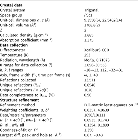
Single-crystal XRD data were collected using an Xcalibur S charge-coupled device (CCD) diffractometer (Faculty of Geology, Moscow State University, Moscow, Russia) operated at 40 kV and 50 mA using MoKα radiation. The data were integrated and corrected for absorption using a multi-scan model and the CrysAlisPro program (CrysAlisPro, 2014). The crystal structure was solved and refined with the ShelX (Version 2016, ShelX, Göttingen, Germany) program package using direct methods (Sheldrick, Reference Sheldrick2015). Crystal data, data collection information and structure refinement details for erssonite are given in Table 1. The hydrogen atoms were localised from residual electron-density maps and refined using a Riding model with U eq values restrained as 1.2 of the donor O atom.
Crystal data, data collection information and structure refinement parameters for erssonite.

*  $w = 1/[ \delta ^2F_0^2 + ( {aP} ) ^2 + bP$, where
 $P = ( {\max ( {F_0^2 , \;\;0} ) + 2F_c^2 } ) /3$
Results
Occurrence, general appearance and physical properties
The new mineral was discovered in a sample collected at the dumps of the Långban mine, Filipstad district, Värmland county, Bergslagen ore province, Sweden. The associated minerals are dolomite, calcite, magnetite–magnesioferrite solid-solution members, phlogopite, chrysotile, pyroaurite and norbergite.
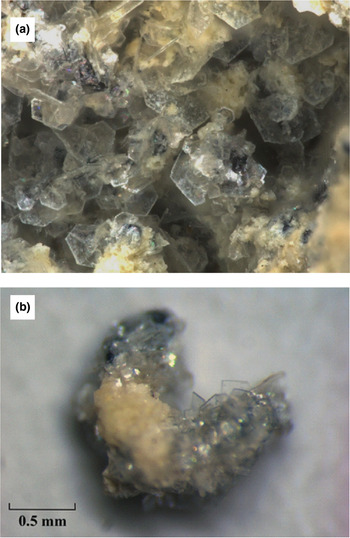
Erssonite occurs as platy hexagonal crystals flattened on [0001], up to 0.5 mm across and up to 10 μm thick, separate or forming aggregates that cover areas up to a few millimetres across (Fig. 1). The observed crystal forms are {0001} (the major form) and {10 $\bar{1}$0} (narrow lateral faces).
Lamellar crystals of erssonite: (a) Field of view, width 2 mm photo: A. Ersson and (b) erssonite in association with yellow dolomite, photo: Anatoly.V. Kasatkin.

The mineral is transparent and colourless with vitreous lustre. Fluorescence was not observed. Erssonite is malleable; separate crystals are flexible and non-elastic. A perfect, mica-like cleavage on {0001} is observed.
Density could not be measured because of the miniscule thickness of crystals and insufficient amount of pure material for volumetric measurements. Density calculated on the basis of the empirical formula and unit-cell volume refined from single-crystal XRD data is equal to 2.02 g⋅cm–3.
Raman spectroscopy
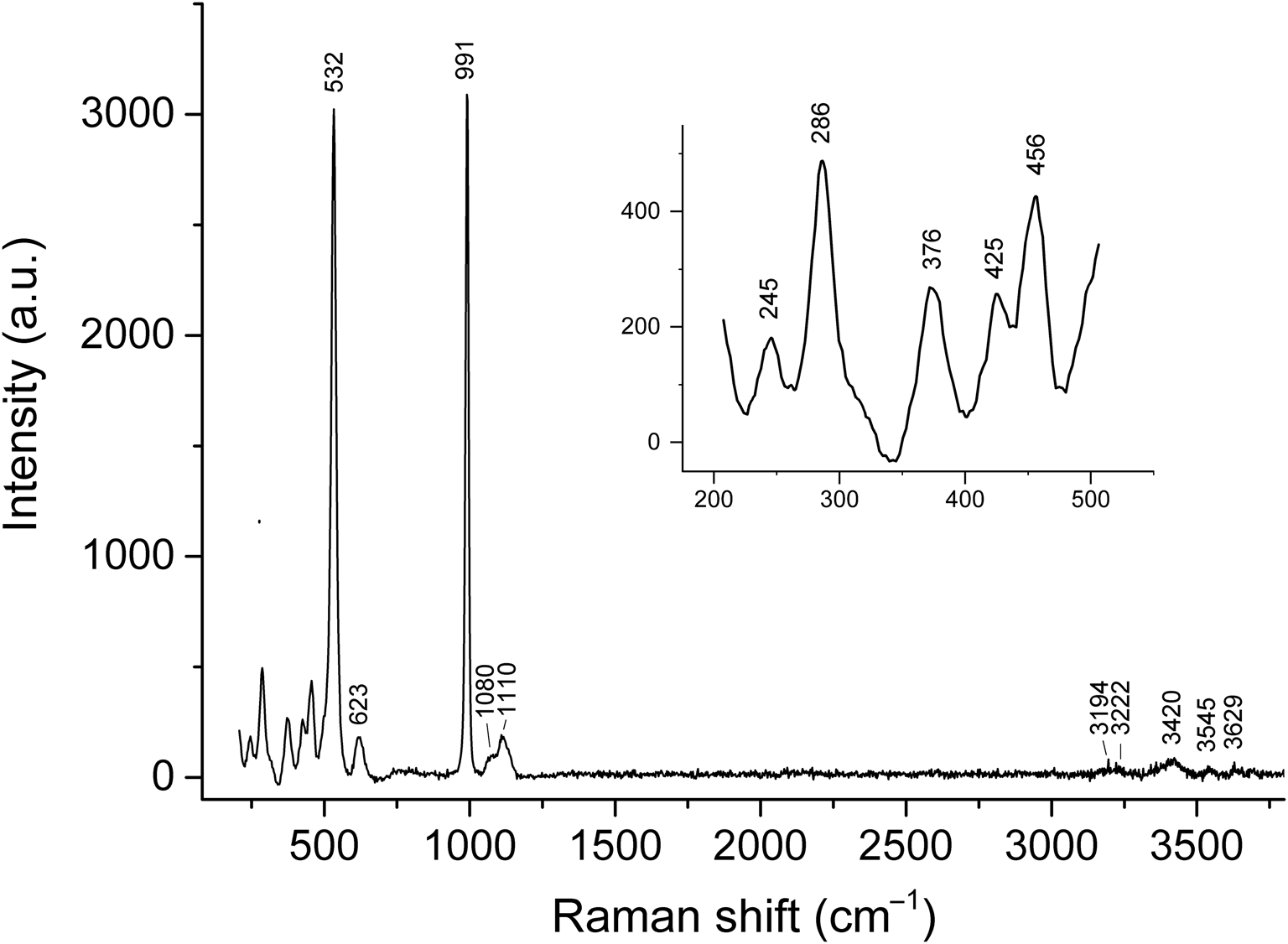
The Raman spectrum of erssonite is given in Fig. 2 and the assignment of Raman bands is as follows.
The Raman spectrum of erssonite.

Bands in the range 3190–3630 cm–1 are attributed to O–H stretching vibrations (Zhitova et al., Reference Zhitova, Pekov, Chukanov, Yapaskurt and Bocharov2020). Bands at 1110 and 1080 cm–1 correspond to asymmetric stretching vibrations of SO42– [the F 2(ν3) mode] (Frost et al., Reference Frost, Theiss, López and Scholz2014). A band at 991 cm–1 corresponds with symmetric stretching vibrations of SO42– [the A 1(ν1) mode] (Frost et al., Reference Frost, Theiss, López and Scholz2014). A band at 623 cm–1 is attributed to bending vibrations of SO42– [the F 2(ν4) mode] (Frost et al., Reference Frost, Theiss, López and Scholz2014) and a band at 532 cm–1 is assigned to Mg–O stretching vibrations, possibly overlapping with a band of Al–O stretching vibrations (Zhitova et al., Reference Zhitova, Pekov, Chukanov, Yapaskurt and Bocharov2020). The range of 400–500 cm–1 corresponds to overlapping bands of Mg–O and Fe3+–O stretching vibrations and bending vibrations of SO42– [the E(ν2) mode]. The bands below 400 cm–1 correspond to lattice modes.
The narrow single band of non-degenerate symmetric stretching vibrations of SO42– observed at 991 cm–1 indicates the presence of only one kind of sulfate anion in the structure of erssonite. The presence of two bands (at 1110 and 1080 cm–1) related to the degenerate stretching mode of SO42– in the Raman spectrum of erssonite reflects a distortion of the SO4 tetrahedron, which is confirmed by the single-crystal X-ray structural analysis (see below).
Bands of CO32– are absent in the Raman spectrum of erssonite.
Chemical data
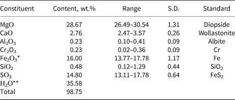
Nineteen spot EDS analyses were obtained for the determination of Mg, Ca, Al, Cr, Fe, Si and S. The contents of other elements with atomic numbers higher than that of carbon are below detection limits. H2O was not analysed because of the paucity of material. H2O content was calculated on the basis of the structure data (see below) and taking into account the charge balance requirement. CO2 was not determined because, according to both Raman spectroscopic and structural data, carbonate anions are absent in erssonite. Analytical data are given in Table 2.
Chemical composition of erssonite.

* Based on the structure data and charge-balance requirement; all iron is considered as Fe3+.
** Calculated, based on the structure data (see below).
S.D. – standard deviation
The empirical formula based on 38 O atoms is H41.48Ca0.52Mg7.47Fe3+2.11Al0.05Cr0.03S1.94Si0.08O38. Taking the structure data into account (see below), the simplified formula can be written as follows: (Ca,Mg,Fe)Mg7Fe3+2(OH)18(SO4)2⋅12H2O. The ideal formula is CaMg7Fe3+2(OH)18(SO4)2⋅12H2O or, taking into account structure data: {Mg7Fe3+2(OH)18}{[Ca(H2O)6](SO4)2(H2O)6}.
The Gladstone–Dale compatibility index 1 – (K p/K c) (Mandarino, Reference Mandarino2007) calculated using single-crystal unit-cell parameters is equal to 0.020 (rated as excellent).
X-ray diffraction and crystal structure
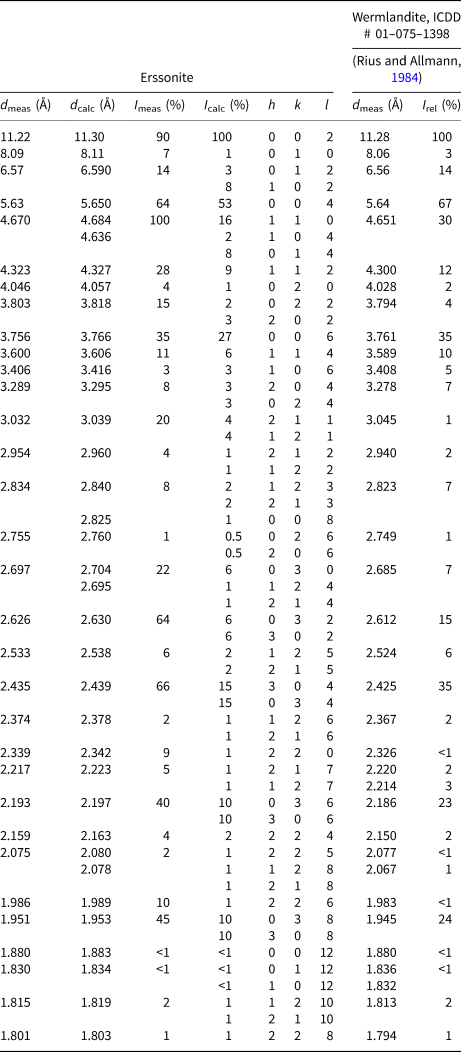
Powder XRD data are provided in Table 3. The obtained powder XRD pattern of erssonite is close to that from the JCPDS-ICDD card 01–075–1398 (powder diffraction files from the International Centre for Diffraction Data, https://www.icdd.com/, Rius and Allmann, Reference Rius and Allmann1984), which corresponds to wermlandite (trigonal, space group P  $\bar{3}$c1). The unit cell parameters of erssonite were refined from the powder data using the Pawley method and Topas software (Bruker, 2009) and are as follows: a = 9.3687(4), c = 22.5982(7) Å and V = 1717.8(2) Å3.
Powder X-ray diffraction data for erssonite and wermlandite.

* A reflection of pyroaurite impurity at d = 7.83 Å with I = 22 is not shown.
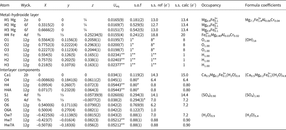
The crystal structure was solved and refined to R 1 = 0.093 for 1020 unique observed reflections with I > 2σ(I). Atom coordinates, site occupancies and displacement parameters are given in Table 4. Selected bond lengths and angles are listed in Table 5. The crystallographic information files have been deposited with the Principal Editor of Mineralogical Magazine and are available as Supplementary material (see below).
Atom coordinates and equivalent isotropic displacement parameters (Å2) and site occupancies for erssonite.

Wyck. – Wyckoff position; * Fixed during refinement in accord with chemical composition; ** U eq values are restrained as 1.2 of donor O atom.
On the basis of the refined site-scattering factors and previously determined chemical composition, the crystal-chemical formula of erssonite is {Mg7.10Fe3+1.80Al0.06Cr0.04(OH)18}1.9+ {[(Ca0.50Mg0.20Fe3+0.10)(H2O)4.80]1.7+(SO4)1.80(H2O)5.4}3.6–}.
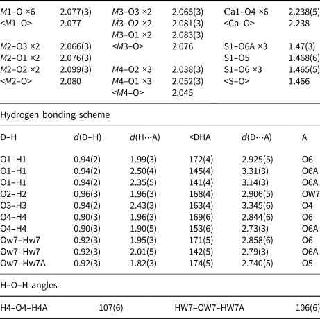
Selected bond lengths (Å) and angles (°) in the crystal structure of erssonite.

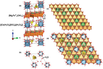
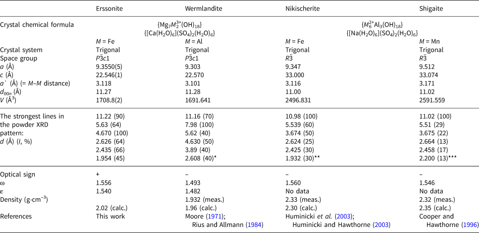
In terms of crystal structure, erssonite (Fig. 3) is isotypic to wermlandite. The structure contains two types of layers: (1) a positively charged metal–hydroxide layer with the ideal composition {Mg7Fe3+2(OH)18}2+ and (2) a negatively charged interlayer with the ideal composition {[Ca(H2O)6][SO4]2(H2O)6}2–. The metal–hydroxide layer contains four cation sites having octahedral coordination and occupied by species-defining di- and trivalent cations. The trivalent cations play a charge-balancing role and compensates the negative charge of the interlayer. The specific feature of wermlandite-group minerals is the presence of a relatively large cation (Na, Ca or Sr) in the interlayer space that results in the fact that the wermlandite-group minerals are characterised by a larger interlayer distance with the d value of ~11 Å (Mills et al., Reference Mills, Christy, Génin, Kameda and Colombo2012, see Table 6) in comparison with the value of d 00n = 7.8 Å in hydrotalcite with a carbonate interlayer (Zhitova et al., Reference Zhitova, Krivovichev, Pekov and Greenwell2019). The trivalent cations may be distributed in ordered or disordered fashion within the metal–hydroxide layers. Wermlandite-group minerals are distinguished by the predominant di- and trivalent cations of the metal–hydroxide layer as well as the nature of anions and cations in the interlayer.
(a) The crystal structure of erssonite; (b) the atomic arrangement of interlayer gallery (xy projection, z = 0); (c) metal–hydroxide layer with Fe and Mg ordering by 3 × 3 superstructure (xy projection, z = ¼) and (d) interlayer superimposed to metal–hydroxide layer. Notes: Mg are Mg(OH)6 octahedra, Fe are Fe3+(OH)6 octahedra, Ca are Ca(H2O)6 octahedra, S are SO4 tetrahedra, and H2O are H2O molecules.

Comparative data of erssonite and closely related wermlandite-group minerals.

* Taken from Moore (Reference Moore1971); ** Taken from Huminicki et al. (Reference Huminicki, Hawthorne, Grice, Roberts and Jambor2003); ***Calculated by Vesta program (Momma, and Izumi, Reference Momma and Izumi2011) for the crystal structure provided by Cooper and Hawthorne (Reference Cooper and Hawthorne1996).
The metal–hydroxide layer contains four symmetrically independent sites: M1–M3 predominantly occupied by Mg (for ~90%) with minor Fe (~10%) and a M4 site occupied by Fe3+0.55 Mg0.40Al0.03Cr0.02. The bulk composition of the metal–hydroxide layer is [Mg7.1Fe3+1.8Al0.06Cr0.04(OH)18]1.9+. The interlayer anion in erssonite is an SO42– group, which is a distorted tetrahedron (a trigonal pyramid with disordered basal O atoms). The occupancy of the S site is 90%, and the occupancies of corresponding O atoms have been fixed in accord with that of the S site giving [(SO4)1.80]3.60–. The occupancy of the Ca1 interlayer metal site has been done by assigning remaining cations in accord with site scattering, which resulted in the site having the population [(Ca0.50Mg0.20Fe3+0.10)Σ0.80]1.7+ and coordinated by (H2O)4.80. The occupancy of the interlayer water site Ow7 (Fig. 3b) is ~90% giving 5.4 H2O molecules per formula unit. Based on the refined site-scattering factors and previously determined chemical composition, the crystal-chemical formula of erssonite is
{Mg7.10Fe3+1.80Al0.06Cr0.04(OH)18}1.9+{[(Ca0.5Mg0.2Fe3+0.1)
(H2O)4.8]1.7+(SO4)1.8(H2O)5.4}3.6–}.
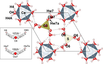
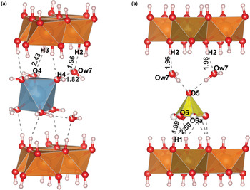
As erssonite contains considerable amounts of OH and H2O (the total content is 35.58 wt.%; Table 2), the mineral exhibits an extensive hydrogen bonding scheme (Table 5). Hydrogen bonding connects metal–hydroxide layers to interlayer components and provides linkage between interlayer species: Ca(H2O)6, SO4 and H2O. In the interlayer the following hydrogen atoms have been localised: H4, H4A of H2O molecules coordinating the interlayer Ca site; and Hw7, Hw7A attached to Ow7 of interlayer H2O molecules (Fig. 4). Interlayer H2O molecules form hydrogen bonding to sulfate and Ca-centred polyhedra. In the metal–hydroxide layer three hydrogen sites have been found: H1, H2 and H3 attached to O1, O2 and O3, respectively, at the distance 0.94–0.96 Å (Table 5). The H1, H2 and H3 atoms are directed nearly perpendicular to the plane of metal–hydroxide layers pointing to the interlayer. The H1 atom forms hydrogen bonds to the basal O6 and O6A atoms of the SO4 disordered tetrahedra at the distances 1.99 and 2.46 Å, respectively. The H2 atom forms hydrogen bonds to the Ow7 atom of the interlayer water molecule at the distance 1.96 Å. The H3 atom connects to the O4 atom of the Ca(H2O)6 octahedra with H3–O4 distance being 2.44 Å (Fig. 5).
The arrangement of interlayer components: SO4 tetrahedra, Ca(H2O)6 octahedra and H2O molecules in the crystal structure of erssonite. The hydrogen bonds are shown as dashed lines.

The hydrogen bonding between octahedral layer and interlayer components: (a) Ca(H2O)6 octahedra and (b) interlayer H2O molecules and SO4 tetrahedra, interlayer H2O molecules.

Discussion
Erssonite is a low-temperature hydrothermal mineral that occurs in small drusy cavities (formed after the leaching of pre-existing calcite and dolomite) in a magnetite-bearing dolomitic rock. In some cases, erssonite occurs directly on the magnetite substrate as very thin crusts and minute crystal aggregates. The Palaeoproterozoic Långban mineralisation is generally interpreted to represent a metamorphosed, deformed and overprinted, originally volcanic–hydrothermal seafloor deposit (e.g. Boström et al., Reference Boström, Rydell and Joensuu1979; Jonsson and Broman, Reference Jonsson and Broman2002, and references therein). We suggest that the formation of erssonite was related to geologically late (significantly post-dating Svecokarelian regional amphibolite facies metamorphism at 1.85–1.80 Ga), low-temperature fluid circulation, leading to localised leaching, transport and re-precipitation of metals, forming minerals such as erssonite. Widespread remobilisation of metals and subsequent crystallisation of diverse minerals stable at low-P conditions and low to very low temperatures affected the Långban deposit, leading to the formation of many exotic assemblages (Jonsson and Broman, Reference Jonsson and Broman2002; Nysten et al., Reference Nysten, Holtstam, Jonsson, Holtstam and Langhof1999, and references therein). We suppose that the formation of erssonite could be related to an even later stage of fluid activity. This assumption is in agreement with the lack of Pb, Mn and As that characterises the main stage of fissure mineral formation at Långban (Jonsson and Boyce, Reference Jonsson, Boyce and Stanley1999; Jonsson and Broman, Reference Jonsson and Broman2002). The elements needed for the formation of erssonite, mainly Mg, Fe and Ca were all readily available from the major earlier-formed minerals of the host assemblage (essentially, a magnetite-bearing carbonate rock), whereas S may have been supplied through the leaching of sparsely disseminated sulfides (chiefly pyrite), which are observed as sporadic minute grains in this type of dolomitic rock.
As noted previously, erssonite is the third hydrotalcite supergroup mineral first discovered in Långban after pyroaurite (2H and 3R polytypes) and wermlandite (Holtstam and Langhof, Reference Holtstam and Langhof1999; Mills et al., Reference Mills, Christy, Génin, Kameda and Colombo2012). Other LDHs at this locality include two Mg-Al-CO3 members: hydrotalcite, Mg6Al2(CO3)(OH)16⋅4H2O and quintinite, Mg4Al2(OH)12(CO3)⋅3H2O, as well as brugnatellite, Mg6Fe3+(CO3)(OH)13⋅4H2O (Nysten et al., Reference Nysten, Holtstam, Jonsson, Holtstam and Langhof1999; Strand, Reference Strand2016). It is noteworthy that brugnatellite has a questionable IMA status because the mineral is commonly confused with pyroaurite (Mills et al., Reference Mills, Christy, Génin, Kameda and Colombo2012). It is of interest that all these minerals have Mg as the main M 2+ cation and differ in (1) the Al3+/Fe3+ proportion in the layers of octahedra (erssonite, pyroaurite and brugnatellite are Fe3+-dominant minerals, in the wermlandite holotype M 3+ = Al0.57Fe3+0.43, while hydrotalcite and quintinite are Al3+-dominant, or (2) the interlayer content is (CO3)2– in pyroaurite, hydrotalcite, quintinite and brugnatellite, and [Ca(H2O)6(SO4)2]2– in erssonite and wermlandite. Wide variations of the composition of the brucite-type layer and interlayer are typical for associated hydrotalcite-supergroup members. For example, LDH minerals from the Terektinsky Ridge, Gorny Altai, Russia, belonging to the stichtite–pyroaurite–iowaite–woodallite solid-solution system and occurring in close associations with each other, are characterised by significantly different Fe3+:Cr3+ and CO32–:Cl– proportions (Zhitova et al., Reference Zhitova, Pekov, Chukanov, Yapaskurt and Bocharov2020).
Comparative data of erssonite and related wermlandite-group minerals are given in Table 6. Erssonite and wermlandite are isotypic and both are known only as two-layer trigonal modifications (2T polytype) that crystallise in space group P  $\bar{3}$c1. The comparison of erssonite to wermlandite by the unit cell parameters shows that these minerals are almost identical by the interlayer distance of 11.27–11.28 Å, reflecting identical interlayer compositions. The comparison of bond lengths between erssonite and wermlandite shows that M1–O, M2–O and M3–O (all predominantly occupied by M 2+) are nearly equivalent in bond distances (Table 7) with average M 2+–O values being 2.08 and 2.07 Å for erssonite and wermlandite, respectively. The M1, M2 and M3 octahedra in both crystal structures exhibit distortion of the bond lengths and angles (Table 5) with the exception of the M1 site in erssonite that shows only angular distortion (Table 5). As erssonite is an Fe3+-analogue of wermlandite, the main chemical difference between the minerals lies in the occupancy of the M4 position (predominantly occupied by M 3+). The <M4–O> distance is 2.01 Å for wermlandite having an Al0.57Fe0.43 occupancy of the M4 site (Rius and Allmann, Reference Rius and Allmann1984). In the crystal structure of erssonite the <M4–O> distance is 2.04 Å and the assigned occupancy is Fe3+0.55Mg0.40Al0.03Cr0.02 (Tables 4, 5). The difference in the <M4–O> bond lengths agrees with the assigned chemical composition of M4 sites with larger cations occupying the M4 site in erssonite in comparison to wermlandite.
Calculated and observed M–O distances in the crystal structures of erssonite and wermlandite and assigned occupancies of cation sites.

<M–O>obs = observed distances; <M–O>calc = calculated M–O distances from assigned site occupancies.
The other two minerals closely related to erssonite are nikischerite and shigaite. According to X-ray structural analysis (Cooper and Hawthorne, Reference Cooper and Hawthorne1996; Huminicki and Hawthorne, Reference Huminicki and Hawthorne2003), these minerals have the interlayer composition [Na(H2O)6](SO4)2(H2O)6, with interlayer Na instead of Ca (as in erssonite and wermlandite). In general, these minerals are similar to wermlandite and erssonite and belong to the wermlandite group. However, a closer look shows that they are crystal-chemically distinct. Nikischerite and shigaite have the M 2+:M 3+ ratio of 6:3, a univalent interlayer cation and crystallise in a rhombohedral three-layer unit cell (3R polytype). The interlayer distance of nikischerite and shigaite is 11.00–11.02 Å, which is significantly smaller than in wermlandite and erssonite. The a unit-cell parameter of erssonite (with M 9 = Mg7Fe3+2) is almost identical to that of nikischerite (with M 9 = Fe2+6Al3), whereas shigaite has a larger a value due to the dominance of Mn2+ among the M cations. It is worth noting that synthetic Na-dominant wermlandite-like synthetic phases have been reported with the following d values (in Å): 11.03 for ‘shigaite’, 11.14 for ‘natroglaucocerinite’ and 11.01 for ‘motukoreaite’. The natural motukoreaite from the Kłodawa Salt Dome (Central Poland) has been reported as a three-layer polytype with an interlayer distance of 11.17–11.19 Å (Wachowiak and Pieczka, Reference Wachowiak and Pieczka2016).
Comparisons of wermlandite-group minerals with determined crystal structures reveal a tendency that species with a univalent cation in the interlayer have an M 2+:M 3+ ratio of 6:3 and are 3R polytypes, whereas erssonite and wermlandite have an M 2+:M 3+ ratio of 7:2, a divalent cation in the interlayer and occur as 2T polytypes. This tendency is also suggested in the case of the recently studied motukoreaite (according to powder XRD) of Wachowiak and Pieczka (Reference Wachowiak and Pieczka2016), as well as for the synthetic analogues of shigaite, natroglaucocerinite and motukoreaite (according to DFT calculations) (Moraes et al., Reference Moraes, Wypych and Leitão2019).
Acknowledgements
The authors are grateful to two anonymous reviewers, Structural Editor Peter Leverett and editors of Mineralogical Magazine for handling the manuscript. This work was carried out in accordance with the state task of Russian Federation, state registration number ААAА-А19-119092390076-7. The authors thank the X-ray Diffraction Centre of Saint-Petersburg State University for instrumental and computational resources.
Supplementary material
To view supplementary material for this article, please visit https://doi.org/10.1180/mgm.2021.70